Abstract
Ribosomes stalled on aberrant mRNAs engage quality control mechanisms that degrade the partially translated nascent polypeptide. Ubiquitination of the nascent protein is mediated by the E3 ligase Listerin via a mechanism involving ribosome subunit dissociation. Here, we reconstitute ribosome-associated ubiquitination with purified factors to define the minimal components and essential steps in this process. We find that the primary role of the ribosome splitting factors Hbs1, Pelota, and ABCE1 is to permit Listerin access to the nascent chain. Listerin alone can discriminate 60S- from 80S-nascent chain complexes to selectively ubiquitinate the former. Splitting factors can be bypassed by artificially removing the 40S subunit, suggesting that mere steric hindrance impedes Listerin recruitment. This was illustrated by a cryo-EM reconstruction of the 60S-Listerin complex that identifies a binding interface that clashes with the 40S ribosomal subunit. These results reveal the mechanistic logic of the core steps in a ribosome-associated quality control pathway.
🔬 Techniques
💻 Software
🏭 Microscope Brands
💻 Software Details
🏛️ Research Organizations (ROR)
Affiliated research institutions:
📋 Methods
Reagents and General Biochemistry Constructs encoding
VHP-β, β, Hbs1, Hbs1-DN, Pelota, and eIF6 have been described ( Shao et al., 2013 ). Standard procedures were used to generate tagged substrate constructs and expression constructs of WT and ΔRING Listerin and ABCE1. Rabbit polyclonal antibodies against Hbs1 and ABCE1 were raised with peptide antigens (Cambridge Research Biomedicals). All other antibody reagents and in vitro translation-based methods have been described ( Hessa et al., 2011 , Mariappan et al., 2010 , Shao and Hegde, 2011 , Shao et al., 2013 , Sharma et al., 2010 , Stefanovic and Hegde, 2007 ). Details can be found in Supplemental Experimental Procedures .
Ubiquitination and Ribosome Splitting Assays
Ubiquitination reactions of F-VHP-β and splitting reactions of F-β 80S RNCs (5 nM) were at 32°C for 5–20 min with the components indicated in individual figures. Final concentrations of purified components were as follows: 50 nM splitting factors, 250 nM eIF6, 75 nM E1, 250 nM of E2 (UbcH5a), 10 μM of His- or HA-tagged ubiquitin, and 0.3 nM to 50 nM Listerin, as indicated in the individual figure legends. Ubiquitination reactions were analyzed directly by SDS-PAGE or denatured and subjected to ubiquitin pull-downs. Splitting reactions were centrifuged in a TLA120.1 rotor at 70,000 rpm for 30 min and equal amounts of the total, supernatant, and pellet fractions analyzed by SDS-PAGE.
Cryo-Electron Microscopy and Image Analysis
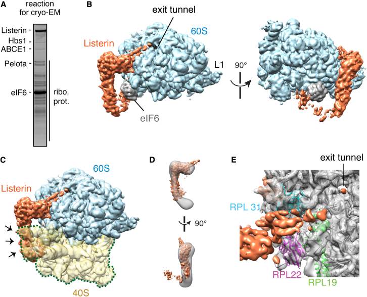
The reaction shown in Figure 6 A was incubated for 15 min at 32°C and immediately vitrified on R2/2 Cu 400 mesh grids (Quantifoil) using an FEI Vitrobot.
Automated data collection
(EPU software, FEI) was conducted on a Titan Krios operated at 300 kV at 104,478× magnification with 1-s exposures ranging in defocus values from 2 to 3.5 μm. Particles were picked with e2boxer ( Tang et al., 2007 ) and then classified and refined using Relion ( Scheres, 2012 ). For comparisons in the displayed figures, all structures were low pass filtered to 8 Å. Figures were visualized and generated using Chimera ( Pettersen et al., 2004 ).
Show full methods section
Reagents and General Biochemistry Constructs encoding
VHP-β, β, Hbs1, Hbs1-DN, Pelota, and eIF6 have been described ( Shao et al., 2013 ). Standard procedures were used to generate tagged substrate constructs and expression constructs of WT and ΔRING Listerin and ABCE1. Rabbit polyclonal antibodies against Hbs1 and ABCE1 were raised with peptide antigens (Cambridge Research Biomedicals). All other antibody reagents and in vitro translation-based methods have been described ( Hessa et al., 2011 , Mariappan et al., 2010 , Shao and Hegde, 2011 , Shao et al., 2013 , Sharma et al., 2010 , Stefanovic and Hegde, 2007 ). Details can be found in Supplemental Experimental Procedures .
Ubiquitination and Ribosome Splitting Assays
Ubiquitination reactions of F-VHP-β and splitting reactions of F-β 80S RNCs (5 nM) were at 32°C for 5–20 min with the components indicated in individual figures. Final concentrations of purified components were as follows: 50 nM splitting factors, 250 nM eIF6, 75 nM E1, 250 nM of E2 (UbcH5a), 10 μM of His- or HA-tagged ubiquitin, and 0.3 nM to 50 nM Listerin, as indicated in the individual figure legends. Ubiquitination reactions were analyzed directly by SDS-PAGE or denatured and subjected to ubiquitin pull-downs. Splitting reactions were centrifuged in a TLA120.1 rotor at 70,000 rpm for 30 min and equal amounts of the total, supernatant, and pellet fractions analyzed by SDS-PAGE.
Cryo-Electron Microscopy and Image Analysis
The reaction shown in Figure 6 A was incubated for 15 min at 32°C and immediately vitrified on R2/2 Cu 400 mesh grids (Quantifoil) using an FEI Vitrobot.
Automated data collection
(EPU software, FEI) was conducted on a Titan Krios operated at 300 kV at 104,478× magnification with 1-s exposures ranging in defocus values from 2 to 3.5 μm. Particles were picked with e2boxer ( Tang et al., 2007 ) and then classified and refined using Relion ( Scheres, 2012 ). For comparisons in the displayed figures, all structures were low pass filtered to 8 Å. Figures were visualized and generated using Chimera ( Pettersen et al., 2004 ).
Supplemental Information Document S1. Figures S1–S5, Table S1, and Supplemental Experimental Procedures Document S2. Article plus Supplemental Information
📊 Figures
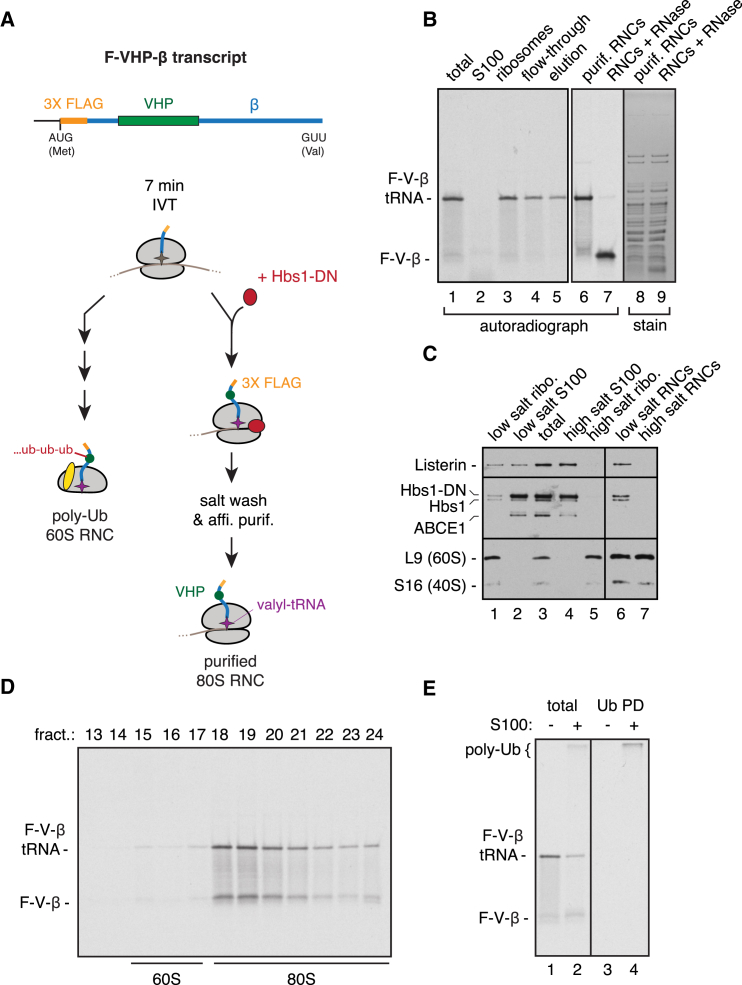
Figureu00a01
Purified Stalled 80S RNCs Are Ubiquitinated Inu00a0Vitro (A) Scheme for purifying stalled 80S RNCs housing the model substrate F-VHP-u03b2. (B) F-VHP-u03b2 was inu00a0vitro translated with 35 S-methio...
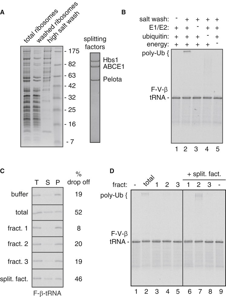
Figureu00a02
Uncoupling Ribosome Splitting and Ubiquitination Activities (A) Coomassie-stained gels of fractions from a high salt extraction of native reticulocyte ribosomes (left) and purified splitting factors (...
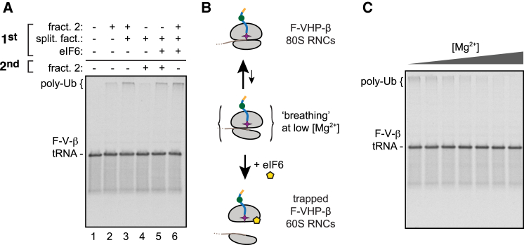
Figureu00a03
Ribosome Splitting Precedes Ubiquitination (A) Purified 35 S-labeled 80S F-VHP-u03b2 RNCs were subjected to a two-stage ubiquitination reaction with a preincubation (1 st ) for 10u00a0min with the ind...
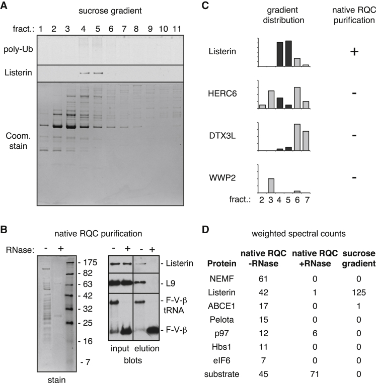
Figureu00a04
Identification of Ubiquitination Machinery (A) Fractions from the final sucrose gradient step of a purification scheme (see Figureu00a0S3 A) were analyzed for RNC ubiquitination activity (top), Lister...
Figureu00a05
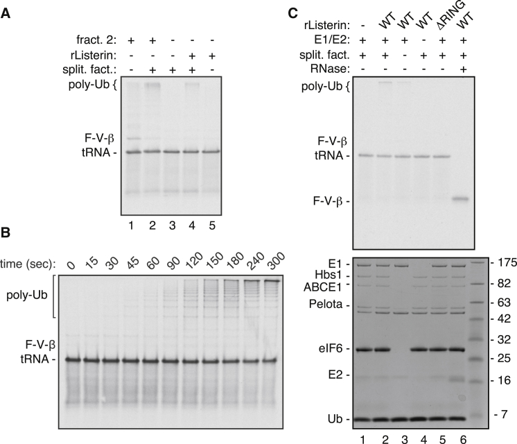
Minimal Machinery for Stalled RNC Ubiquitination (A) Ubiquitination reactions of 35 S-labeled 80S F-VHP-u03b2 RNCs with the indicated components comparing fraction 2 of the ribosome salt wash with pur...
Figureu00a06
Listerin Specifically Recognizes 60S Subunits (A) Coomassie stain of reaction used for cryo-EM analysis containing u223c120u00a0nM F-VHP-u03b2 RNCs, equimolar splitting factors (Hbs1, ABCE1, and Pelot...
Figure images are served from the NIH/NLM PubMed Central Open Access Subset or Europe PMC; copyright remains with the publishers and authors.
💬 Discussion
0 commentsNo comments yet. Be the first to start a discussion!
Leave a Comment