Abstract
Oral squamous cell carcinoma (OSCC) is the most prevalent form of cancer of lips and oral cavity, and its diagnostic delay, caused by misdiagnosis at the early stages, is responsible for high mortality ratios. Biopsy and histopathological assessment are the gold standards for OSCC diagnosis, but they are time-consuming, invasive, and do not always enable the patient's compliance, mainly in cases of follow-up with the need for more biopsies. The use of adjunctive noninvasive imaging techniques improves the diagnostic approach, making it faster and better accepted by patients. The present review aims to focus on the most consolidated diagnostic techniques, such as vital staining and tissue autofluorescence, and to report the potential role of some of the most promising innovative techniques, such as narrow-band imaging, high-frequency ultrasounds, optical coherence tomography, and in vivo confocal microscopy. According to their contribution to OSCC diagnosis, an ideal three-step diagnostic procedure is proposed, to make the diagnostic path faster, better, and more accurate.
🔬 Techniques
🧪 Sample Preparation
🔬 Cell Lines
🏭 Microscope Brands
📊 Figures
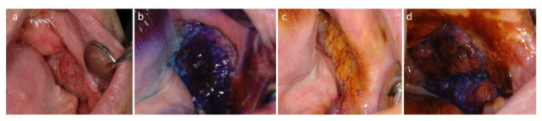
Figure 1
OSCC of the edentulous mucosa: ( a ) clinical presentation; ( b ) TB positivity (royal blue); ( c ) LI scarcely dyes the lesion, and brown-orange staining is limited to the periphery of the lesion; ( ...
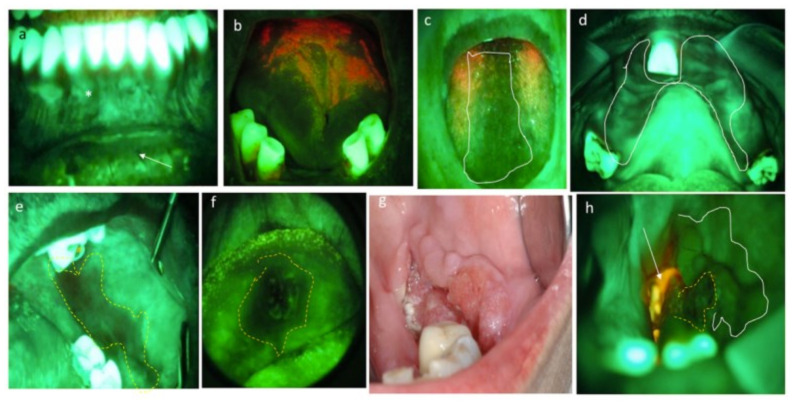
Figure 2
( a ) RF at healthy mucosa: brighter at the gingiva (masticatory mucosa) than lip mucosa (covering mucosa). ( b ) PF, in red, due to the presence of bacterial plaque. ( c , d ) Reduced fluorescence du...
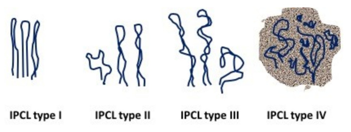
Figure 3
Oral IPCL classification according to Takano et al. [ 52 ] (Figure designed by Maria Contaldo).
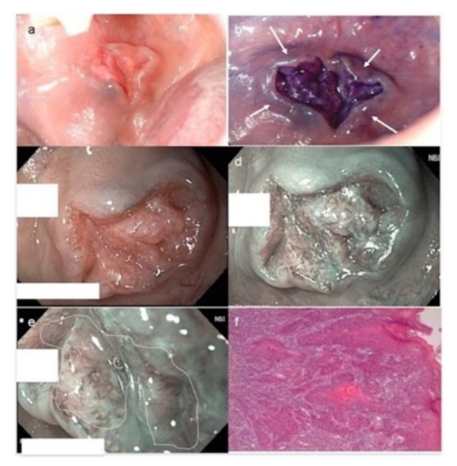
Figure 4
OSCC moderately differentiated. ( a ) Clinical photograph of a fissured exophytic lesion of the cheek. ( b ) Positivity to royal blue staining after TB test. Note bluish areas at the periphery of the ...
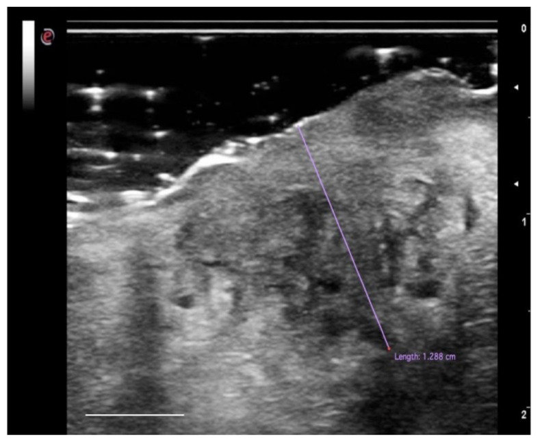
Figure 5
High-definition US imaging (frequency 18 MHz) of a suspicious lesion of the tongue margin. The high-definition ultrasound image shows a hypoechoic area with a thickness of 1.288 cm (TT), highlighted w...
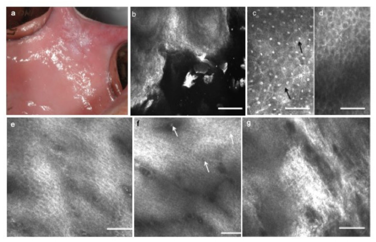
Figure 6
Leucoplakia of the cheek with dysplasia. ( a ) Clinical presentation of leucoplakia of the left cheek in a 41 year old male, smoker. ( b ) On RCM imaging, the superficial keratinization is expressed b...
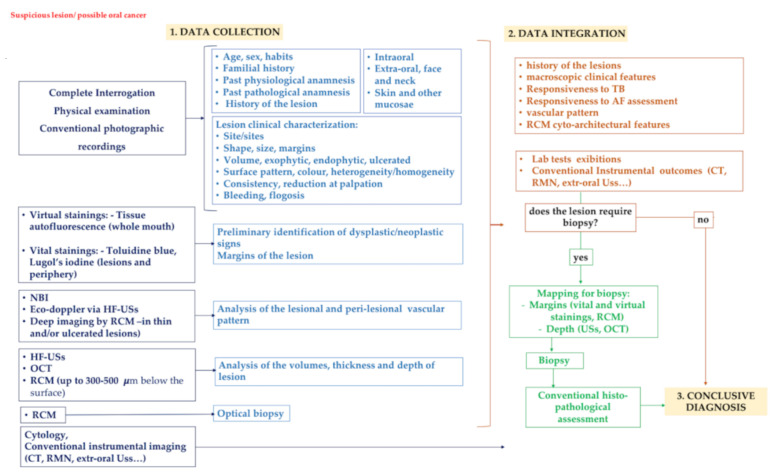
Figure 7
The proposed three-step diagnostic algorithm in oral pathology considering three consecutive phases: 1, data collection; 2, data integration; 3, conclusive diagnosis. (1) During the first phase, a ser...
Figure images are served from the NIH/NLM PubMed Central Open Access Subset or Europe PMC; copyright remains with the publishers and authors.
💬 Discussion
0 commentsNo comments yet. Be the first to start a discussion!
Leave a Comment