Abstract
Leishmania is a single-cell eukaryotic parasite of the Trypanosomatidae family, whose members cause an array of tropical diseases. The often fatal outcome of infections, lack of effective vaccines, limited selection of therapeutic drugs, and emerging resistant strains, underline the need to develop strategies to combat these pathogens. The Trypanosomatid ribosome has recently been highlighted as a promising therapeutic target due to structural features that are distinct from other eukaryotes. Here, we present the 2.8-Å resolution structure of the Leishmania donovani large ribosomal subunit (LSU) derived from a cryo-EM map, further enabling the structural observation of eukaryotic rRNA modifications that play a significant role in ribosome assembly and function. The structure illustrates the unique fragmented nature of leishmanial LSU rRNA and highlights the irregular distribution of rRNA modifications in Leishmania, a characteristic with implications for anti-parasitic drug development.
🔬 Techniques
🧬 Organisms
🏭 Microscope Brands
💻 Software Details
💾 Data Repositories
🏛️ Research Organizations (ROR)
Affiliated research institutions:
📋 Methods
91S ribosomes were purified from L. donovani promastigotes by gradual centrifugation steps that included a 1.1M sucrose cushion followed by a 15–30% sucrose gradient. The samples were vitrified and data were manually recorded on a Titan Krios electron microscope (FEI) equipped with a K2 Summit direct electron detector (Gatan, Inc.). 3D reconstruction was performed using RELION 1.3 ( Scheres, 2012 ). Model building was executed in COOT ( Emsley et al., 2010 ) and was later refined by phenix.real_space_refine implemented in PHENIX ( Afonine et al., 2013 ). See Supplemental Experimental Procedures for a detailed description of methods.
Supplementary Material 1 2
📊 Figures
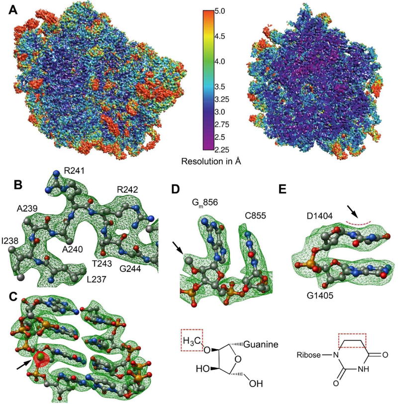
Figure 1
Cryo-EM map and modeling of L. donovani LSU
(A) Surface (left) and cross-section (right) representations of the cryo-EM density map colored according to local resolution distribution. (Bu2013C) Snapshots of map vs. model from segments of protei...
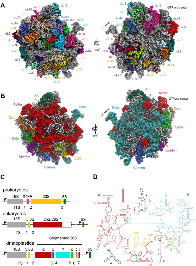
Figure 2
Structure of L. donovani LSU
(A) Protein distribution from two LSU views: solvent facing side (left) and subunit interface (right). Ribosomal proteins are shown as variably colored cartoon representations and rRNA as light grey r...
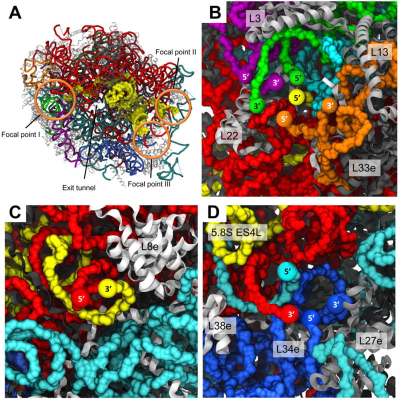
Figure 3
Focal points of LSU rRNA segmentation
26S rRNA segment terminals in the leishmanial ribosome converge to three focal points, all located on the solvent exposed side of the LSU. (A) LSU surface with localization of the three focal points i...
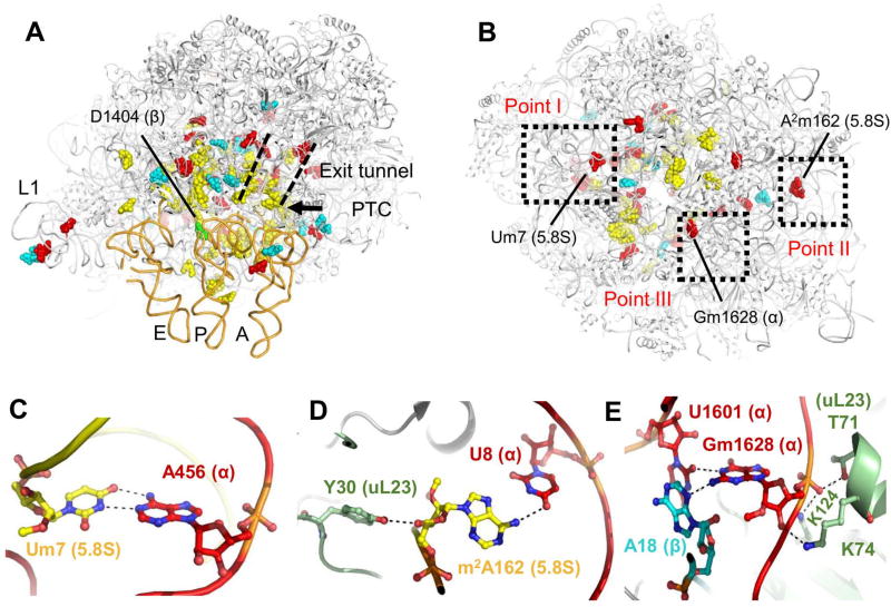
Figure 4
LSU rRNA modifications
Leishmania LSU rRNA is heavily modified by multiple 2u2019-O methyl groups and pseudouridins. Modified residues are mainly localized at functional ribosomal sites such as the PTC, the L1 stalk, the pr...
Figure images are served from the NIH/NLM PubMed Central Open Access Subset or Europe PMC; copyright remains with the publishers and authors.
💬 Discussion
0 commentsNo comments yet. Be the first to start a discussion!
Leave a Comment