Abstract
AbstractNon-steroidal anti-inflammatory drugs (NSAIDs) that act as cyclo-oxygenase (COX) inhibitors are commonly used in the treatment of a range of headache disorders, although their mechanism of action is unclear. Indomethacin is of particular interest given its very special effect in some primary headaches. Here the in vivo technique of intravital microscopy in rats has been utilised as a model of trigeminovascular nociception to study the potential mechanism of action of indomethacin. Dural vascular changes were produced using electrical (neurogenic) dural vasodilation (NDV), calcitonin gene-related peptide (CGRP) induced dural vasodilation and nitric oxide (NO) induced dural vasodilation using NO donors. In each of these settings the effect of intravenously administered indomethacin (5 mg kg−1), naproxen (30 mg kg−1) and ibuprofen (30 mg kg−1) was tested. All of the tested drugs significantly inhibited NDV (between 30 and 52%). Whilst none of them was able to inhibit CGRP-induced dural vasodilation, only indomethacin reduced NO induced dural vasodilation (35 ± 7%, 10 min post administration). We conclude NSAIDs inhibit release of CGRP after NDV without an effect on CGRP directly. Further we describe a differentiating effect of indomethacin inhibiting nitric oxide induced dural vasodilation that is potentially relevant to understanding its unique action in disorders such as paroxysmal hemicrania and hemicrania continua.
🔬 Techniques
🧪 Sample Preparation
💻 Software Details
🏛️ Research Organizations (ROR)
Affiliated research institutions:
📋 Methods
General procedures
All experiments were conducted under license of the University of California, San Francisco Institutional Animal Care and Use Committee and conforming to the National Institutes of Health Guide for the Care and Use of Laboratory Animals. For experiments male Sprague–Dawley rats (280–390 g) were anesthetized with sodium pentobarbital (60 mg kg −1 ; i.p.). The left femoral artery and veins of both sides were cannulated for blood pressure monitoring (CT-1000+ ALM 932, CWE Inc., Ardmore, PA, USA) and further administration of anesthetics (pentobarbital 25–30 mg kg −1 h −1 ) and drug administration, respectively. The animals were ventilated with oxygen enriched air, 2–3 ml, 80–100 strokes min −1 (Small rodent ventilator––Model 683, Harvard Instruments, Kent, U.K.) after cannulation with a tracheal tube. End Tidal CO 2 was monitored (Capstar––100, CWE Inc., Ardmore, PA, USA) and kept between 3.5 and 4.5%. A thermostatically controlled homeothermic blanket system was used to keep the body temperature in physiological range. The blood pressure (BP), end tidal CO 2 , and temperature were electronically displayed. Animals were fixed in a stereotactic frame for the surgery and recordings. The withdrawal reflex after paw pinch and testing of the corneal reflexes were recorded allowing monitoring of the depth of anesthesia. Data for all experiments were displayed and saved on a personal computer using an online data analysis system (Power 1401plus, CED and Spike5 software, UK). At the end of the experiments animals were euthanized by a lethal dose of pentobarbital.
Show full methods section
General procedures
All experiments were conducted under license of the University of California, San Francisco Institutional Animal Care and Use Committee and conforming to the National Institutes of Health Guide for the Care and Use of Laboratory Animals. For experiments male Sprague–Dawley rats (280–390 g) were anesthetized with sodium pentobarbital (60 mg kg −1 ; i.p.). The left femoral artery and veins of both sides were cannulated for blood pressure monitoring (CT-1000+ ALM 932, CWE Inc., Ardmore, PA, USA) and further administration of anesthetics (pentobarbital 25–30 mg kg −1 h −1 ) and drug administration, respectively. The animals were ventilated with oxygen enriched air, 2–3 ml, 80–100 strokes min −1 (Small rodent ventilator––Model 683, Harvard Instruments, Kent, U.K.) after cannulation with a tracheal tube. End Tidal CO 2 was monitored (Capstar––100, CWE Inc., Ardmore, PA, USA) and kept between 3.5 and 4.5%. A thermostatically controlled homeothermic blanket system was used to keep the body temperature in physiological range. The blood pressure (BP), end tidal CO 2 , and temperature were electronically displayed. Animals were fixed in a stereotactic frame for the surgery and recordings. The withdrawal reflex after paw pinch and testing of the corneal reflexes were recorded allowing monitoring of the depth of anesthesia. Data for all experiments were displayed and saved on a personal computer using an online data analysis system (Power 1401plus, CED and Spike5 software, UK). At the end of the experiments animals were euthanized by a lethal dose of pentobarbital.
Intravital microscopy
The carotid artery was cannulated for all experiments and used to administer the chemical dilators, CGRP and the NO donor sodium nitroprusside (SNP). It has been demonstrated previously that using the intracarotid route of administration reduces significantly the overall concentration of molecules required to induce maximal dural blood vessel dilation, while minimizing cardiovascular effects compared to the more commonly used intravenous route [ 14 ]. After exposing the skull, the parietal bone of the right or left side was thinned using a saline cooled dental drill until the underlying vessels of the dura mater were clearly visible and the skull was left intact (closed window). The area was then covered with mineral oil (37°C) and a branch of the middle meningeal artery (MMA) was then focused on and projected onto a TV monitor using an intravital microscope (Microvision MV2100, UK). On-line monitoring of the blood vessel diameter was achieved through a video dimension analyzer (Living System Instrumentation, USA) [ 17 ]. After a resting period and evaluation of a steady vessel diameter, one of the following protocols was applied. Neurogenic dural vasodilation (NDV) A bipolar stimulation electrode (NE 200, Clark Electromedical) was placed within a range of about 200 μm to the monitored vessel. The bone adjacent to the MMA was stimulated with constant settings for stimulation adjusted to 5 Hz 1 ms for 10 s (Grass Stimulator S88, Grass Instrumentation, USA). For each animal the voltage was increased until a maximal dilation was observed, the resulting current was monitored on an oscilloscope ranging between 100 and 160 μA. All following stimulations were then performed using this voltage [ 18 , 19 ]. Calcitonin gene-related peptide (CGRP)-induced dilation CGRP (1 μg kg −1 dissolved in 0.9% NaCl) was given i.a. over 3 min and demonstrated the maximal dilatory effect. Following a recovery period of 17 min, the infusion of CGRP was then repeated. By following this time pattern it was possible to ensure a sufficient recovery as well as to evaluate vasodilatory responses at the same time points as with nitric oxide induced dilation. The two vascular responses were recorded and summarized as the baseline response. After recovery from the second infusion of CGRP the control or one of the COX inhibitors was injected i.v. The pattern of CGRP-injection followed by a 17 min recovery period was then repeated for four times starting 10 min after the injection of the vehicle or drug. Nitric oxide induced dilation SNP (2 μg kg −1 min −1 dissolved in 0.9% NaCl) was given i.a. over 5 min as similar doses have shown a maximal dilatory effect [ 8 ]. This was followed by a recovery period of 15 min, the injection of SNP was then repeated. The two vascular responses were recorded and summarized as the baseline response. 15 min after the second baseline either control or one of the drugs was injected i.v. The injection of the NO donor SNP followed by a 15 min recovery period was repeated 10 min after the injection of the drug for four times. Each animal only received a single dose of a NSAID. Drugs For intravenous application indomethacin was dissolved in 0.9% NaCl solution (5 mg ml −1 ) and administered at the dose of 5 mg kg −1 . Naproxen and Ibuprofen were dissolved in 0.9% NaCl (30 mg ml −1 ) and the dose for administration was 30 mg kg −1 . An equal volume of saline was used in the control group. All solutions were pH adjusted to pH 8–8.3.
Statistics
Statistical analysis was carried out using SPSS (18.0, IL, USA). For repeated measures an analysis of variance (ANOVA) was performed. In case the assumption of sphericity was violated Greenhouse–Geisser corrections were applied. Bonferroni correction was applied for multiple comparisons if applicable. Statistical significance was set at P < 0.05. In case of P values
📊 Figures
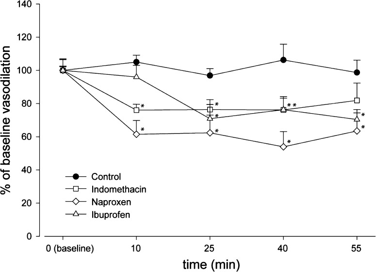
Fig.u00a01
Effect of COX inhibitors on electrical stimulation induced vasodilation of the middle meningeal artery in intravital microscopy. After the baseline response was established one of the drugs (indometha...
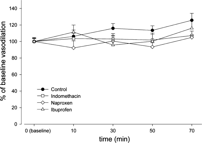
Fig.u00a02
Effect of COX inhibitors on repeated CGRP (0.33u00a0u03bcgu00a0kg u22121 min u22121 over 3u00a0min intra-arterial) induced vasodilations in intravital microscopy. After the baseline response was estab...
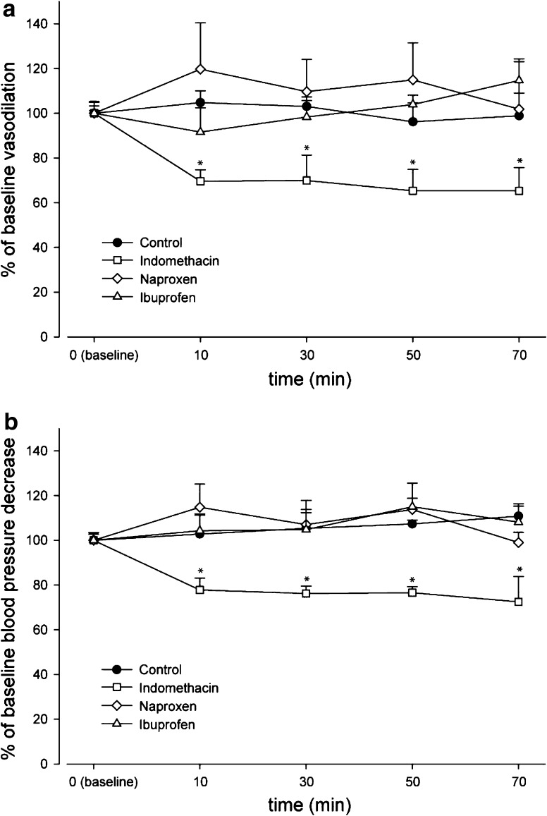
Fig.u00a03
Effect of COX inhibitors on dural vasodilation, induced by repeated sodium nitroprusside infusions, 2u00a0u03bcgu00a0kg u22121 min u22121 over 5u00a0min intra arterial, in intravital microscopy ( a ) ...
Figure images are served from the NIH/NLM PubMed Central Open Access Subset or Europe PMC; copyright remains with the publishers and authors.
💬 Discussion
0 commentsNo comments yet. Be the first to start a discussion!
Leave a Comment