Abstract
BACKGROUND: Large image datasets acquired on automated microscopes typically have some fraction of low quality, out-of-focus images, despite the use of hardware autofocus systems. Identification of these images using automated image analysis with high accuracy is important for obtaining a clean, unbiased image dataset. Complicating this task is the fact that image focus quality is only well-defined in foreground regions of images, and as a result, most previous approaches only enable a computation of the relative difference in quality between two or more images, rather than an absolute measure of quality. RESULTS: We present a deep neural network model capable of predicting an absolute measure of image focus on a single image in isolation, without any user-specified parameters. The model operates at the image-patch level, and also outputs a measure of prediction certainty, enabling interpretable predictions. The model was trained on only 384 in-focus Hoechst (nuclei) stain images of U2OS cells, which were synthetically defocused to one of 11 absolute defocus levels during training. The trained model can generalize on previously unseen real Hoechst stain images, identifying the absolute image focus to within one defocus level (approximately 3 pixel blur diameter difference) with 95% accuracy. On a simpler binary in/out-of-focus classification task, the trained model outperforms previous approaches on both Hoechst and Phalloidin (actin) stain images (F-scores of 0.89 and 0.86, respectively over 0.84 and 0.83), despite only having been presented Hoechst stain images during training. Lastly, we observe qualitatively that the model generalizes to two additional stains, Hoechst and Tubulin, of an unseen cell type (Human MCF-7) acquired on a different instrument. CONCLUSIONS: Our deep neural network enables classification of out-of-focus microscope images with both higher accuracy and greater precision than previous approaches via interpretable patch-level focus and certainty predictions. The use of synthetically defocused images precludes the need for a manually annotated training dataset. The model also generalizes to different image and cell types. The framework for model training and image prediction is available as a free software library and the pre-trained model is available for immediate use in Fiji (ImageJ) and CellProfiler.
🔬 Techniques
💻 Software
✨ Fluorophores
🧪 Sample Preparation
🔬 Cell Lines
💻 Software Details
💻 Code & Software
💾 Data Repositories
🏛️ Research Organizations (ROR)
Affiliated research institutions:
📊 Figures
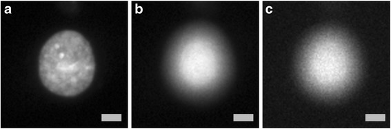
Fig. 1
The training data consists of synthetically defocused Hoechst stain images of U2OS cells. a A real in-focus image of a cell. b A real out-of-focus image of the same cell. c A synthetically defocused i...
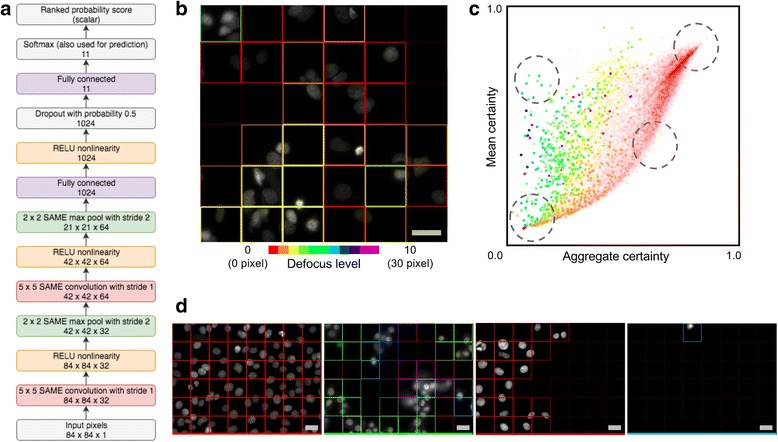
Fig. 2
a Neural network model architecture; a probability distribution over 11 discrete focus classes is predicted for each input 84u2009u00d7u200984 image patch. This distribution can be summarized (see tex...
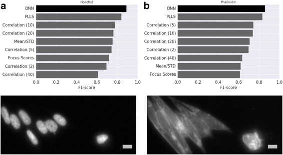
Fig. 3
Accuracy, measured with F-score, on the binary in/out-of-focus classification task compared with various methods in Bray et al. [ 2 ] for Hoechst ( a ) and Phalloidin ( b ) stained U2OS cell images. T...
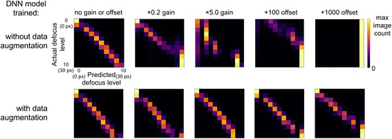
Fig. 4
Prediction of absolute focus quality on training data cell type (U2OS cells), Hoechst stain with varying image brightness and background by applying a multiplicative gain and additive offset (16-bit r...
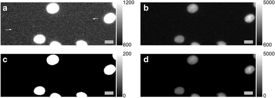
Fig. 5
Image artifact removal applied to all images from BBBC021 dataset of MCF-7 cells [ 12 ]. Example of a contrast adjusted ( a ) and unadjusted ( b ) original image with noise artifact. The artifact cons...
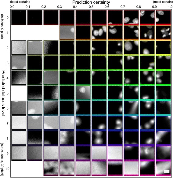
Fig. 6
Prediction of absolute focus quality on an unseen cell type (MCF-7 cells, from BBBC021 dataset [ 12 ]) but familiar stain, Hoechst. An 11u2009u00d7u200910 image montage showing sample patch-level pred...
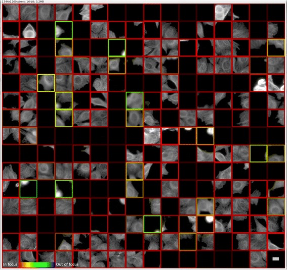
Fig. 7
Prediction of absolute focus quality on an unseen cell type (MCF-7 cells, from BBBC021 dataset [ 12 ]) and unseen stain, Tubulin, using our Fiji (ImageJ) [ 20 ] plugin with pre-trained TensorFlow mode...
Figure images are served from the NIH/NLM PubMed Central Open Access Subset or Europe PMC; copyright remains with the publishers and authors.
💬 Discussion
0 commentsNo comments yet. Be the first to start a discussion!
Leave a Comment