Abstract
We present a correlation-driven molecular dynamics (CDMD) method for automated refinement of atomistic models into cryo-electron microscopy (cryo-EM) maps at resolutions ranging from near-atomic to subnanometer. It utilizes a chemically accurate force field and thermodynamic sampling to improve the real-space correlation between the modeled structure and the cryo-EM map. Our framework employs a gradual increase in resolution and map-model agreement as well as simulated annealing, and allows fully automated refinement without manual intervention or any additional rotamer- and backbone-specific restraints. Using multiple challenging systems covering a wide range of map resolutions, system sizes, starting model geometries and distances from the target state, we assess the quality of generated models in terms of both model accuracy and potential of overfitting. To provide an objective comparison, we apply several well-established methods across all examples and demonstrate that CDMD performs best in most cases.
🔬 Techniques
🧬 Organisms
💻 Software
✨ Fluorophores
🧪 Sample Preparation
💻 Software Details
💾 Data Repositories
🏛️ Research Organizations (ROR)
Affiliated research institutions:
📋 Methods
Preparing maps and starting structures Unless differently specified, VMD ( Humphrey et al., 1996 ) and UCSF Chimera ( Pettersen et al., 2004 ) were used to perform all map and structure manipulations. We used the previously published data sets specified in Table 8 . All maps used in this study and the corresponding deposited reference models were publicly available for download from the EMDB or kindly provided by the corresponding authors. 10.7554/eLife.43542.027 Table 8. Data sets used in this study. System Resolution, Å EMDB Deposited PDB Method Citation Aldolase 2.6 8743 5VY3 Rosetta/Phenix Herzik et al. (2017) Tubulin 4.1 n/a* 3JAS, 6DPV COOT/Refmac Zhang and Nogales (2015) TRPV1 3.3 (2.5–7) 5778 3J5P COOT Liao et al. (2013) TRPV1 (TM)
📊 Figures
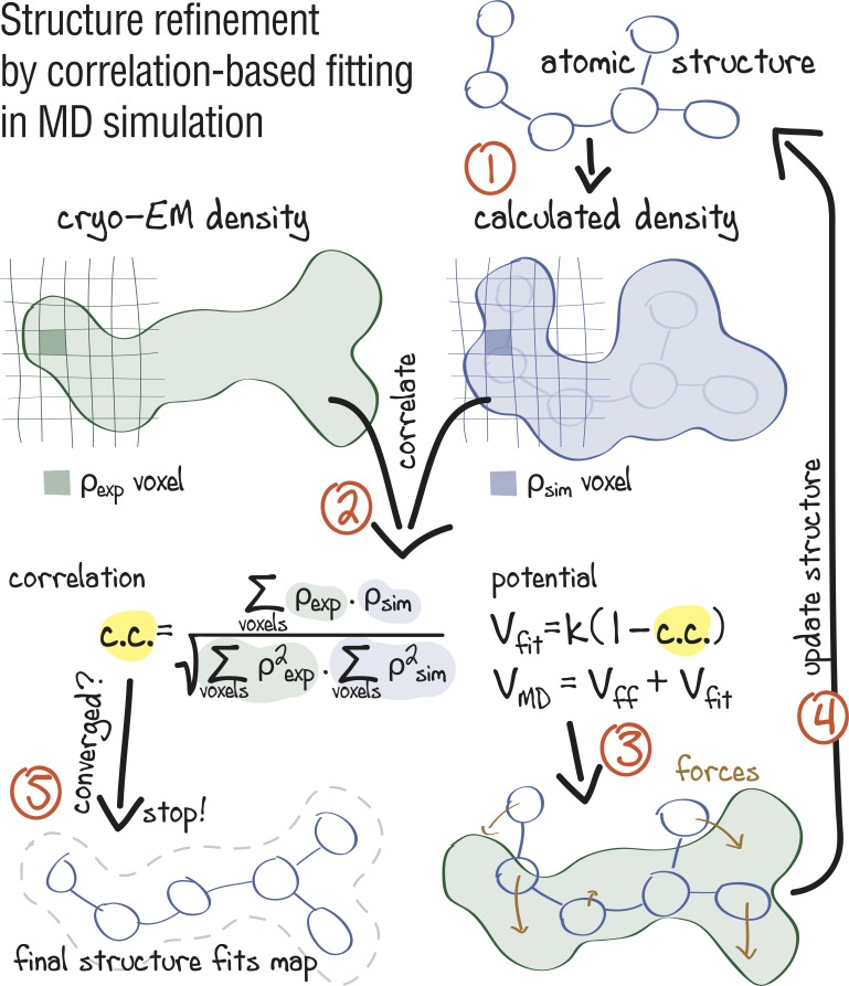
Figure 1.
Approach: using correlation-based refinement in MD simulation to steer the atomic positions of a macromolecule such that they optimally fit a cryo-EM map.
The molecule is subjected to a global biasing potential V fit in addition to the MD force field V ff . The forces resulting from V fit act on every atom to enhance the real-space correlation coefficie...
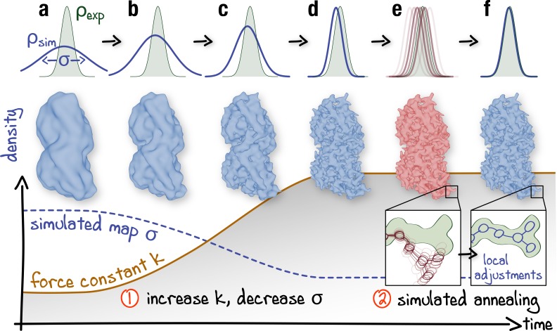
Figure 2.
Schematic representation of the proposed continuous refinement protocol: ( 1 ) a low temperature optimization phase, where V fit is monotonously increased by increasing the force constant k (columns au2013d ), followed by ( 2 ) simulated annealing (columns e, f ).
The local effect of the protocol is exemplified in the upper row for a one-dimensional single-atom case. Simulated densities shown in the middle row were generated using the atomic structure of a tubu...
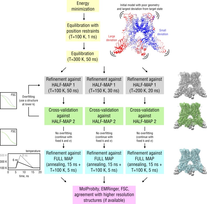
Figure 2u2014figure supplement 1.
Detailed scheme of the proposed continuous refinement protocol subdivided in five stages.
In stage 1 (yellow), the starting structure is subjected to initial equilibration in explicit solvent. Equilibration at T = 300 K for 50 ns is needed to drive the starting structure further away from ...
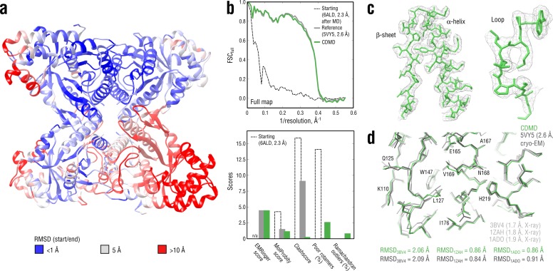
Figure 3.
Refining a distant starting model into a high-resolution map: rabbit muscle aldolase at 2.6 u00c5.
( a ) RMSD (C u03b1 atoms) between the starting and the reference model (5VY5) showing the extent of rearrangements during refinement. ( b ) Reciprocal-space agreement of the starting (black dashed), ...
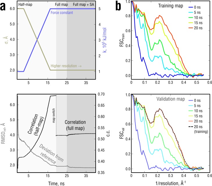
Figure 3u2014figure supplement 1.
Extension of Figure 3 showing the time evolution of various characteristics during refinement.
( a ) The simulated map resolution u03c3 and force constant k were linearly ramped from 0.6 nm and 0.5 u00d7u00a010 5 kJ mol -1 to the target values of 0.2 nm and 5 u00d7u00a010 5 kJ mol -1 , respecti...
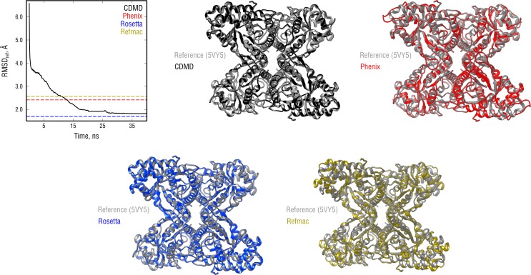
Figure 3u2014figure supplement 2.
Extension of Figure 3 showing the comparison of the radii of convergence across different refinement methods for the aldolase system and using the same distant starting structure.
RMSD to the reference structure for all methods used is shown in the upper left plot. Overlays between the reference (gray) and the refined structures are depicted as ribbons (colors as in the RMSD pl...
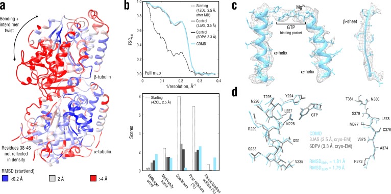
Figure 4.
Refinement of a curved tubulin dimer into a map of the straight, microtubule-like state.
( a ) RMSD (C u03b1 atoms) between the starting and the control model (6DPV) showing the extent of rearrangements between the solution (curved) and the microtubule-like (straight) tubulin conformation...
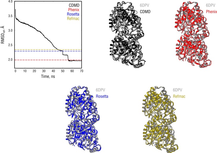
Figure 4u2014figure supplement 1.
Extension of Figure 4 showing the comparison of the radii of convergence across different refinement methods for the tubulin system and using the same distant starting structure.
RMSD to the reference structure for all methods used is shown in the upper left plot. Overlays between the control (6DPV, gray) and the refined structures are depicted as ribbons (colors as in the RMS...
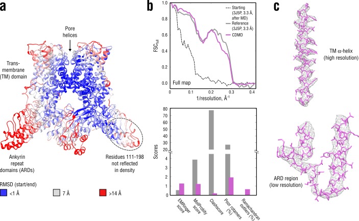
Figure 5.
Refinement of a poor structure of TRPV1 in a distant conformation into a map with highly heterogeneous local resolution.
( a ) RMSD (C u03b1 atoms) between the starting and the reference model (3J5P) showing the extent of rearrangements the TRPV1 structure undergoes during refinement. ( b ) Reciprocal-space agreement of...
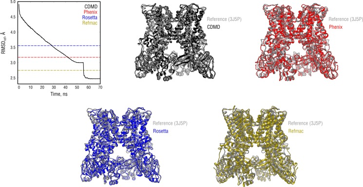
Figure 5u2014figure supplement 1.
Extension of Figure 5 showing the comparison of the radii of convergence across different refinement methods for the TRPV1 system and using the same distant starting structure.
RMSD to the reference structure for all methods used is shown in the upper left plot. Overlays between the reference (3J5P, gray) and the refined structures are depicted as ribbons (colors as in the R...
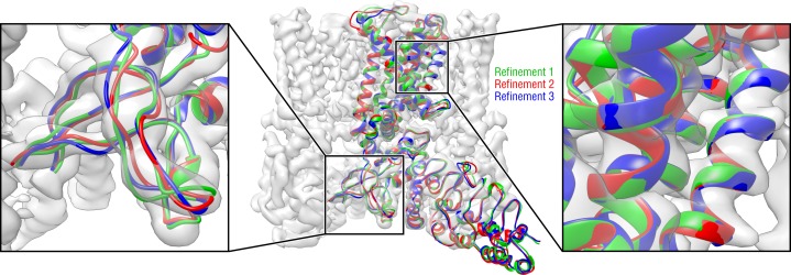
Figure 5u2014figure supplement 2.
Convergence of the TRPV1 refinement both in the higheru00a0resolution TM region and in the loweru00a0resolution ARD region assessed by means of three independent refinement runs using different but similarly distant starting structures.
Only one TRPV1 monomer and no side chains are shown for clarity.
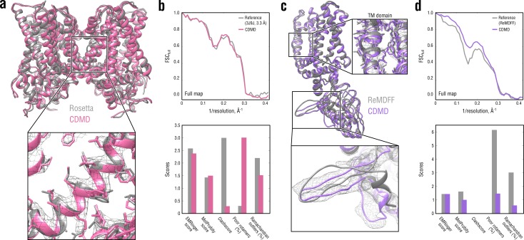
Figure 6.
Comparison of our TRPV1 model with those previously refined using Rosetta ( a,u00a0b ) and ReMDFF ( c,u00a0d ).
Overlays of our model (pink and violet ribbon) with the Rosetta (left, gray ribbon) and ReMDFF (right, gray ribbon) models are shown in ( a ) and ( c ), respectively. Reciprocal-space agreement with t...
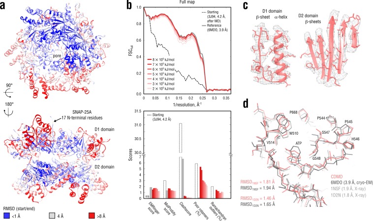
Figure 7.
Refinement of a substrate-free NSF complex in a distant conformation into a medium-resolution map at 3.9 u00c5.
( a ) RMSD (C u03b1 atoms) between the starting and the reference model (6MDO) showing the extent of rearrangements the NSF structure undergoes during refinement. ( b ) Reciprocal-space agreement with...
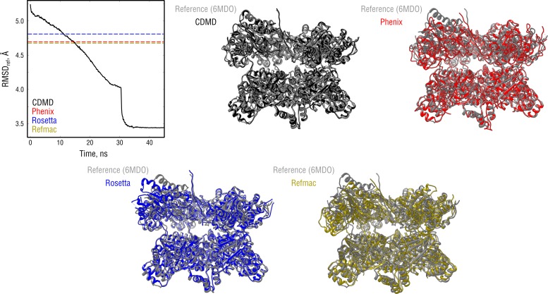
Figure 7u2014figure supplement 1.
Extension of Figure 7 showing the comparison of the radii of convergence across different refinement methods for the NSF system and using the same distant starting structure.
RMSD to the reference structure for all methods used is shown in the upper left plot. Overlays between the reference (6MDO, gray) and the refined structures are depicted as ribbons (colors as in the R...
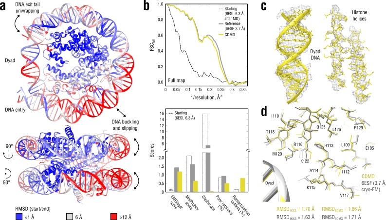
Figure 8.
Refinement of a distant nucleosome structure into a medium-resolution map of the canonical nucleosome state.
( a ) RMSD (DNA and protein backbone) between the starting and the reference model (6ESF) showing the extent of rearrangements during the refinement. ( b ) Reciprocal-space agreement of the starting (...
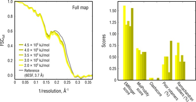
Figure 8u2014figure supplement 1.
Extension of Figure 8b showing the reciprocal-space agreement and stereochemical quality for nucleosome models independently refined using force constants ranging from 2 to 4.5 u00d7 10 5 kJ mol -1 .
The structure shown in Figure 8 was refined using k = 3 u00d7 10 5 kJ mol -1 .
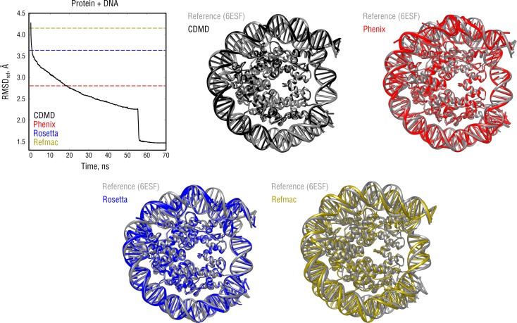
Figure 8u2014figure supplement 2.
Extension of Figure 8 showing the comparison of the radii of convergence across different refinement methods for the nucleosome system and using the same distant starting structure.
RMSD to the reference structure for all methods used is shown in the upper left plot. Overlays between the reference (6ESF, gray) and the refined structures are depicted as ribbons (colors as in the R...
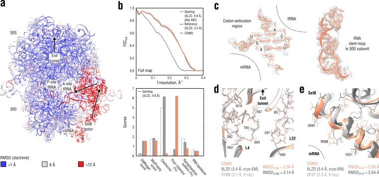
Figure 9.
Refinement of a ribosome complex in the CR state into a 3.4 u00c5 map of the GA state.
( a ) RMSD (RNA and protein backbone) between the starting (CR) and the final (GA) model showing the extent of rearrangements during the refinement. ( b ) Reciprocal-space agreement of the starting (b...
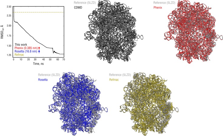
Figure 9u2014figure supplement 1.
Extension of Figure 9 showing the comparison of the radii of convergence across different refinement methods for the ribosome system and using the same distant starting structure.
RMSD to the reference structure for all methods used is shown in the upper left plot. Overlays between the reference (5LZD, gray) and the refined structures are depicted as ribbons (colors as in the R...
Figure 9u2014video 1.
Refinement trajectory for the 70S ribosome.
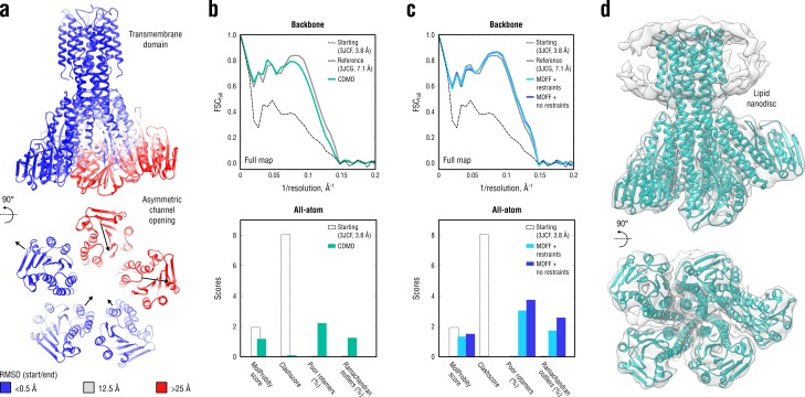
Figure 10.
Refinement of a CorA magnesium transporter in the symmetric closed state into a low-resolution 7.1 u00c5 map of the asymmetric open state.
( a ) RMSD (C u03b1 atoms) between the starting (closed) and the reference (open) model showing the extent of rearrangements in the cytosolic part of the channel during refinement. ( b ) Reciprocal-sp...
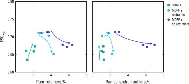
Figure 10u2014figure supplement 1.
Extension of Figure 10 showing map-model agreement vs. rotamer or Ramachandran outliers for all CDMD and MDFF refinements.
Average FSC values (FSC avg ) were calculated as described in Materials and methods. Each dot represents the result of a single independent refinement. MDFF force constants ranged from 0.05 to 0.5 (se...
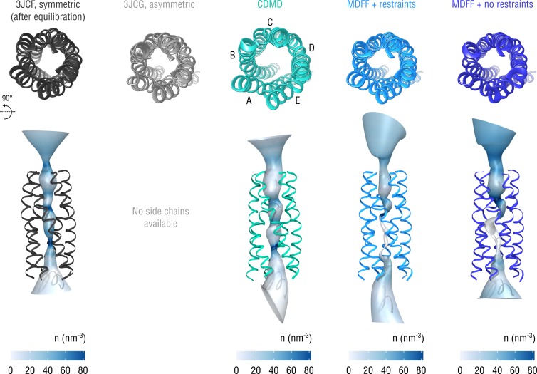
Figure 10u2014figure supplement 2.
Extension of Figure 10 showing the structure of the gating pore.
The pore transmembrane helices (residues 281u2013312) are shown for clarity.
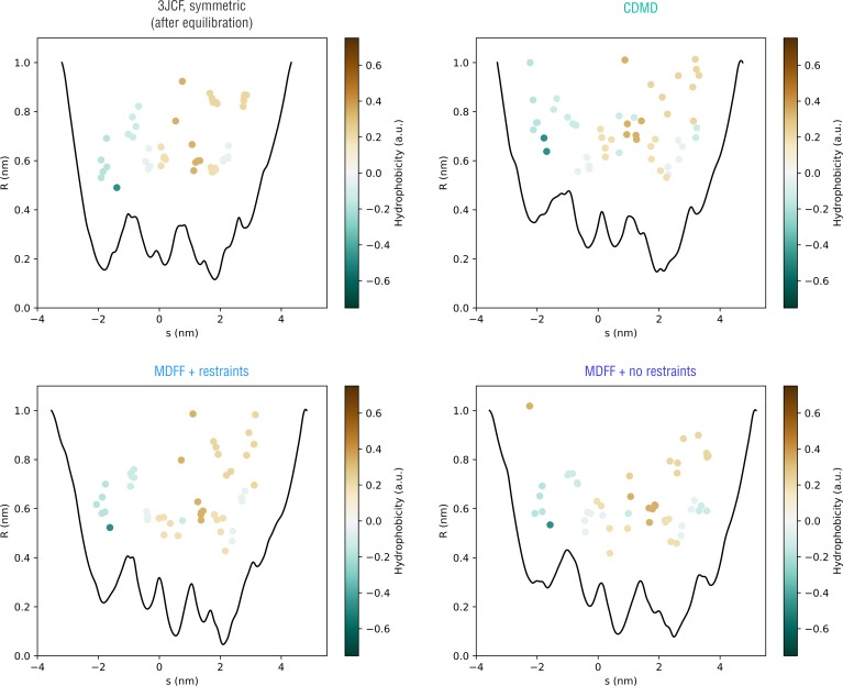
Figure 10u2014figure supplement 3.
Extension of Figure 10 showing how the pore radius changes along the nonlinear gating pathway showin in Figure 10u2014figure supplement 2 (bottom).
Residues facing the gating pathway are shown as dots color-coded by their relative hydrophobicities.
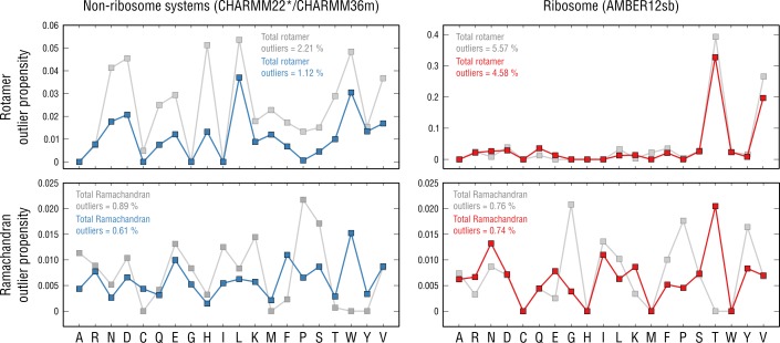
Figure 11.
Per-residueu00a0outlieru00a0analysis.
Per-residue rotamer and Ramachandran outlier propensities calculated from the non-ribosome structures (left panels; CHARMM22*/CHARMM36m force fields were used ( Piana et al., 2011 ; Huang et al., 2017...
Figure images are served from the NIH/NLM PubMed Central Open Access Subset or Europe PMC; copyright remains with the publishers and authors.
💬 Discussion
0 commentsNo comments yet. Be the first to start a discussion!
Leave a Comment