Abstract
Activation of adrenergic receptors (AR) represents the primary mechanism to increase cardiac performance under stress. Activated βAR couple to Gs protein, leading to adenylyl cyclase-dependent increases in secondary-messenger cyclic adenosine monophosphate (cAMP) to activate protein kinase A. The increased protein kinase A activities promote phosphorylation of diversified substrates, ranging from the receptor and its associated partners to proteins involved in increases in contractility and heart rate. Recent progress with live-cell imaging has drastically advanced our understanding of the βAR-induced cAMP and protein kinase A activities that are precisely regulated in a spatiotemporal fashion in highly differentiated myocytes. Several features stand out: membrane location of βAR and its associated complexes dictates the cellular compartmentalization of signaling; βAR agonist dose-dependent equilibrium between cAMP production and cAMP degradation shapes persistent increases in cAMP signals for sustained cardiac contraction response; and arrestin acts as an agonist dose-dependent master switch to promote cAMP diffusion and propagation into intracellular compartments by sequestrating phosphodiesterase isoforms associated with the βAR signaling cascades. These features and the underlying molecular mechanisms of dynamic regulation of βAR complexes with adenylyl cyclase and phosphodiesterase enzymes and the implication in heart failure are discussed.
🔬 Techniques
🧪 Sample Preparation
🔬 Cell Lines
🏛️ Research Organizations (ROR)
Affiliated research institutions:
📊 Figures
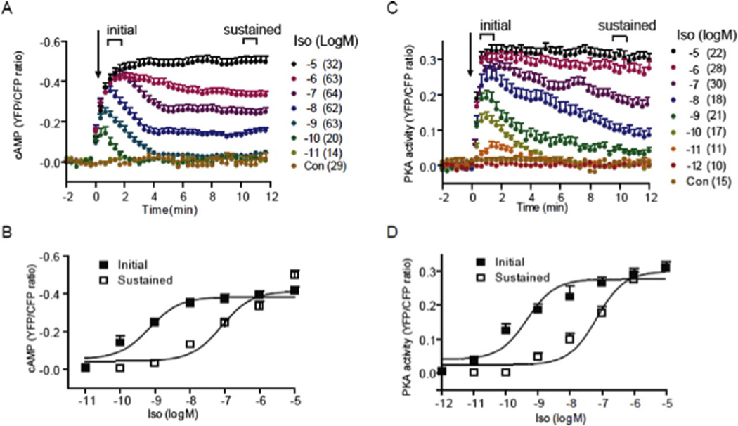
Figure 1
Activation of u03b2ARs induces a dose-dependent increase in cAMP ICUE3 and PKA AKAR3 FRET ratio in cardiac myocyte (Reprint with permission from Molecular Pharmacology). A and B, the cAMP biosensor IC...
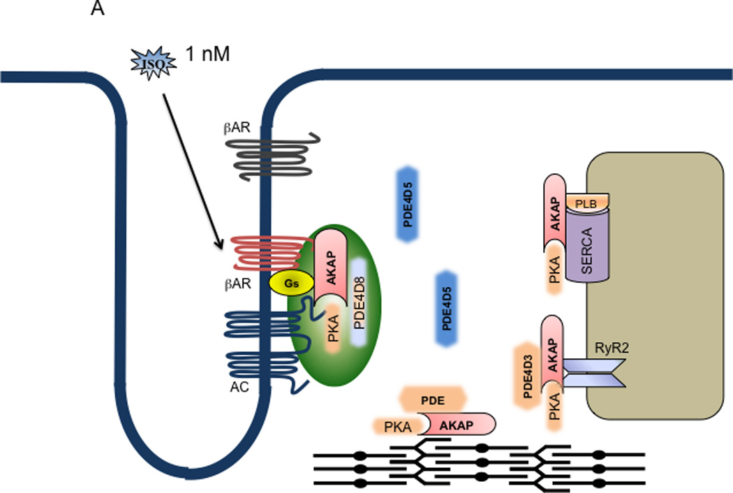
Figure 2
Model of dual mechanistic regulation of cAMP/PKA activities by AC and PDE4D under different doses of adrenergic stimulation. A. At 10 u22129 M isoproterenol, only the G-protein precoupled u03b2AR is a...
Figure images are served from the NIH/NLM PubMed Central Open Access Subset or Europe PMC; copyright remains with the publishers and authors.
💬 Discussion
0 commentsNo comments yet. Be the first to start a discussion!
Leave a Comment