Abstract
Dyneins are motor proteins responsible for transport in the cytoplasm and the beating of axonemes in cilia and flagella. They bind and release microtubules via a compact microtubule-binding domain (MTBD) at the end of a coiled-coil stalk. We address how cytoplasmic and axonemal dynein MTBDs bind microtubules at near atomic resolution. We decorated microtubules with MTBDs of cytoplasmic dynein-1 and axonemal dynein DNAH7 and determined their cryo-EM structures using helical Relion. The majority of the MTBD is rigid upon binding, with the transition to the high-affinity state controlled by the movement of a single helix at the MTBD interface. DNAH7 contains an 18-residue insertion, found in many axonemal dyneins, that contacts the adjacent protofilament. Unexpectedly, we observe that DNAH7, but not dynein-1, induces large distortions in the microtubule cross-sectional curvature. This raises the possibility that dynein coordination in axonemes is mediated via conformational changes in the microtubule.
🔬 Techniques
🧬 Organisms
💻 Software
✨ Fluorophores
🧪 Sample Preparation
🔬 Cell Lines
🏭 Microscope Brands
🧪 Reagent Suppliers
📷 Detectors
💻 Software Details
💾 Data Repositories
🏛️ Research Organizations (ROR)
Affiliated research institutions:
📋 Methods
Protein preparations 6xHis 12-heptad SRS fusion proteins were expressed in SoluBL21 Escherichia coli cells (Invitrogen) from a pet42a vector. The mouse SRS-DYNC1H1 3260-3427 construct is identical to the SRS-MTBD-85:82 used in Carter et al. (2008) . The DNAH7 MTBD and stalk was made as a synthetic gene product (EpochGene). SRS + -DNAH7 2758-2896 was made by cloning the DNAH7 sequence into SRS-DYNC1H1 3260-3427 in the place of the MTBD, delineated by the universally conserved proline residues as in Imai et al. (2015) . Cells were grown in LB media at 37°C until their OD 600 measured 0.4–0.6, at which point they were supplemented with 1 mM IPTG and grown for 16 hr at 16°C. Cultures were spun at 4000x rcf for 15 min, and used directly for purification. Both SRS constructs were purified according to the same protocol. A 1L pellet was resuspended in 50 mL Lysis buffer (50 mM Tris pH8.0, 100 mM NaCl, 1 mM MgCl 2 , 10% Glycerol, 10 mM Imidazole pH8.0, 1 mM DTT, 2 mM PMSF) and lysed by sonication. The lysate was centrifuged at 30,000x rcf in a Ti70 rotor (Beckman) for 30 min and at 4°C. The supernatant was loaded onto a 5 mL NiNTA HisTrap HP Column (GE), washed with 10 column volumes of 10% elution buffer (Lysis buffer with 500 mM Imidazole pH 8.0 and without PMSF) and eluted with a step gradient to 40% elution buffer. Peak fractions were pooled and concentrated in a 15 mL 30kMWCO centrifugal concentrator (Amicon) to a concentration of ~5 mg/mL. Aliquots were snap frozen in liquid nitrogen. ZZ-tagged human cytoplasmic dynein one motor domain (DYNC1H1 1230-4646 ) was cloned into pFastBac and expressed in Sf9 insect cells as in Schmidt et al. (2015) . A 1L pellet was resuspended in 50 mL ZZ-Lysis buffer (as above but without imidazole) and dounce homogenised with 30 strokes. Lysate was centrifuged at 50,0000x rcf in a Ti70 rotor (Beckman) for 60 min and at 4°C. Supernatant was mixed with 2 mL IgG Sepharose 6 Fast Flow resin (GE, equilibrated in ZZ-lysis buffer) on a horizontal roller for 2 hr at 4°C. The mixture was applied to a gravity flow column, and the resin was washed with 150 mL ZZ-Lysis buffer and 150 mL TEV buffer (50 mM Tris pH 7.4, 150 mM KOAc, 2 mM MgAc, 1 mM EGTA, 10% Glycerol, 1 mM DTT). The resin was resuspended in 5 mL TEV buffer, supplemented with 0.1 mg/mL TEV protease and incubated on a horizontal roller at 25°C for 80 min. The sample was reapplied to a gravity flow column, the eluate was collected and concentrated to 6 mg/mL with a 15 mL 100kMWCO centrifugal concentrator (Amicon) and snap frozen in aliquots. Aliquots of each sample were gel filtered prior to each grid freezing session. Thawed sample was spun through a 0.22 um spin filter (Amicon) to remove aggregates and loaded onto a Superose 6 10/300 gel filtration column (GE) equilibrated in GF buffer (25 mM Tris pH8.0, 50 mM NaCl, 1 mM MgCl 2 , 1 mM DTT). Peak fractions were pooled and concentrated in a 4 mL 30MWCO Amicon centrifugal concentrator to 1/10 th of the original volume. The sample was then diluted fivefold in salt-free GF buffer (i.e. without 50 mM NaCl) and reconcentrated. This was repeated twice, resulting in a 25-fold dilution of the NaCl. The sample was further diluted to a final concentration of 2 mg/mL to be used for grid freezing. Lyophilised tubulin was resuspended in MES-NaCl buffer (25 mM MES pH6.5, 70 mM NaCl, 1 mM MgCl 2 , 1 mM DTT) to a concentration of 10 mg/mL and snap frozen in aliquots. For polymerisation, an aliquot was thawed and mixed 1:1 with MES-NaCl buffer supplemented with 6 mM GTP, and incubated at 37°C for 2 hr. 100 μL MES-NaCl buffer supplemented with 20 μM Taxol and pre-warmed to 37°C was added, and the sample was left at room temperature overnight. Before use, the microtubules were spun at 20,000x rcf for 10 min, and resuspended in MES-NaCl buffer with taxol. Grid preparation Quantifoil R1.2/1.3 Au300 grids were glow-discharged for 40 s. 4 μL 0.4 mg/mL microtubules was added to the grid and incubated at room temperature for 1 min. This was removed by side blotting, 4 μL of dynein was added and the grid was incubated for a further 2 min. Manual side blotting was repeated, and after the second MTBD application the grid was taken into the humidity chamber of a Vitrobot Mark II set to 100% humidity and 22°C. After 2 min, the grid was double-side blotted for 4 s and plunged into liquid ethane.
Show full methods section
Protein preparations 6xHis 12-heptad SRS fusion proteins were expressed in SoluBL21 Escherichia coli cells (Invitrogen) from a pet42a vector. The mouse SRS-DYNC1H1 3260-3427 construct is identical to the SRS-MTBD-85:82 used in Carter et al. (2008) . The DNAH7 MTBD and stalk was made as a synthetic gene product (EpochGene). SRS + -DNAH7 2758-2896 was made by cloning the DNAH7 sequence into SRS-DYNC1H1 3260-3427 in the place of the MTBD, delineated by the universally conserved proline residues as in Imai et al. (2015) . Cells were grown in LB media at 37°C until their OD 600 measured 0.4–0.6, at which point they were supplemented with 1 mM IPTG and grown for 16 hr at 16°C. Cultures were spun at 4000x rcf for 15 min, and used directly for purification. Both SRS constructs were purified according to the same protocol. A 1L pellet was resuspended in 50 mL Lysis buffer (50 mM Tris pH8.0, 100 mM NaCl, 1 mM MgCl 2 , 10% Glycerol, 10 mM Imidazole pH8.0, 1 mM DTT, 2 mM PMSF) and lysed by sonication. The lysate was centrifuged at 30,000x rcf in a Ti70 rotor (Beckman) for 30 min and at 4°C. The supernatant was loaded onto a 5 mL NiNTA HisTrap HP Column (GE), washed with 10 column volumes of 10% elution buffer (Lysis buffer with 500 mM Imidazole pH 8.0 and without PMSF) and eluted with a step gradient to 40% elution buffer. Peak fractions were pooled and concentrated in a 15 mL 30kMWCO centrifugal concentrator (Amicon) to a concentration of ~5 mg/mL. Aliquots were snap frozen in liquid nitrogen. ZZ-tagged human cytoplasmic dynein one motor domain (DYNC1H1 1230-4646 ) was cloned into pFastBac and expressed in Sf9 insect cells as in Schmidt et al. (2015) . A 1L pellet was resuspended in 50 mL ZZ-Lysis buffer (as above but without imidazole) and dounce homogenised with 30 strokes. Lysate was centrifuged at 50,0000x rcf in a Ti70 rotor (Beckman) for 60 min and at 4°C. Supernatant was mixed with 2 mL IgG Sepharose 6 Fast Flow resin (GE, equilibrated in ZZ-lysis buffer) on a horizontal roller for 2 hr at 4°C. The mixture was applied to a gravity flow column, and the resin was washed with 150 mL ZZ-Lysis buffer and 150 mL TEV buffer (50 mM Tris pH 7.4, 150 mM KOAc, 2 mM MgAc, 1 mM EGTA, 10% Glycerol, 1 mM DTT). The resin was resuspended in 5 mL TEV buffer, supplemented with 0.1 mg/mL TEV protease and incubated on a horizontal roller at 25°C for 80 min. The sample was reapplied to a gravity flow column, the eluate was collected and concentrated to 6 mg/mL with a 15 mL 100kMWCO centrifugal concentrator (Amicon) and snap frozen in aliquots. Aliquots of each sample were gel filtered prior to each grid freezing session. Thawed sample was spun through a 0.22 um spin filter (Amicon) to remove aggregates and loaded onto a Superose 6 10/300 gel filtration column (GE) equilibrated in GF buffer (25 mM Tris pH8.0, 50 mM NaCl, 1 mM MgCl 2 , 1 mM DTT). Peak fractions were pooled and concentrated in a 4 mL 30MWCO Amicon centrifugal concentrator to 1/10 th of the original volume. The sample was then diluted fivefold in salt-free GF buffer (i.e. without 50 mM NaCl) and reconcentrated. This was repeated twice, resulting in a 25-fold dilution of the NaCl. The sample was further diluted to a final concentration of 2 mg/mL to be used for grid freezing. Lyophilised tubulin was resuspended in MES-NaCl buffer (25 mM MES pH6.5, 70 mM NaCl, 1 mM MgCl 2 , 1 mM DTT) to a concentration of 10 mg/mL and snap frozen in aliquots. For polymerisation, an aliquot was thawed and mixed 1:1 with MES-NaCl buffer supplemented with 6 mM GTP, and incubated at 37°C for 2 hr. 100 μL MES-NaCl buffer supplemented with 20 μM Taxol and pre-warmed to 37°C was added, and the sample was left at room temperature overnight. Before use, the microtubules were spun at 20,000x rcf for 10 min, and resuspended in MES-NaCl buffer with taxol. Grid preparation Quantifoil R1.2/1.3 Au300 grids were glow-discharged for 40 s. 4 μL 0.4 mg/mL microtubules was added to the grid and incubated at room temperature for 1 min. This was removed by side blotting, 4 μL of dynein was added and the grid was incubated for a further 2 min. Manual side blotting was repeated, and after the second MTBD application the grid was taken into the humidity chamber of a Vitrobot Mark II set to 100% humidity and 22°C. After 2 min, the grid was double-side blotted for 4 s and plunged into liquid ethane.
Cryo-electron microscopy
Cytoplasmic dynein 1 MTBD-SRS grids were imaged on our in-house Titan Krios microscope, and DNAH7 MTBD grids were imaged on Krios III at Diamond eBIC. For cytoplasmic dynein, 1995 1.5 s exposures were collected with a pixel size of 1.04 Å 2 and a flux of 40e - /Å 2 s on a Falcon III detector in linear mode. For DNAH7, 4641 1.5 s exposures were collected with a pixel size of 1.085 Å 2 and a flux of 45e - /Å 2 s. Dynein motor domain decorated microtubules were imaged on a Polara microscope, with 2455 1.5 s exposures collected with a pixel size of 1.34 Å 2 and a flux of 37e - /Å 2 s on a Falcon III detector in linear mode. In each case, images were acquired with a defocus ranging between −1.5 μm and −4.5 μm semi-automatically in EPU.
Image processing
All processings were performed inside the Relion 3.0 pipeline ( Zivanov et al., 2018 ). Details are given for processing the SRS-DYNC1H1 3260-3427 data, followed by modifications to this workflow used for the other datasets. The unaligned raw movies were aligned and dose weighted in Relion’s implementation of MotionCorr2 using 4 × 4 patches ( Zheng et al., 2017 ). CTF determination was performed with Gctf on dose-weighted micrographs ( Zhang, 2016 ). Manual picking and 2D classification was performed to generate references for autopicking. Start and end coordinates of 30 microtubules from five micrographs were extracted into 82 Å segments (box size 512), resulting in ~650 particles which were classified into five classes. These were used as references for autopicking on all the micrographs, using the following parameters: mask diameter 497 Å, in-plane angular sampling 1°, lowpass references 20 Å, picking threshold 0.04, minimum inter-particle distance 79 Å, maximum stddev noise 1.4, shrink factor 0.5, helical picking, tube diameter 400 Å, helical rise 82 Å, number of asymmetrical units 1, maximum curvature 0.4. Particles were extracted (4x binned) and entered for 2D classification into 100 classes. Classes were rejected if they were obviously not microtubules (carbon, ice etc), if they appeared blurred or poorly aligned, if they had low levels of decoration and if they showed signs of non-13-PF architectures ( Figure 1—figure supplement 1C ). A 3D reference was made by docking a model of dynein MTBD decorated tubulin into density for a 13-PF microtubule (PDB 3J1T and EMD 6351 respectively). The PDB was converted to electron density in EMAN2 ( pdb2mrc) . 3D classification of unbinned particles into three classes was used to separate out the remaining sample heterogeneity. The single good class was entered into a 3D refinement using the following parameters: initial angular sampling of 0.9°, and initial offset range and step sizes of 3 and 1 pixels, respectively. C1 symmetry, inner tube diameter 100 Å, outer tube diameter 400 Å, angular search range tilt 15°, psi 10°, tilt prior fixed, range factor of local averaging 4, helical symmetry with one asymmetric unit, initial rise 82 Å, initial twist 0°, central Z length 40%, local searches of symmetry, rise search 78–86 Å, step size 1 Å, no search for twist. A solvent mask and 13-fold local symmetry were applied during refinement. For local symmetry, a mask was made by docking copies of PDB 3J1T into the protofilament to the left of the seam (if the MT is being viewed plus-end up). The PDB protofilament was then converted to electron density with the EMAN program pdb2mrc . This was converted into a mask with relion_mask_create. relion_local_symmetry requires a STAR file containing the translational and rotational operators needed to move the original mask onto each successive protofilament. The psi angle, rotating around the microtubule long axis, is given as multiples of −27.69° (360°/13). The centre of rotation is the centre of the microtubule lumen, so the only translation needed is the rise between adjacent protofilaments. For a three-start helix, there is a rise of 1.5 dimers through 360°. The refined helical rise between dimers in the same protofilament as measured by Relion was 82.29 Å. As such, the _rlnOriginZ parameter increases by multiples of 9.495 Å (82.293 * 1.5/13). Local symmetry was applied during refinement with the additional argument --local_symmetry. Following completion of refinement, local symmetry was applied to both unfiltered half maps. Postprocessing and resolution assessment was performed with three tubulin dimers docked along a single protofilament as previously ( Zhang et al., 2015 , Zhang et al., 2018 ). Refinement of the dynein motor domain also followed this protocol. For the DNAH7 structure, initial 3D classification did not result in a coherent class. Instead all good particles following 2D classification were entered into 3D refinement, resulting in a map with blurred features. Following this, 3D classification into eight classes using the orientations used in the refinement (i.e. with no image alignment) was performed. Local symmetry operators were found for the resulting map with the search command in relion_localsym (see https://www2.mrc-lmb.cam.ac.uk/relion/index.php?title=Local_symmetry ). The seam was less well defined in the DNAH7 structure, presumably due to local curvature being a stronger feature than seam position in some particles during refinement. As a result, three seam-adjacent profilaments were not included for symmetrisation. For DNAH7 MTBD sub-classification, signal subtraction was performed on the refined DNAH7 structure. Using the refined DNAH7 model to make a mask, everything but one MTBD and the two tubulin dimers it contacts was subtracted from the raw particles. This was performed for two protofilament pairs (6/7 and 8/9). These particles were subjected to masked 3D classification without alignments (15 classes, T = 100, 25 iterations, limit resolution E-step to 15 Å). For classes A and B, another 3D classification (eight classes, T = 20, 25 iterations, 0.9° local angular searches) was performed with the original unsubtracted particles. The EB3 dataset was downloaded from EMPIAR (ID 10030) and processed as for the dynein MTBD with modifications. 3D classification was skipped since the microtubules in this dataset almost exclusively have 13 protofilaments ( Zhang and Nogales, 2015 ). EB3 does not bind across the seam, which means that applying regular 13-fold symmetry was not appropriate. A separate mask was created for the tubulin and EB3 densities. The tubulin mask and EB3 masks were applied with 13- and 12-fold symmetry, respectively. Local resolution estimation was performed in relion_postprocess . Model building For cytoplasmic dynein 1 a low-affinity crystal structure (PDB 3ERR) was used as the starting model for refinement. For human DNAH7, a sequence alignment to C. reinhardtii flagellar dynein c was used to generate a homology model to the low-affinity NMR structure (PDB 2RR7) in Modeller ( Sali and Blundell, 1993 ). The homology model was used as an initial model. The models were fit in their respective maps using Chimera ( Pettersen et al., 2004 ). A tubulin dimer was also docked in to the density using PDB 5SYF (for SRS + -DNAH7 2758-2896 the tubulin dimer being contacted by the flap was added as well). Coot Real Space refine zone ( Emsley and Cowtan, 2004 ) was used to manually fit the model to the density, followed by whole model refinement using Refmac5 in the CCP-EM suite ( Brown et al., 2015 ; Burnley et al., 2017 ). These two steps were performed iteratively until the model to map measures were maximised. For model to map FSC curves ( Figure 2—figure supplement 2B ), phenix.mtriage was used ( Adams et al., 2010 ). All model visualisations were performed in Chimera ( Moores et al., 2004 ).
Analysis of microtubule distortion
Protofilament angles were measured by docking a tubulin dimer pdb model (5SYF) into two adjacent protofilaments of the relevant reconstruction in Chimera. The relative rotation was measured with the ‘measure rotation’ command. Ellipticity was measured with the Matlab fit_ellipse script deposited in the Mathworks file exchange ( https://www.mathworks.com/matlabcentral/fileexchange/3215-fit_ellipse ) . x,y coordinates for each protofilament were obtained from maximum intensity projections of each class, binarised with the same threshold in FIJI ( Schindelin et al., 2012 ). The centre of mass of each protofilament was used as the coordinate. Decoration level was determined by the volume of the zoned MTBD density in chimera at set thresholds.
Additional files 10.7554/eLife.47145.021 Transparent reporting form
📊 Figures
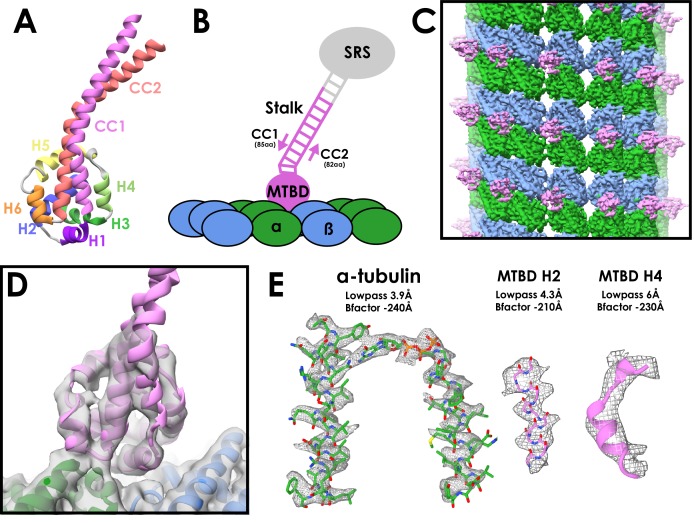
Figure 1.
Cryo-EM Structure of the cytoplasmic dynein-1 microtubule-binding domain.
( A )u00a0Crystal structure of the cytoplasmic dynein-1 MTBD in the low-affinity u03b2u00a0+registry (PDB 3ERR) coloured by helix. ( B ) Schematic of the MTBD constructs used for structure determinati...
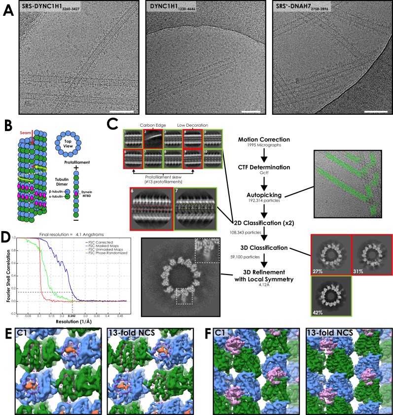
Figure 1u2014figure supplement 1.
Processing workflow of the SRS-DYNC1H1 3260-3427 structure.
( A ) Representative cryo-EM images from each of the three datasets presented in this work.u00a0White bar corresponds to 100 nm ( B ) Cartoon depicting a 13-protofilament microtubule with a seam. u03b...
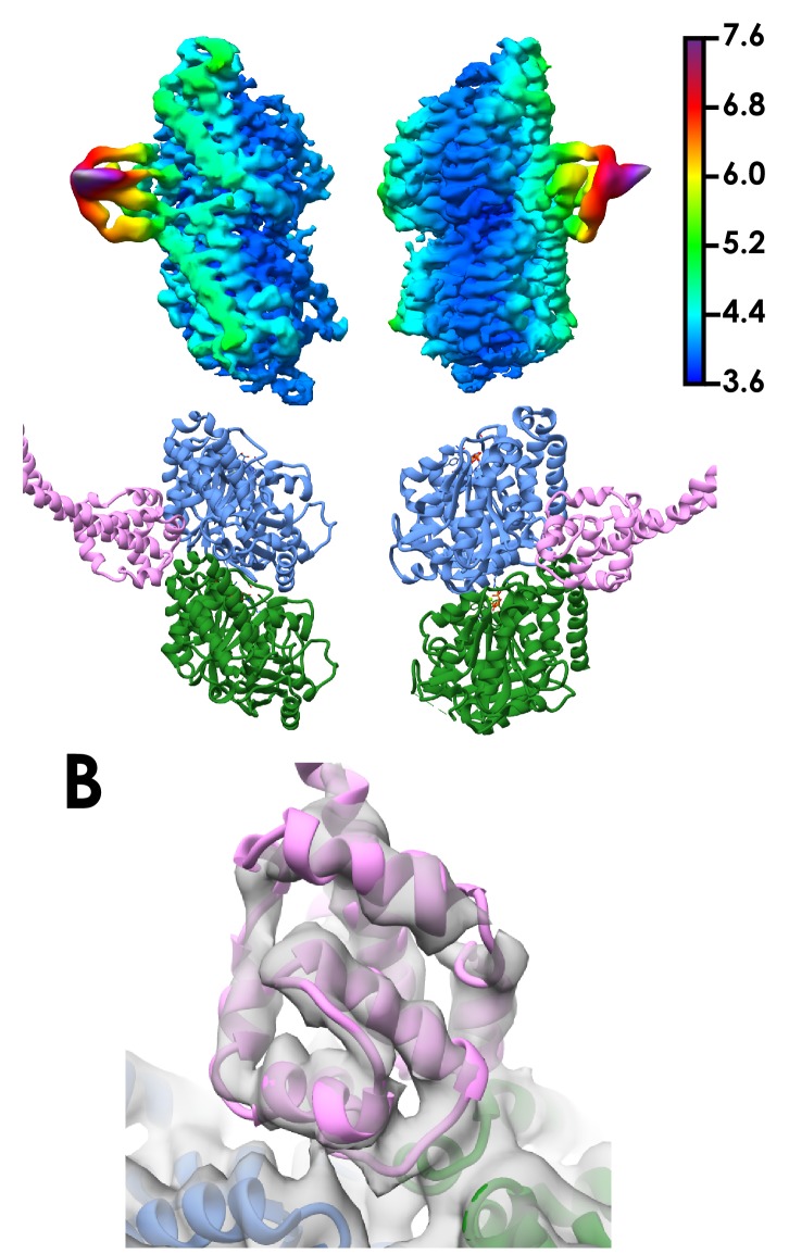
Figure 1u2014figure supplement 2.
Local resolution of the cytoplasmic dynein-1 MTBD SRS structure.
( A ) Density corresponding to one tubulin dimer and the cytoplasmic dynein-1 MTBD coloured and filtered according to local resolution as determined in relion_postprocess.u00a0Density (top) and the co...
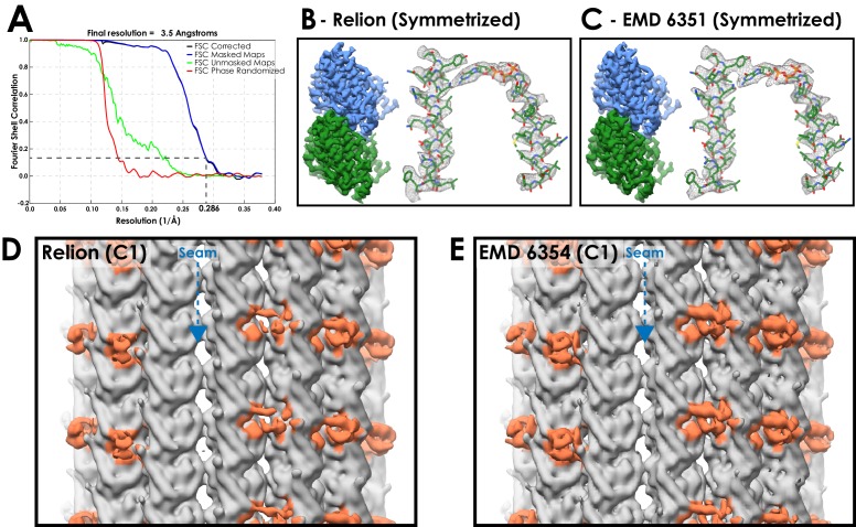
Figure 1u2014figure supplement 3.
A comparison between microtubule reconstructions using Relion or previous methods.
( A ) FSC curves for EMPIAR dataset 10030 (EB3 decorated microtubules) processed in the Relion pipeline following symmetrization (FSC 0.143 cut-off, gold-standard).u00a0( B ) Density from the Relion r...
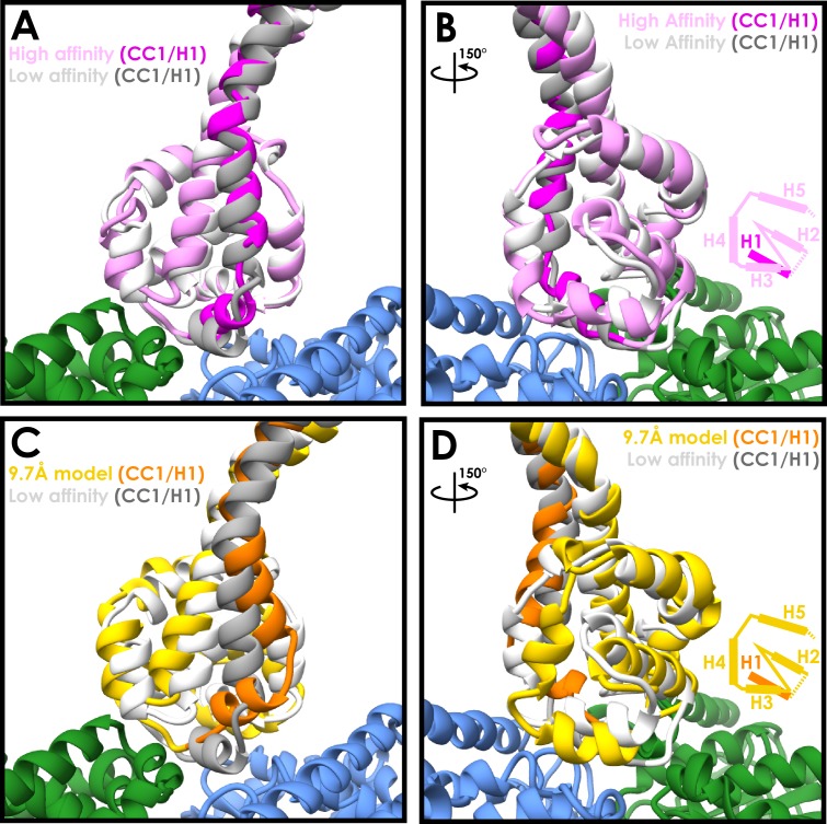
Figure 2.
Similarities between the high- and low-affinity states of the cytoplasmic dynein-1 MTBD.
( A ) A comparison between the newly refined cytoplasmic dynein-1 MTBD model (pink) and the low-affinity state crystal structure (PDB 3ERR, docked to the same density, white).u00a0CC1 and H1 (highligh...
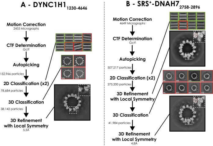
Figure 2u2014figure supplement 1.
Processing pipeline for DYNC1H1 1230-4646 andu00a0SRS + -DNAH7 2758-2896 structures.
( A ) Processing pipeline for DYNC1H1 1230-4646 structure.u00a0( B ) Processing pipeline for SRS + -DNAH7 2758-2896 structure. 3D classification directly after 2D classification did not result in a go...
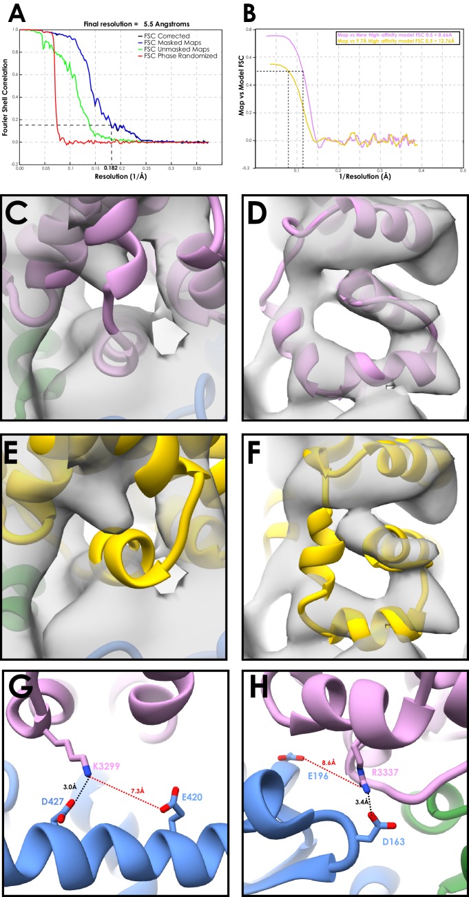
Figure 2u2014figure supplement 2.
Validation of the SRS-DYNC1H1 3260-3427 model with the dynein motor domain.
( A ) FSC curve for the DYNC1H1 1230-4646 decorated microtubule structure (FSC 0.143 cut-off, gold-standard).u00a0( B ) Model to map FSC curves for our new high-affinity cytoplasmic dynein-1 model or ...
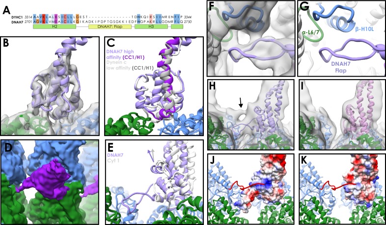
Figure 3.
Structure of the DNAH7 MTBD.
( A ) Partial sequence alignment of human cytoplasmic dynein-1 (DYHC1) and human Axonemal Dynein 7 (DNAH7).u00a0DNAH7 has a 13-residue insert between H2 and H3 compared to cytoplasmic dynein called th...
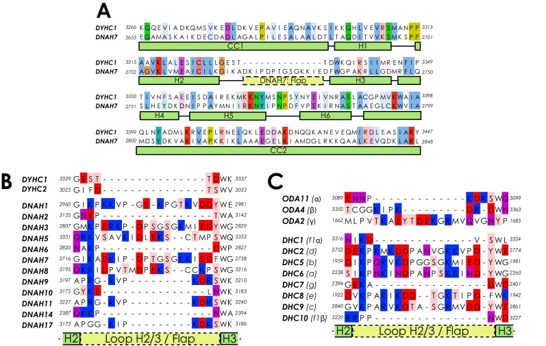
Figure 3u2014figure supplement 1.
Sequence alignments of dynein microtubule-binding domains.
( A ) Full sequence alignment for the MTBD of human cytoplasmic dynein-1 (DYHC1) and human DNAH7.u00a0( B ) A sequence alignment of the region between H2 and H3 of the MTBDs of the human dynein heavy ...
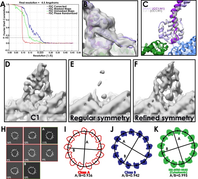
Figure 3u2014figure supplement 2.
Assessment of the DNAH7 MTBD structure.
( A ) FSC curve for the final, symmetrized (with refined operators) DNAH7 map (FSC 0.143 cut-off, gold-standard).u00a0( B ) The refined DNAH7 model docked into density, at a low threshold to visualise...
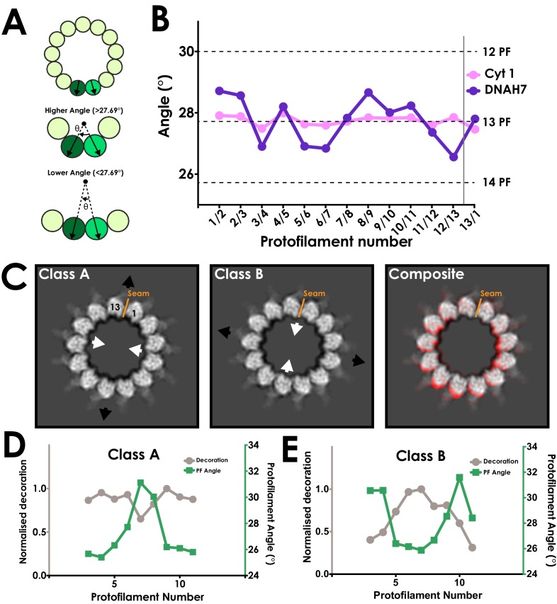
Figure 4.
DNAH7u00a0binding causes cross-sectional deformations in the microtubule.
( A ) Schematic representing measurement of protofilament angles.u00a0A PDB model for tubulin (PDB 5SYF) was docked into adjacent protofilaments, and the rotation to superimpose the two models was mea...
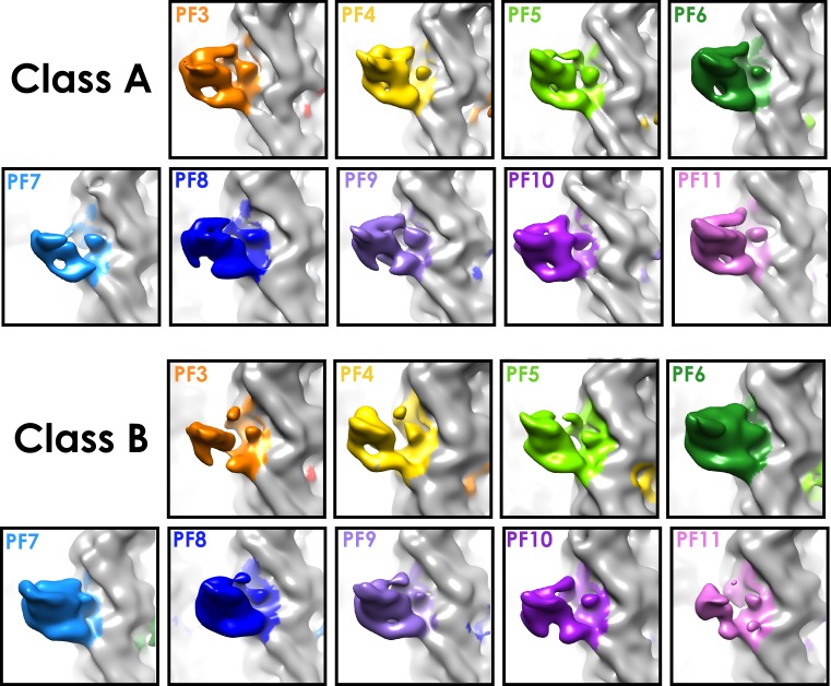
Figure 4u2014figure supplement 1.
Decoration levels in classes A and B.
Density for each protofilament used for decoration measurements in Figure 4D and E .
Video 1.
A morph between Classes A and B, depicting their cross-sectional distortion.
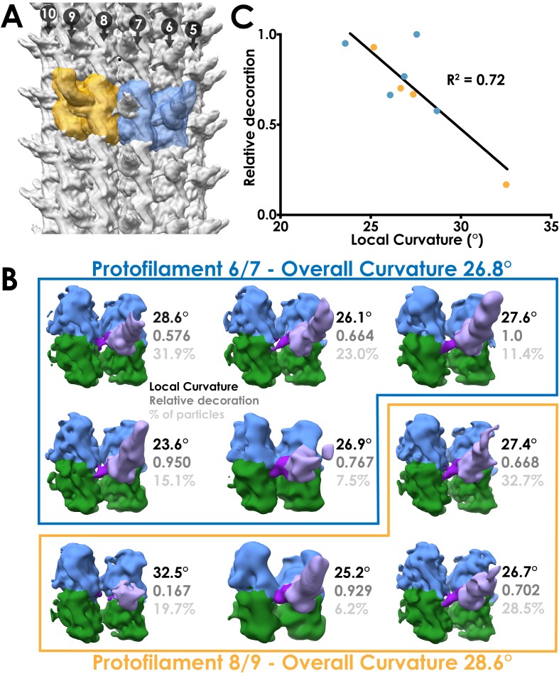
Figure 5.
Local sub-classification of DNAH7 decorated microtubules.
( A ) The two masks used for focused sub-classification of the DNAH7 dataset, encompassing one MTDD and two tubulin dimers (blue/orange).u00a0One mask (blue, protofilaments 6/7) corresponded to the re...
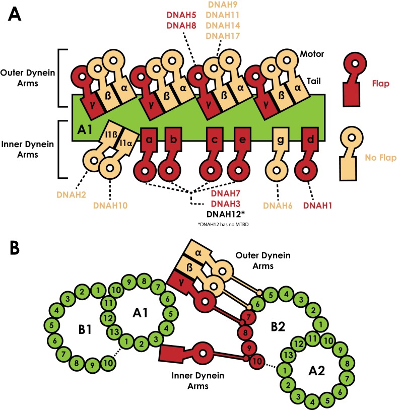
Figure 6.
Distribution of dyneins in the axoneme.
( A ) Schematic view of the positions the dynein heavy chain tails dock on the A-tubule of the microtubule doublet (green) in the 96 nm axonemal repeat, based on previous structural data on C.u00a0rei...
Figure images are served from the NIH/NLM PubMed Central Open Access Subset or Europe PMC; copyright remains with the publishers and authors.
💬 Discussion
0 commentsNo comments yet. Be the first to start a discussion!
Leave a Comment