Abstract
Colorectal cancer (CRC) is the second most common cancer in women and the third most common in men, with an increasing incidence. Pathology diagnosis complemented with prognostic and predictive biomarker information is the first step for personalized treatment. The increased diagnostic load in the pathology laboratory, combined with the reported intra- and inter-variability in the assessment of biomarkers, has prompted the quest for reliable machine-based methods to be incorporated into the routine practice. Recently, Artificial Intelligence (AI) has made significant progress in the medical field, showing potential for clinical applications. Herein, we aim to systematically review the current research on AI in CRC image analysis. In histopathology, algorithms based on Deep Learning (DL) have the potential to assist in diagnosis, predict clinically relevant molecular phenotypes and microsatellite instability, identify histological features related to prognosis and correlated to metastasis, and assess the specific components of the tumor microenvironment.
🔭 Microscopes
🏭 Microscope Brands
🏛️ Research Organizations (ROR)
Affiliated research institutions:
📋 Methods
2.1.
Search Strategy
We systematically searched PubMed from inception to 31 December 2021 for primary studies developing a DL model for the histopathological interpretation of large bowel biopsy tissues and CRC. For this purpose, we used the following algorithm: (convolutional neural networks OR CNN OR deep learning) AND ((cancer AND (colon OR colorectal OR intestin* OR bowel)) OR (adenocarcinoma AND (colon OR colorectal OR intestin* OR bowel)) OR (carcinoma AND (colon OR colorectal OR intestin* OR bowel)) OR (malignan* AND (colon OR colorectal OR intestin* OR bowel))) AND (biop* OR microscop* OR histolog* OR slide* OR eosin OR histopatholog*). The search was conducted on 14 January 2022. 2.2.
Study Eligibility Criteria
The study was conducted according to the PRISMA guidelines and registered to PROSPERO 2020. Eligible articles were considered based on the following criteria. We included studies presenting the development of at least one DL model for the histopathological assessment of large bowel slides and CRC. Eligible applications of the DL models included diagnosis, tumor tissue classification, tumor microenvironment analysis, prognosis, survival and metastasis risk evaluation, tumor mutational burden characterization and, finally, microsatellite instability detection. We excluded articles that presented in vitro models, used endoscopic or radiological images instead of histological sections, and involved non-photonic microscopy. Furthermore, eligible articles should report original studies and not reviews/meta-analyses, concern humans and be written in English. Additionally, articles referring to organs other than the large bowel and benign entities were deemed ineligible. 2.3. Study Selection All citations collected by the previously mentioned methodology were independently screened by four researchers, who were properly trained before the process started, using the online software Rayyan. Three of the researchers were scientifically capable of evaluating the medical aspect of the query and one of them was a CNN expert, able to assess the technical part. During the screening period, the researchers would meet regularly to discuss disagreements and continue training. Conflicts were resolved by consensus. The full texts of potentially eligible articles were later retrieved for further evaluation. 2.4.
Show full methods section
2.1.
Search Strategy
We systematically searched PubMed from inception to 31 December 2021 for primary studies developing a DL model for the histopathological interpretation of large bowel biopsy tissues and CRC. For this purpose, we used the following algorithm: (convolutional neural networks OR CNN OR deep learning) AND ((cancer AND (colon OR colorectal OR intestin* OR bowel)) OR (adenocarcinoma AND (colon OR colorectal OR intestin* OR bowel)) OR (carcinoma AND (colon OR colorectal OR intestin* OR bowel)) OR (malignan* AND (colon OR colorectal OR intestin* OR bowel))) AND (biop* OR microscop* OR histolog* OR slide* OR eosin OR histopatholog*). The search was conducted on 14 January 2022. 2.2.
Study Eligibility Criteria
The study was conducted according to the PRISMA guidelines and registered to PROSPERO 2020. Eligible articles were considered based on the following criteria. We included studies presenting the development of at least one DL model for the histopathological assessment of large bowel slides and CRC. Eligible applications of the DL models included diagnosis, tumor tissue classification, tumor microenvironment analysis, prognosis, survival and metastasis risk evaluation, tumor mutational burden characterization and, finally, microsatellite instability detection. We excluded articles that presented in vitro models, used endoscopic or radiological images instead of histological sections, and involved non-photonic microscopy. Furthermore, eligible articles should report original studies and not reviews/meta-analyses, concern humans and be written in English. Additionally, articles referring to organs other than the large bowel and benign entities were deemed ineligible. 2.3. Study Selection All citations collected by the previously mentioned methodology were independently screened by four researchers, who were properly trained before the process started, using the online software Rayyan. Three of the researchers were scientifically capable of evaluating the medical aspect of the query and one of them was a CNN expert, able to assess the technical part. During the screening period, the researchers would meet regularly to discuss disagreements and continue training. Conflicts were resolved by consensus. The full texts of potentially eligible articles were later retrieved for further evaluation. 2.4.
Data Extraction
To facilitate the data extraction process, we specially designed a spreadsheet form, which all researchers could access to import data from all the eligible articles. From each paper, we extracted information on first author, year and journal of publication, PubMed ID, title, aim of medical research, technical method, classification details, dataset and performance metrics.
📊 Figures
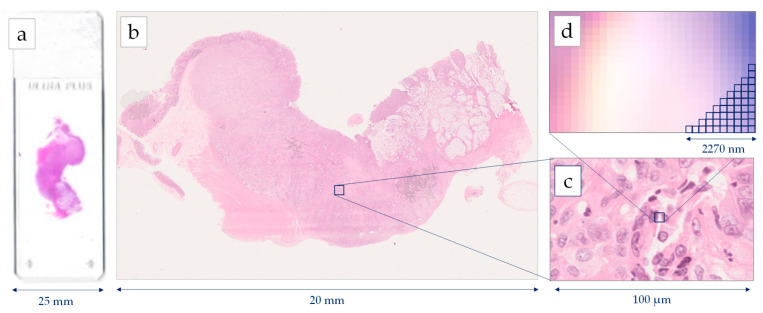
Figure 1
Image generation using a Hamamatsu NanoZoomer whole slide scanner: ( a ) histological slide 75 mm u00d7 25 mm, ( b ) Whole Slide Image (WSI), ( c ) cell level in 40u00d7 magnification, ( d ) pixel lev...
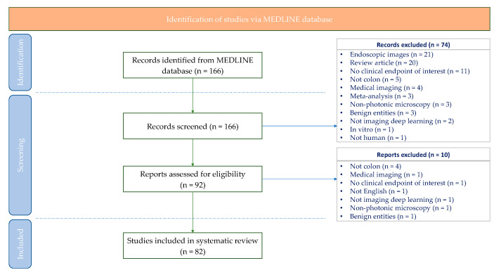
Figure 2
Systematic review flow-chart illustrating systematic search and screening strategy, including number of studies meeting eligibility criteria and number of excluded studies. Last search carried out on ...
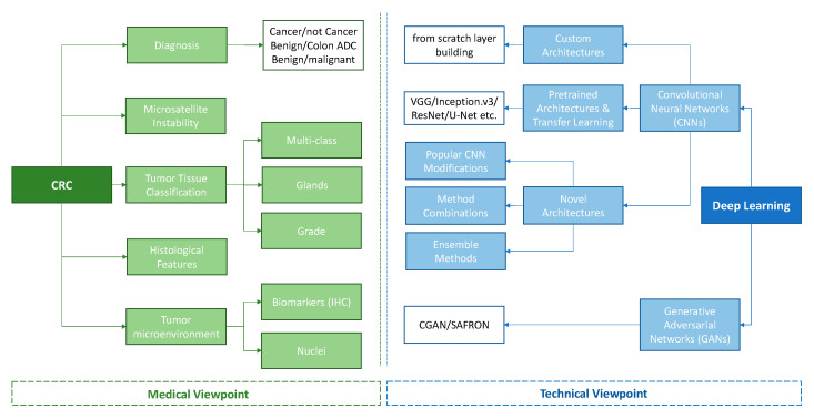
Figure 3
Tree diagram for the categorization of the studies.
Figure images are served from the NIH/NLM PubMed Central Open Access Subset or Europe PMC; copyright remains with the publishers and authors.
💬 Discussion
0 commentsNo comments yet. Be the first to start a discussion!
Leave a Comment