Abstract
Resuscitation from hemorrhage induces profound pathophysiologic alterations and activates inflammatory cascades able to initiate neutrophil accumulation in a variety of tissues. This process is accompanied by acute organ damage (e.g., lungs and liver). We have previously demonstrated that significant leukocyte-endothelium interactions occur very early in other forms of ischemia/reperfusion (i.e., splanchnic ischemia/reperfusion and traumatic shock) which are largely mediated by increased expression of the adhesion molecule, P-selectin, on the vascular endothelium. Here we postulated that increased endothelial expression of P-selectin in the microvasculature would play an essential role in initiating the inflammatory signaling of hemorrhagic shock. Using intravital microscopy, we found that hemorrhagic shock significantly increased the number of rolling and adherent leukocytes in the mouse splanchnic microcirculation. In contrast, mice genetically deficient in P-selectin, or wild-type mice given either an anti-P-selectin monoclonal antibody or a recombinant soluble P-selectin glycoprotein ligand (PSGL)-1 immunoglobulin, exhibited markedly attenuated leukocyte-endothelium interaction after hemorrhagic shock. Thus, activation of P-selectin protein on the microvascular endothelium is essential for the initial upregulation of the inflammatory response occurring in hemorrhagic shock. Moreover, endogenous levels of PSGL-1 mRNA were significantly increased in the lung, liver, and small intestine of wild-type mice subjected to hemorrhagic shock. Since PSGL-1 promotes adhesive interactions largely through P-selectin expressed on the vascular endothelium, this result further supports the crucial role played by P-selectin in the recruitment of leukocytes during hemorrhagic shock.
🔬 Techniques
🧪 Sample Preparation
🔬 Cell Lines
🏭 Microscope Brands
🧪 Reagent Suppliers
🏛️ Research Organizations (ROR)
Affiliated research institutions:
📋 Methods
Hemorrhagic Shock Protocol. This study was performed in accordance with the National Institute of Health guidelines for the use of experimental animals, and all animal protocols have been approved by the Institutional Animal Care and Use Committee (IACUC) of Thomas Jefferson University. Wild-type (C57BL/6) mice and P-selectin–deficient (C57BL/ 6J-Sel) mice were obtained from The Jackson Laboratory . Male and female mice (8–14 wk old and 20–30 g body wt) were anesthetized with sodium pentobarbital (120 mg/kg) injected intraperitoneally. A tracheotomy was performed to maintain a patent airway throughout the experiment. The left carotid artery was cannulated for continuous blood pressure monitoring, and the right jugular vein was cannulated for blood withdrawal and fluid or antibody administration. Mice were subjected to hemorrhage by withdrawal of blood to allow mean arterial blood pressure (MABP) 1 to be maintained at 40 mmHg for 45 min. The mean bleedout volume was 0.72 ± 0.12 ml and 0.81 ± 0.09 ml for the wild-type and P-selectin–deficient mouse, respectively. Blood was collected in a heparinized (5 U) syringe and kept at 37°C until reinfusion. Mice were then resuscitated by infusion of the shed blood and intravenous injection of 0.5 ml 0.9% NaCl alone or with either an anti–P-selectin mAb or rs.PSGL.Ig. Mice were killed by exsanguination 45 min after resuscitation, after completion of intravital microscopy studies. Control wild-type mice underwent cannulation and anesthesia for the same period of time as hemorrhaged mice, but were not bled ( n = 6). Wild-type and P-selectin–deficient mice were randomly assigned to one of four experimental hemorrhage groups: (i) wild-type mice receiving saline ( n = 7); (ii) P-selectin–deficient mice receiving saline ( n = 6); (iii) wild-type mice receiving 1 mg/kg anti–P-selectin mAb (RB40.34; PharMingen ) ( n = 6); and (iv) wild-type mice receiving 1 mg/kg high affinity mutant rs.PSGL.Ig (rsPSGL.47mutFc; Genetics Institute, Inc.) ( n = 6). The total number of circulating white blood cells in all experimental groups of mice was determined by hemocytometric count of smears of blood, which was obtained through the jugular vein cannula. Intravital Microscopy of Mouse Peri-intestinal Venules. All intravital microscopy experiments were conducted in anesthetized mice, which were surgically prepared as reported in the above hemorrhagic shock protocol section. Intravital microscopy was performed on mouse peri-intestinal venules, after exteriorization of a loop of ileal tissue via a midline laparotomy. The ileum was placed in a temperature-controlled fluid-filled plexiglas chamber and transilluminated for brightfield observation of the peri-intestinal microcirculation according to a previously described procedure ( 15 ). The ileum and mesentery were superfused throughout the experiment with a buffered Tyrode solution (pH 7.4, 37 ± 1°C). Preparations were allowed to stabilize for 15 min. Observations of rolling and adherent leukocytes were made with a Microphot microscope and a 40× salt water–immersion lens ( Nikon Corp. ). Images were projected by a high-resolution color video camera (DC-330; DAGE-MTI, Inc.) onto a color Sony high resolution video monitor (Multiscan 200-sf), and the image was recorded with a videocassette recorder. All images were then analyzed using computerized imaging software (Phase 3 Image System; Media Cybernetics) on a Pentium based IBM-compatible computer (Micron Millenia Mxe; Micron Electronics Inc.). Red blood cell velocity was determined on-line using an optical Doppler velocimeter ( 16 ) obtained from the Microcirculation Research Institute, College Station, TX. This method gives an average red blood cell velocity, which is digitally displayed on a meter, and allows for the calculation of shear rates ( 17 ). The number of rolling and adhered leukocytes was determined off-line by playback analysis of the videotape. Histologic Assessment of Neutrophil Infiltration and Immunohistochemistry of P-selectin. The number of neutrophils (PMNs) infiltrating into lung, liver, and intestine was determined in both wild-type and P-selectin–deficient mice. At the end of the 45 min reinfusion period, mice were killed and the pulmonary circulation was flushed by injecting PBS into the right ventricle. Lungs were injected with formalin through the trachea, then removed and placed into 4% formalin. Similarly, the splanchnic organs were perfused through the superior mesenteric artery and fixed in vivo by infusion of formalin ( 15 ). Lungs, liver, and intestine were then cut into blocks and dehydrated in graded acetone washes at 4°C. Tissue blocks were embedded in plastic (Immunobed; Polysciences, Inc.), and 4-μm-thick sections were cut and transferred to Vectabond-coated slides (Vector Laboratories, Inc.). The slides were soaked in ethanol for 10 min to remove the plastic embedding material and to allow staining of the tissue. After the 10-min ethanol wash, the tissue sections were stained with hematoxylin and eosin and examined blindly. The number of PMNs was counted and tallied. Five fields from each of two slides were counted from each organ, and three mice were studied per group. In addition, grading of the histopathologic changes in the lung (i.e., neutrophil infiltration, interstitial edema, and intraparenchymal hemorrhage) was performed on a 0 to 3 scale (with 0 being normal and 3 being the most severe abnormality). Immunohistochemical localization of P-selectin was determined in ileal samples after intravital microscopy was completed, according to previously described methods ( 15 ). mAb PB1.3 only recognizes surface expressed P-selectin ( 18 ). Quantification of P-selectin was accomplished using the avidin-biotin immunoperoxidase technique (Vectastain ABC Reagent; Vector Labs.) as previously described by Weyrich et al. ( 18 ). 50 venules were analyzed per tissue section, 20 sections were examined per group, and the percentage of positive staining venules was tallied.
Show full methods section
Hemorrhagic Shock Protocol. This study was performed in accordance with the National Institute of Health guidelines for the use of experimental animals, and all animal protocols have been approved by the Institutional Animal Care and Use Committee (IACUC) of Thomas Jefferson University. Wild-type (C57BL/6) mice and P-selectin–deficient (C57BL/ 6J-Sel) mice were obtained from The Jackson Laboratory . Male and female mice (8–14 wk old and 20–30 g body wt) were anesthetized with sodium pentobarbital (120 mg/kg) injected intraperitoneally. A tracheotomy was performed to maintain a patent airway throughout the experiment. The left carotid artery was cannulated for continuous blood pressure monitoring, and the right jugular vein was cannulated for blood withdrawal and fluid or antibody administration. Mice were subjected to hemorrhage by withdrawal of blood to allow mean arterial blood pressure (MABP) 1 to be maintained at 40 mmHg for 45 min. The mean bleedout volume was 0.72 ± 0.12 ml and 0.81 ± 0.09 ml for the wild-type and P-selectin–deficient mouse, respectively. Blood was collected in a heparinized (5 U) syringe and kept at 37°C until reinfusion. Mice were then resuscitated by infusion of the shed blood and intravenous injection of 0.5 ml 0.9% NaCl alone or with either an anti–P-selectin mAb or rs.PSGL.Ig. Mice were killed by exsanguination 45 min after resuscitation, after completion of intravital microscopy studies. Control wild-type mice underwent cannulation and anesthesia for the same period of time as hemorrhaged mice, but were not bled ( n = 6). Wild-type and P-selectin–deficient mice were randomly assigned to one of four experimental hemorrhage groups: (i) wild-type mice receiving saline ( n = 7); (ii) P-selectin–deficient mice receiving saline ( n = 6); (iii) wild-type mice receiving 1 mg/kg anti–P-selectin mAb (RB40.34; PharMingen ) ( n = 6); and (iv) wild-type mice receiving 1 mg/kg high affinity mutant rs.PSGL.Ig (rsPSGL.47mutFc; Genetics Institute, Inc.) ( n = 6). The total number of circulating white blood cells in all experimental groups of mice was determined by hemocytometric count of smears of blood, which was obtained through the jugular vein cannula. Intravital Microscopy of Mouse Peri-intestinal Venules. All intravital microscopy experiments were conducted in anesthetized mice, which were surgically prepared as reported in the above hemorrhagic shock protocol section. Intravital microscopy was performed on mouse peri-intestinal venules, after exteriorization of a loop of ileal tissue via a midline laparotomy. The ileum was placed in a temperature-controlled fluid-filled plexiglas chamber and transilluminated for brightfield observation of the peri-intestinal microcirculation according to a previously described procedure ( 15 ). The ileum and mesentery were superfused throughout the experiment with a buffered Tyrode solution (pH 7.4, 37 ± 1°C). Preparations were allowed to stabilize for 15 min. Observations of rolling and adherent leukocytes were made with a Microphot microscope and a 40× salt water–immersion lens ( Nikon Corp. ). Images were projected by a high-resolution color video camera (DC-330; DAGE-MTI, Inc.) onto a color Sony high resolution video monitor (Multiscan 200-sf), and the image was recorded with a videocassette recorder. All images were then analyzed using computerized imaging software (Phase 3 Image System; Media Cybernetics) on a Pentium based IBM-compatible computer (Micron Millenia Mxe; Micron Electronics Inc.). Red blood cell velocity was determined on-line using an optical Doppler velocimeter ( 16 ) obtained from the Microcirculation Research Institute, College Station, TX. This method gives an average red blood cell velocity, which is digitally displayed on a meter, and allows for the calculation of shear rates ( 17 ). The number of rolling and adhered leukocytes was determined off-line by playback analysis of the videotape. Histologic Assessment of Neutrophil Infiltration and Immunohistochemistry of P-selectin. The number of neutrophils (PMNs) infiltrating into lung, liver, and intestine was determined in both wild-type and P-selectin–deficient mice. At the end of the 45 min reinfusion period, mice were killed and the pulmonary circulation was flushed by injecting PBS into the right ventricle. Lungs were injected with formalin through the trachea, then removed and placed into 4% formalin. Similarly, the splanchnic organs were perfused through the superior mesenteric artery and fixed in vivo by infusion of formalin ( 15 ). Lungs, liver, and intestine were then cut into blocks and dehydrated in graded acetone washes at 4°C. Tissue blocks were embedded in plastic (Immunobed; Polysciences, Inc.), and 4-μm-thick sections were cut and transferred to Vectabond-coated slides (Vector Laboratories, Inc.). The slides were soaked in ethanol for 10 min to remove the plastic embedding material and to allow staining of the tissue. After the 10-min ethanol wash, the tissue sections were stained with hematoxylin and eosin and examined blindly. The number of PMNs was counted and tallied. Five fields from each of two slides were counted from each organ, and three mice were studied per group. In addition, grading of the histopathologic changes in the lung (i.e., neutrophil infiltration, interstitial edema, and intraparenchymal hemorrhage) was performed on a 0 to 3 scale (with 0 being normal and 3 being the most severe abnormality). Immunohistochemical localization of P-selectin was determined in ileal samples after intravital microscopy was completed, according to previously described methods ( 15 ). mAb PB1.3 only recognizes surface expressed P-selectin ( 18 ). Quantification of P-selectin was accomplished using the avidin-biotin immunoperoxidase technique (Vectastain ABC Reagent; Vector Labs.) as previously described by Weyrich et al. ( 18 ). 50 venules were analyzed per tissue section, 20 sections were examined per group, and the percentage of positive staining venules was tallied.
Quantification of P-selectin and PSGL-1 mRNA
Expression by Ribonuclease Protection Assays. Immediately after intravital microscopy was performed, mouse lungs, liver, and small intestine were removed for total RNA extraction. Total RNA was extracted from these tissues using the acid guanidium-phenol-chloroform extraction method described by Chomczynski and Sacchi ( 19 ). The concentration of RNA suspended in ribonuclease-free water was determined spectrophotometrically. The plasmid containing the full-length mouse P-selectin cDNA was provided by Professor Dietmar Vestweber (Institut fur Zellbiologie, ZMBE, Westfalische Wilhelms Universitat Munster, Munster, Germany). The plasmid for synthesis of mouse tissue P-selectin probe for ribonuclease protection assays was created by recloning of a PstI–AvrII fragment of mouse P-selectin (from 1878 to 2270; sequence data available from EMBL/GenBank/ DDBJ under accession number M87861 ) into a PstI–SpeI site of pBluescript II SK + vector (Stratagene). This plasmid was digested with XhoI and used as a template for in vitro transcription of a 474-base radiolabeled antisense probe containing a 393-base protected fragment using T3 RNA-polymerase ( Boehringer Mannheim ) in the presence of [ 32 P]UTP ( Amersham Corp. ). Mouse PSGL-1 cDNA was synthesized by reverse transcriptase PCR using mouse lung total RNA and oligo(dT), and was amplified using forward primer (5′-CCTGGGAATTCACCTGCCCC-3′) and reverse primer (5′-GAGAGTGGAGCTAGCAAAGG-3′). These oligonucleotides correspond to amino acid sequences 267–283 and 394–388 of mouse PSGL-1, respectively (sequence data available from EMBL/GenBank/DDBJ under accession number X91144 ). A 384-bp PCR fragment was cloned using PCR 2.1-TOPO Cloning Kit (a gift from Invitrogen Corp.). The Sst I–XbaI fragment of this plasmid was recloned in pTRIPLEscript vector (pTRIamp 18; Ambion). This plasmid was digested with XbaI to make, with T7 polymerase, a 570-base radiolabeled antisense probe that contained a 384-base protected fragment. All constructs used in this investigation were verified by sequencing the insert in the plasmid. These sequences were found to be 100% identical to the published sequences. The expression of PSGL-1 mRNA was analyzed by reverse transcriptase PCR analysis. Amplified PCR products were analyzed by electrophoresis on a 1.5% agarose gel. The intensity of each P-selectin and PSGL-1 mRNA band was normalized for GAPDH (data not shown).
Mouse β-actin antisense
RNA probes were used to evaluate total RNA, which in turn were used for P-selectin and PSGL-1 mRNA expression analysis. The mouse β-actin plasmid for the synthesis of the antisense RNA probe was received from Ambion. For the ribonuclease protection assay, 2 μg of total RNA for detection of β-actin mRNA, and 60 μg for detection of PSGL-1 mRNA in the lung, liver, and small intestine have been used accordingly to previously described procedures ( 20 ). The intensity of each P-selectin and PSGL-1 mRNA band was normalized for β-actin mRNA levels. Statistical Analyses. All values for data listed in the text and figures are presented as means ± SEM of n independent experiments. Data were compared by analysis of variance using post-hoc analysis with Fisher's correct t test. P ≤ 0.05 was considered significant in all cases.
Hemorrhagic Shock Protocol. This study was performed in accordance with the National Institute of Health guidelines for the use of experimental animals, and all animal protocols have been approved by the Institutional Animal Care and Use Committee (IACUC) of Thomas Jefferson University. Wild-type (C57BL/6) mice and P-selectin–deficient (C57BL/ 6J-Sel) mice were obtained from The Jackson Laboratory . Male and female mice (8–14 wk old and 20–30 g body wt) were anesthetized with sodium pentobarbital (120 mg/kg) injected intraperitoneally. A tracheotomy was performed to maintain a patent airway throughout the experiment. The left carotid artery was cannulated for continuous blood pressure monitoring, and the right jugular vein was cannulated for blood withdrawal and fluid or antibody administration. Mice were subjected to hemorrhage by withdrawal of blood to allow mean arterial blood pressure (MABP) 1 to be maintained at 40 mmHg for 45 min. The mean bleedout volume was 0.72 ± 0.12 ml and 0.81 ± 0.09 ml for the wild-type and P-selectin–deficient mouse, respectively. Blood was collected in a heparinized (5 U) syringe and kept at 37°C until reinfusion. Mice were then resuscitated by infusion of the shed blood and intravenous injection of 0.5 ml 0.9% NaCl alone or with either an anti–P-selectin mAb or rs.PSGL.Ig. Mice were killed by exsanguination 45 min after resuscitation, after completion of intravital microscopy studies. Control wild-type mice underwent cannulation and anesthesia for the same period of time as hemorrhaged mice, but were not bled ( n = 6). Wild-type and P-selectin–deficient mice were randomly assigned to one of four experimental hemorrhage groups: (i) wild-type mice receiving saline ( n = 7); (ii) P-selectin–deficient mice receiving saline ( n = 6); (iii) wild-type mice receiving 1 mg/kg anti–P-selectin mAb (RB40.34; PharMingen ) ( n = 6); and (iv) wild-type mice receiving 1 mg/kg high affinity mutant rs.PSGL.Ig (rsPSGL.47mutFc; Genetics Institute, Inc.) ( n = 6). The total number of circulating white blood cells in all experimental groups of mice was determined by hemocytometric count of smears of blood, which was obtained through the jugular vein cannula.
📊 Figures
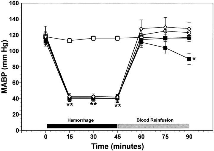
Figure 1
Time course of MABP over the course of hemorrhage and reinfusion for the five experimental groups of mice. Wild-type and P-selectinu2013deficient (P-selectin u2212/u2212 ) mice were subjected to hemor...
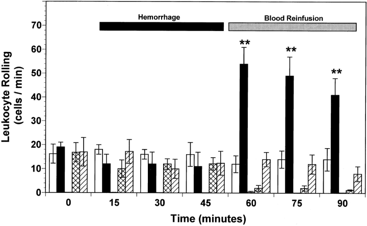
Figure 2
Leukocyte rolling observed in peri-intestinal venules of wild-type mice, P-selectinu2013deficient (P-selectin u2212/u2212 ) mice, and wild-type mice given either antiu2013P-selectin mAb or rs.PSGL.Ig,...
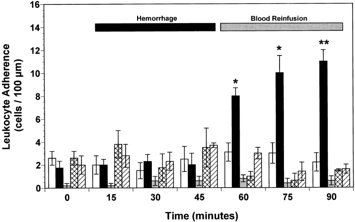
Figure 3
Leukocyte adherence observed in peri-intestinal venules of wild-type mice, P-selectinu2013deficient (P-selectin u2212/u2212 ) mice, and wild-type mice given either antiu2013P-selectin mAb or rs.PSGL.I...
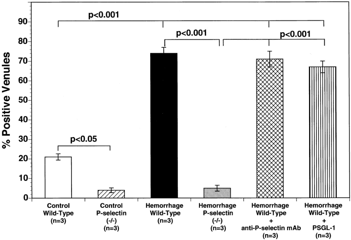
Figure 4
Immunohistochemistry of mouse ileal microvessels: percentage of vessels staining positive for P-selectin in the five experimental groups of mice. Bar heights represent mean values, and brackets indica...
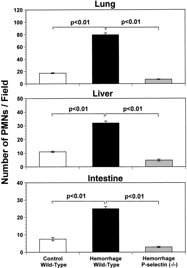
Figure 5
Quantification of histological staining of neutrophils in lung (top), liver (center) and small intestine (bottom) of wild-type and P-selectinu2013deficient (P-selectin u2212/u2212 ) mice subjected to ...
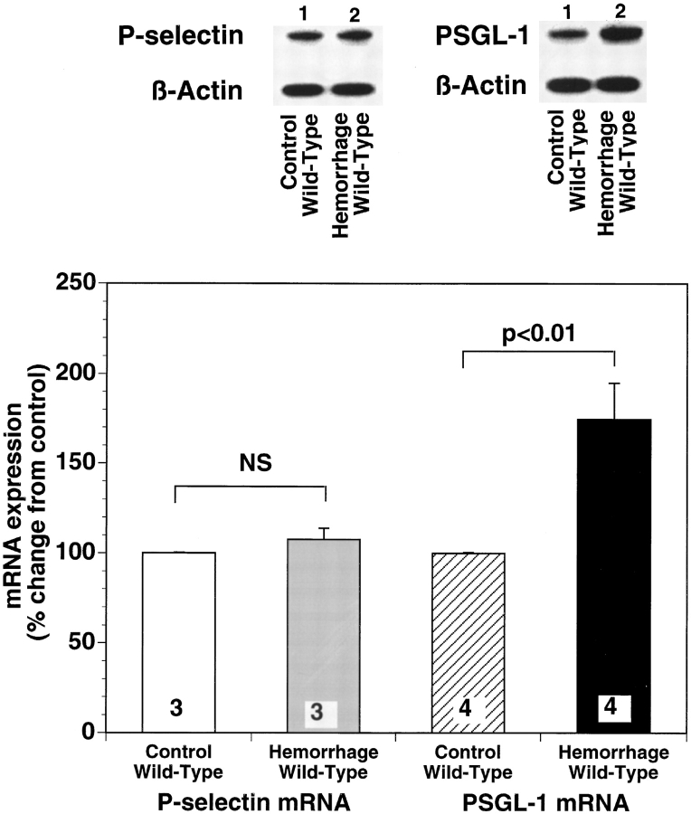
Figure 6
Representative autoradiograph of a polyacrylamide gel used in typical ribonuclease protection assay comparing PSGL-1 and P-selectin mRNA expression in lungs of sham-operated control and hemorrhaged wi...
Figure images are served from the NIH/NLM PubMed Central Open Access Subset or Europe PMC; copyright remains with the publishers and authors.
💬 Discussion
0 commentsNo comments yet. Be the first to start a discussion!
Leave a Comment