Abstract
It has been challenging to generate in vitro models of alveolar lung diseases, as the stable culture of alveolar type 2 (AT2) cells has been difficult. Methods of generating and expanding AT2 cells derived from induced pluripotent stem cells (iPSCs) have been established and are expected to be applicable to disease modeling. Hermansky-Pudlak syndrome (HPS) is an autosomal recessive disorder characterized by dysfunction of lysosome-related organelles, such as lamellar bodies (LBs), in AT2 cells. From an HPS type 2 (HPS2) patient, we established disease-specific iPSCs (HPS2-iPSCs) and their gene-corrected counterparts. By live cell imaging, the LB dynamics were visualized and altered distribution, enlargement, and impaired secretion of LBs were demonstrated in HPS2-iPSC-derived AT2 cells. These findings provide insight into the AT2 dysfunction in HPS patients and support the potential use of human iPSC-derived AT2 cells for future research on alveolar lung diseases.
🔬 Techniques
✨ Fluorophores
🧪 Sample Preparation
🔬 Cell Lines
🏭 Microscope Brands
🏛️ Research Organizations (ROR)
Affiliated research institutions:
📋 Methods
Comparison of the Methods of NKX2-1 + Cell Isolation
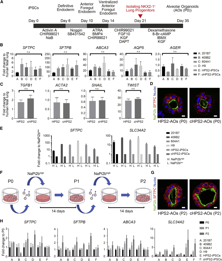
The isolation of NKX2-1 + lung progenitor cells is a critical step in the generation of lung epithelial cells from hPSCs. We compared the isolation efficiency of previously reported sorting methods, carboxypeptidase M (CPM) and CD47 combined with or without CD26 ( Figure S2 A) ( Gotoh et al., 2014 , Hawkins et al., 2017 ). At day 21 of our induction protocol ( Figure 2 A), CPM high cells contained the most NKX2-1 + cells in all hPSC lines ( Figures S2 B and S2C). Since there was no interaction between the cell lines and sorting methods analyzed by two-way ANOVA, the samples from different cell lines were all analyzed together. As a result, CPM-based sorting was able to isolate more NKX2-1 + cells than CD47-based methods ( Figure S2 D). Figure 2 Generation of iPSC-derived AOs and Their Expansion Based on the Expression of NaPi2b (A) Schematic overview of the generation of hPSC-derived AOs. (B) qRT-PCR of AT1 and AT2 markers in AOs (P0) (mean ± SEM, n = 3 independent experiments). Kruskal-Wallis with Dunn's multiple comparisons test was used. n.s., not significant. (C) qRT-PCR of fibrosis-related markers in AOs (P0) (mean ± SEM, n = 5 independent experiments). The Mann-Whitney test was used. n.s., not significant. (D) Confocal IF staining of AOs (P0) derived from HPS2-iPSCs and cHPS2-iPSCs. Scale bars, 10 μm. (E) qRT-PCR of SFTPC and SLC34A2 in NaPi2b high and NaPi2b low cells sorted from AOs (P0) (mean ± SEM, n = 3 independent experiments). (F) Schematic overview of the subculture of NaPi2b high cells in AOs. NaPi2b high cells were passaged every 2 weeks. (G) Confocal IF staining of AOs (P2) subcultured using anti-NaPi2b antibodies. Scale bars, 10 μm. (H) qRT-PCR of AT2 markers in subcultured AOs (P0–P2) (mean ± SEM, n = 3 independent experiments). See also Figures S2 and S3 .
Show full methods section
Comparison of the Methods of NKX2-1 + Cell Isolation
The isolation of NKX2-1 + lung progenitor cells is a critical step in the generation of lung epithelial cells from hPSCs. We compared the isolation efficiency of previously reported sorting methods, carboxypeptidase M (CPM) and CD47 combined with or without CD26 ( Figure S2 A) ( Gotoh et al., 2014 , Hawkins et al., 2017 ). At day 21 of our induction protocol ( Figure 2 A), CPM high cells contained the most NKX2-1 + cells in all hPSC lines ( Figures S2 B and S2C). Since there was no interaction between the cell lines and sorting methods analyzed by two-way ANOVA, the samples from different cell lines were all analyzed together. As a result, CPM-based sorting was able to isolate more NKX2-1 + cells than CD47-based methods ( Figure S2 D). Figure 2 Generation of iPSC-derived AOs and Their Expansion Based on the Expression of NaPi2b (A) Schematic overview of the generation of hPSC-derived AOs. (B) qRT-PCR of AT1 and AT2 markers in AOs (P0) (mean ± SEM, n = 3 independent experiments). Kruskal-Wallis with Dunn's multiple comparisons test was used. n.s., not significant. (C) qRT-PCR of fibrosis-related markers in AOs (P0) (mean ± SEM, n = 5 independent experiments). The Mann-Whitney test was used. n.s., not significant. (D) Confocal IF staining of AOs (P0) derived from HPS2-iPSCs and cHPS2-iPSCs. Scale bars, 10 μm. (E) qRT-PCR of SFTPC and SLC34A2 in NaPi2b high and NaPi2b low cells sorted from AOs (P0) (mean ± SEM, n = 3 independent experiments). (F) Schematic overview of the subculture of NaPi2b high cells in AOs. NaPi2b high cells were passaged every 2 weeks. (G) Confocal IF staining of AOs (P2) subcultured using anti-NaPi2b antibodies. Scale bars, 10 μm. (H) qRT-PCR of AT2 markers in subcultured AOs (P0–P2) (mean ± SEM, n = 3 independent experiments). See also Figures S2 and S3 .
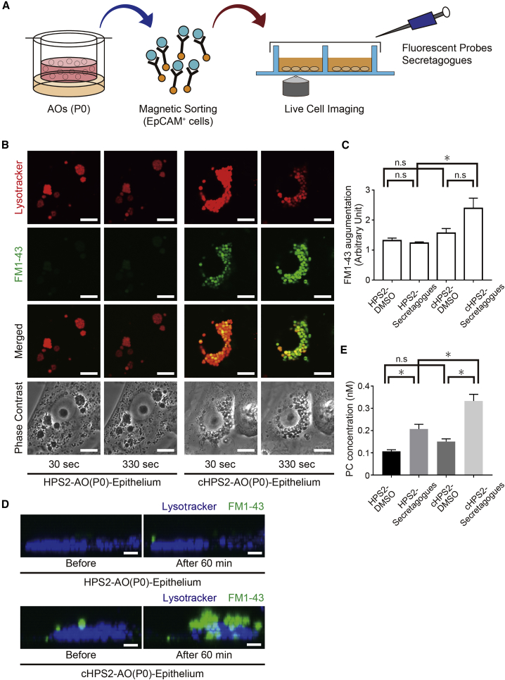
Experimental Procedures Live Cell Imaging After AOs or reseeded cells were stained with fluorescent probes, all of the samples were examined under an FV10i-LIV confocal microscope (Olympus) with a 60× objective under 5% CO 2 at 37°C. For the LB secretion assay, the medium was supplemented with a secretagog cocktail so that the final concentration of each component was 5 μM forskolin, 15 μM ATP, 150 nM ionomycin, and 150 nM phorbol 12-myristate 13-acetate. For further details, see Supplemental Experimental Procedures .
Ethics
The use of H9 hESCs was approved by the Ministry of Education, Culture, Sports, Science and Technology (MEXT), Japan. The animal experiments were approved by the Animal Research Committee of Kyoto University. The use of human lung samples was approved by the Ethics Committee of Kyoto University Graduate School and Faculty of Medicine.
Statistical Analyses
All error bars indicate the SEM. Quantified data represent the findings of three or more independent experiments. The statistical tests used are shown in each legend. All statistical analyses were performed using the Prism7 software program (GraphPad).
Supplemental Information Document S1. Supplemental Experimental Procedures, Figures S1–S3, and Tables S1–S4 Document S2. Article plus Supplemental Information
📊 Figures
Figureu00a01
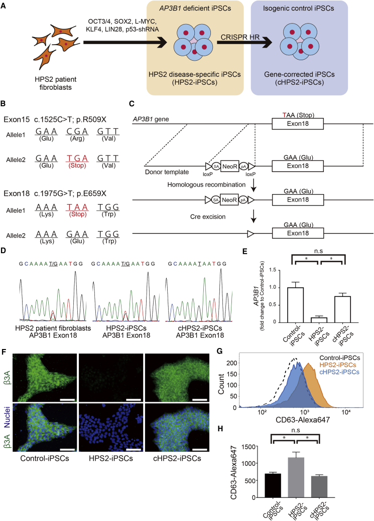
Generation of HPS2-iPSCs and cHPS2-iPSCs (A) Schematic overview of the generation of HPS2-iPSCs and cHPS2-iPSCs. (B) Different mutations in each allele of the patient fibroblasts. (C) Strategy for cor...
Figureu00a02
Generation of iPSC-derived AOs and Their Expansion Based on the Expression of NaPi2b (A) Schematic overview of the generation of hPSC-derived AOs. (B) qRT-PCR of AT1 and AT2 markers in AOs (P0) (mean ...
Figureu00a03
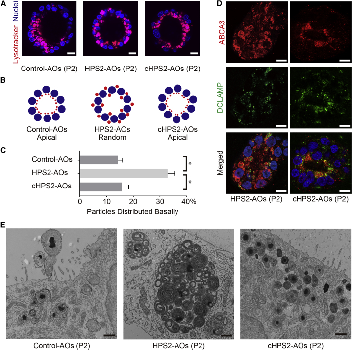
Morphological Features of HPS2-AOs and cHPS2-AOs (A) Live cell imaging of AOs (P2) derived from control iPSCs, HPS2-iPSCs, and cHPS2-iPSCs, respectively. Scale bars, 10u00a0u03bcm. (B) Schematic illus...
Figureu00a04
LB Secretion Assay for AT2 Cells Derived from HPS2-iPSCs and cHPS2-iPSCs (A) Schematic overview of the LB secretion assay. (B) Confocal images of HPS2-AO(P0) and cHPS2-AO(P0) epithelial cells at 30 an...
Figure images are served from the NIH/NLM PubMed Central Open Access Subset or Europe PMC; copyright remains with the publishers and authors.
💬 Discussion
0 commentsNo comments yet. Be the first to start a discussion!
Leave a Comment