Abstract
ImportanceImprovements in skin appearance resulting from treatment with fractionated picosecond‐lasers have been noted, but optimizing the treatment efficacy depends on a thorough understanding of the specific skin response. The development of non‐invasive laser imaging techniques in conjunction with laser therapy can potentially provide feedback for guidance and optimizing clinical outcome.ObjectiveThe purpose of this study was to demonstrate the capability of multiphoton microscopy (MPM), a high‐resolution, label‐free imaging technique, to characterize in vivo the skin response to a fractionated non‐ablative picosecond‐laser treatment.Design, Setting, and ParticipantsTwo areas on the arm of a volunteer were treated with a fractionated picosecond laser at the Dermatology Clinic, UC Irvine. The skin response to treatment was imaged in vivo with a clinical MPM‐based tomograph at 3 hours and 24 hours after treatment and seven additional time points over a 4‐week period.Main Outcomes and MeasuresMPM revealed micro‐injuries present in the epidermis. Pigmented cells were particularly damaged in the process, suggesting that melanin is likely the main absorber for laser induced optical breakdown.ResultsDamaged individual cells were distinguished as early as 3 hours post pico‐laser treatment with the 532 nm wavelength, and 24 hours post‐treatment with both 532 and 1064 nm wavelengths. At later time points, clusters of cellular necrotic debris were imaged across the treated epidermis. After 24 hours of treatment, inflammatory cells were imaged in the proximity of epidermal micro‐injuries. The epidermal injuries were exfoliated over a 4‐week period.Conclusions and RelevanceThis observational and descriptive pilot study demonstrates that in vivo MPM imaging can be used non‐invasively to provide label‐free contrast for describing changes in human skin following a fractionated non‐ablative laser treatment. The results presented in this study represent the groundwork for future longitudinal investigations on an expanded number of subjects to understand the response to treatment in different skin types with different laser parameters, critical factors in optimizing treatment outcome. Lasers Surg. Med. 49:555–562, 2017. © 2017 The Authors. Lasers in Surgery and Medicine Published by Wiley Periodicals, Inc.
🔬 Techniques
🔬 Cell Lines
🏭 Microscope Brands
🔴 Lasers
📷 Detectors
🏛️ Research Organizations (ROR)
Affiliated research institutions:
📋 Methods
Fractionated Picosecond Treatment Laser (PicoWay) The treatment was based on a fractionated non‐ablative picosecond Nd:YAG laser (PicoWay Resolve, Candela). This is a dual wavelength laser system delivering 1064 nm, 450 ps pulses with maximum microbeam energy per pulse of 3 mJ and 532 nm, 375 ps pulses with 1.5 mJ maximum microbeam energy per pulse. The PicoWay delivers an array of 100 microbeams per 6 mm × 6 mm area using a holographic diffractive beam‐splitter technology. In this pilot study, we employed the PicoWay laser to treat each of two areas of a volunteer forearm with the different wavelengths provided by the laser, 532 and 1064 nm. We used the maximum energy/pulse available for each wavelength and a single pass treatment. Multiphoton Microscopy (MPM)‐Based Clinical Tomograph (MPTflex) MPM is an imaging technique based on laser‐scanning microscopy. In MPM imaging of skin tissue, the contrast mechanisms are based on second harmonic generation (SHG) from collagen fibers and two‐photon excited fluorescence (TPEF) from reduced nicotinamide adenine dinucleotide (NADH), flavin adenine dinucleotide (FAD), keratin, melanin, and elastin fibers. Importantly, these contrast mechanisms rely on the intrinsic optical properties of endogenous tissue biomolecules without using specific fluorescent labels. The nonlinear light‐matter interaction through the aforementioned mechanisms allows MPM to provide 3D sub‐micron resolution images of tissue. In this study, we used an MPM‐based clinical tomograph (MPTflex, JenLab GmbH, Jena, Germany) for in vivo imaging of the treated skin area. As previously described in other studies where this system has been used 4 , 5 , this tomograph consists of a compact, turn‐key femtosecond laser (MaiTai Ti:Sappire oscillator, sub–100 fs, 80 MHz, tunable 690–1020 nm; Spectra Physics, Mountain View, CA), an articulated arm with near‐infrared optics, and beam scanning module. The system has two photomultiplier tube (PMT) detectors employed for parallel acquisition of TPEF and SHG signals. A customized metallic ring taped on the subject's skin attaches magnetically to the objective holder in the articulated arm, minimizing motion artifacts. The excitation wavelength used for this study was 790 nm. The TPEF signal was detected over the spectral range of 410–650 nm while the SHG signal was detected over a narrow spectral bandwidth 385–405 nm through emission filters placed in the TPEF and SHG detection channels, respectively. We used a Zeiss objective (40X, 1.3NA, oil immersion) for focusing into the tissue.
Show full methods section
Fractionated Picosecond Treatment Laser (PicoWay) The treatment was based on a fractionated non‐ablative picosecond Nd:YAG laser (PicoWay Resolve, Candela). This is a dual wavelength laser system delivering 1064 nm, 450 ps pulses with maximum microbeam energy per pulse of 3 mJ and 532 nm, 375 ps pulses with 1.5 mJ maximum microbeam energy per pulse. The PicoWay delivers an array of 100 microbeams per 6 mm × 6 mm area using a holographic diffractive beam‐splitter technology. In this pilot study, we employed the PicoWay laser to treat each of two areas of a volunteer forearm with the different wavelengths provided by the laser, 532 and 1064 nm. We used the maximum energy/pulse available for each wavelength and a single pass treatment. Multiphoton Microscopy (MPM)‐Based Clinical Tomograph (MPTflex) MPM is an imaging technique based on laser‐scanning microscopy. In MPM imaging of skin tissue, the contrast mechanisms are based on second harmonic generation (SHG) from collagen fibers and two‐photon excited fluorescence (TPEF) from reduced nicotinamide adenine dinucleotide (NADH), flavin adenine dinucleotide (FAD), keratin, melanin, and elastin fibers. Importantly, these contrast mechanisms rely on the intrinsic optical properties of endogenous tissue biomolecules without using specific fluorescent labels. The nonlinear light‐matter interaction through the aforementioned mechanisms allows MPM to provide 3D sub‐micron resolution images of tissue. In this study, we used an MPM‐based clinical tomograph (MPTflex, JenLab GmbH, Jena, Germany) for in vivo imaging of the treated skin area. As previously described in other studies where this system has been used 4 , 5 , this tomograph consists of a compact, turn‐key femtosecond laser (MaiTai Ti:Sappire oscillator, sub–100 fs, 80 MHz, tunable 690–1020 nm; Spectra Physics, Mountain View, CA), an articulated arm with near‐infrared optics, and beam scanning module. The system has two photomultiplier tube (PMT) detectors employed for parallel acquisition of TPEF and SHG signals. A customized metallic ring taped on the subject's skin attaches magnetically to the objective holder in the articulated arm, minimizing motion artifacts. The excitation wavelength used for this study was 790 nm. The TPEF signal was detected over the spectral range of 410–650 nm while the SHG signal was detected over a narrow spectral bandwidth 385–405 nm through emission filters placed in the TPEF and SHG detection channels, respectively. We used a Zeiss objective (40X, 1.3NA, oil immersion) for focusing into the tissue.
Study Design
This observational and descriptive pilot study included one volunteer (skin type II). Two areas on the subject forearm, approximately 1 cm apart from each other, were treated with the PicoWay fractionated laser, each area corresponding to different wavelengths of the treatment laser: 532 and 1064 nm. The skin response to treatment was imaged in vivo at nine time points post‐treatment for a period of 4 weeks: 3 hours, 24 hours, 48 hours, 72 hours, 7 day, 9 day, 11 day, 21 day, and 28 day. All in vivo measurements were conducted according to an approved institutional review board protocol (HS#2008–6307) with written informed consent obtained from the subject. We used the MPTflex tomograph for in vivo imaging of the treated skin area. For a better understanding and visualization of the changes in human skin following treatment, we acquired the MPM images of the treated areas using two scanning modalities: (1) xy scanning, which provides images of horizontal optical sections. We obtained z‐stacks of images at different depths by moving the objective in the z direction, thus scanning from the stratum corneum to the superficial dermis. The field of view for each optical section was about 200 × 200 µm 2 and the step between optical sections, 5 µm. As the optical section is limited to a small scan field, the overall investigation required the acquisition of several stacks of images at different locations across the treatment area. We acquired about three image stacks for each lesion. (2) xz scanning, which provides cross‐sectional, “vertical histology‐like” images from the stratum corneum to superficial dermis. The xy sections were 512 × 512 pixels images acquired at ∼6 seconds/frame. The xz sections were 1024 × 1024 pixels images acquired at ∼30 seconds/frame.
📊 Figures
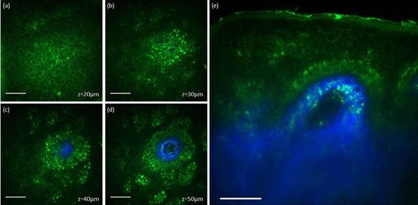
Figure 1
In vivo MPM images of normal human skin. (au2013d) Enu2010face MPM images (XY scans) showing keratinocytes (green fluorescence) and normal pigmented cells (bright green fluorescence) in the epidermis ...
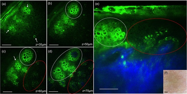
Figure 2
In vivo MPM images of human skin 3 hours postu2010treatment, 532u2009nm. (au2013d) Enu2010face MPM images (XY scans) showing keratinocytes in the epidermis and individual damaged cells (white arrows) ...
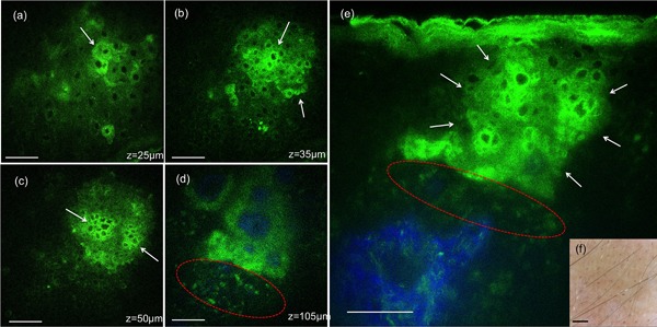
Figure 3
In vivo MPM images of human skin 24 hours postu2010treatment, 532u2009nm. (au2013d) Enu2010face MPM images (XY scans) showing keratinocytes in the epidermis, clusters of individual damaged cells (whit...
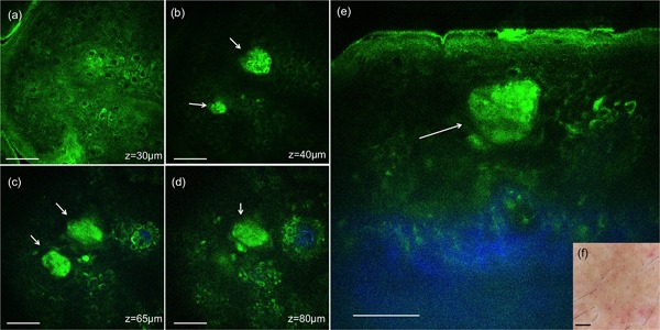
Figure 4
In vivo MPM images of human skin 24 hours postu2010treatment, 1064u2009nm. (au2013d) Enu2010face MPM images (XY scans) showing keratinocytes in the epidermis and clusters of individual damaged cells (...
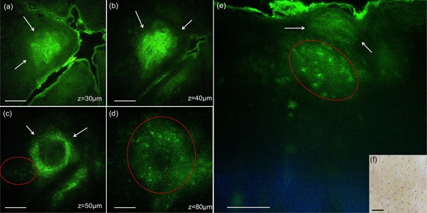
Figure 5
In vivo MPM images of human skin 7 days postu2010treatment, 532u2009nm. (au2013d) Enu2010face MPM images (XY scans) showing clusters of cellular necrotic debris in the epidermis (white arrows) at z =u...
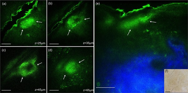
Figure 6
In vivo MPM images of human skin 7 days postu2010treatment, 1064u2009nm. (au2013d) Enu2010face MPM images (XY scans) showing clusters of cellular necrotic debris in the epidermis (white arrows) at z =...
Figure images are served from the NIH/NLM PubMed Central Open Access Subset or Europe PMC; copyright remains with the publishers and authors.
💬 Discussion
0 commentsNo comments yet. Be the first to start a discussion!
Leave a Comment