Abstract
Spindle orientation and nuclear migration are crucial events in cell growth and differentiation of many eukaryotes. Here we show that KIP3, the sixth and final kinesin-related gene in Saccharomyces cerevisiae, is required for migration of the nucleus to the bud site in preparation for mitosis. The position of the nucleus in the cell and the orientation of the mitotic spindle was examined by microscopy of fixed cells and by time-lapse microscopy of individual live cells. Mutations in KIP3 and in the dynein heavy chain gene defined two distinct phases of nuclear migration: a KIP3-dependent movement of the nucleus toward the incipient bud site and a dynein-dependent translocation of the nucleus through the bud neck during anaphase. Loss of KIP3 function disrupts the unidirectional movement of the nucleus toward the bud and mitotic spindle orientation, causing large oscillations in nuclear position. The oscillatory motions sometimes brought the nucleus in close proximity to the bud neck, possibly accounting for the viability of a kip3 null mutant. The kip3 null mutant exhibits normal translocation of the nucleus through the neck and normal spindle pole separation kinetics during anaphase. Simultaneous loss of KIP3 and kinesin-related KAR3 function, or of KIP3 and dynein function, is lethal but does not block any additional detectable movement. This suggests that the lethality is due to the combination of sequential and possibly overlapping defects. Epitope-tagged Kip3p localizes to astral and central spindle microtubules and is also present throughout the cytoplasm and nucleus.
🔬 Techniques
🧬 Organisms
💻 Software
✨ Fluorophores
🧪 Sample Preparation
🏭 Microscope Brands
🧪 Reagent Suppliers
📷 Detectors
🔎 Objectives
💻 Software Details
🏛️ Research Organizations (ROR)
Affiliated research institutions:
📋 Methods
Strains, Media, and Genetic Techniques Genotypes and sources of the strains and plasmids used in this study are listed in Table I . Strains were constructed by standard genetic methods ( Rose et al., 1990 ) and as described in the text. The yeast media used were SD minimal medium and sporulation medium supplemented with adenine, uracil, and appropriate amino acids; SC complete medium containing 5-fluoroorotic acid (5-FOA) 1 (1 mg/ml); and YPD rich medium ( Rose et al., 1990 ). The frequency of chromosome loss was assayed by scoring kip3/kip3 and wild-type diploid strains for loss of heterozygosity at the mating type locus of chromosome III. The diploid strains were mated to MAT a and MATα tester strains on YPD plates and replica printed to score prototrophic triploid colonies ( Spencer et al., 1990 ). Sensitivity to benomyl was tested on YPD medium containing 1% DMSO and the desired concentration of benomyl (E.I. du Pont de Nemours, Wilmington, DE).
Strains expressing the Nuf2p–green fluorescent protein
(GFP) fusion protein for localization of spindle poles ( Kahana et al., 1995 ) were constructed by integrating the NUF2–GFP gene fusion at the NUF2 locus using plasmid pJK67 (a gift of J. Kahana and P. Silver, Dana Farber Cancer Institute, Boston, MA). The NUF2–GFP gene fusion is expressed from the NUF2 promoter, and the GFP gene contains S65T and V163A mutations. KIP3 Deletion Construction, Gene Recovery, and Epitope Tagging A precise deletion of the KIP3 open reading frame ( kip3Δ ) was constructed by PCR ( Baudin et al., 1993 ). The 3′ ends of primers 1 and 2 contained sequences for PCR amplification of the HIS3 or TRP1 genes present in pRS403 or pRS404 ( Sikorski and Hieter, 1989 ), and the 5′ ends of the primers contained sequences immediately flanking the KIP3 gene. Primer 1 was of sequence 5′- ACTTGAGTTTTCTTTCCAGCTGTATACTATTGACACTAAC GAATTC GATTGTACTGAGAGTGCACC- 3′ , and primer 2 was of sequence 5′- GAAAGAAGTTATATTCGAT-AGTTTACGTAGGATATGTAT G GAATTC CTGTGCGGTAT T TC A- CACC-3′. (Sequences flanking KIP3 are underlined, and EcoRI restriction sites are in bold type.) The KIP3 locus in a diploid yeast strain was replaced by this PCR product using the one-step gene replacement procedure ( Rothstein, 1983 ). Correct recombinants were identified by PCR amplification and restriction digest analysis of genomic DNA. The primers used were primer 3, 5′-CCG GGATCC GACTCTTCTAATTGGTCTCT-3′ and primer 4, 5′-GGC GTCGAC GTCTCCTGAGAAACGTTTT-3′. (BamHI and SalI restriction sites are in bold type.) To recover the KIP3 gene, we screened a library of S. cerevisiae genomic DNA fragments cloned into the vector YCp50 ( URA3 CEN ARS ) by colony hybridization using a radiolabeled probe. The probe was a 556-bp sequence adjacent to the 5′ end of KIP3 that was prepared from a plasmid carrying the kip3Δ1::HIS3 allele using the EcoRI and BamHI sites introduced by primers 1 and 3. We identified a single plasmid that carried the full-length KIP3 gene on a 10–15-kb genomic DNA insert (pDR605) out of ∼4,100 colonies screened. For epitope tagging of KIP3 , the myc tag coding sequence ( Evan et al., 1985 ) was introduced into the KIP3 coding sequence as follows. A 3.8-kb fragment containing the entire KIP3 open reading frame, but lacking the termination codon, was generated by PCR using the Expand High Fidelity PCR system ( Boehringer Mannheim Corp. , Indianapolis, IN). This fragment was cloned into a vector containing six tandem copies of the myc tag (pB896 [ Li, 1997 ]). The resulting construct contained 1,419 bases of 5′ noncoding sequence, the entire KIP3 open reading frame, and the sequence encoding the myc epitopes just 5′ of the termination codon. To eliminate the possibility of PCR-generated errors, a 2.25-kb fragment of wild-type KIP3 was used to replace most of the PCR-generated clone. The remaining PCR-generated KIP3 sequences were verified using DNA sequencing. KIP3-6MYC (pB956) appears to be functional, as it fully complements the lethality of kip3Δ kar3Δ and kip3Δ dyn1Δ strains using the plasmid shuffle test described below. Tests of Synthetic Lethality To test whether the kip3Δ mutation was lethal in combination with other mutations, we generally constructed double mutants in the presence of a plasmid-based wild-type copy of one of the genes and then determined whether the strain remained viable after plasmid loss. The presence of the wild-type gene during strain construction prevented the potential isolation of aneuploid strains because of genomic instability. Synthetic lethality of kip3 and dyn1 mutations was tested by replacing the wild-type DYN1 gene with the dyn1Δ::HIS3 deletion allele (from plasmid p2-1H, a gift of E. Yeh and K. Bloom, University of North Carolina, Chapel Hill, NC) in the kip3Δ strain DS667, which carries the KIP3 URA3 plasmid pDR605, using one-step gene replacement. Correct deletion of DYN1 was confirmed by restriction analysis and Southern blotting of genomic DNA. Five independently constructed double mutants were inviable when plated on 5-FOA medium, which selects against cells carrying the URA3 plasmid ( Boeke et al., 1987 ), indicating that kip3 is synthetically lethal with dyn1. Synthetic lethality of kip3 with kar3 was tested by crossing a kip3Δ:: HIS3 strain with strain DS276 containing kar3Δ102::LEU2 and a KAR3 plasmid. The resultant diploid strain was sporulated, and the tetrads were dissected. The spores were 83% viable, and all 11 viable kip3 kar3 spores carried the KAR3 plasmid. An additional 16 inviable spores were inferred to carry both the kip3 and kar3 mutations based on analysis of markers in the remaining spores of each tetrad; these spores may have been inviable because they did not inherit the KAR3 plasmid. The viable kip3 kar3 strains were sensitive to 5-FOA at 30°C, indicating that the KAR3 plasmid was essential and that kip3 is synthetically lethal with kar3. In a second test of synthetic lethality, a diploid strain homozygous for kip3 and heterozygous for kar3 was sporulated, and tetrads were dissected. Two spores in each tetrad were kip3 KAR3 and viable, and two spores inferred to be kip3 kar3 germinated and divided two to six times before ceasing growth, confirming lethality of the double mutant. Synthetic lethality of kip3 with kip1 and cin8 was tested by crossing a kip3Δ::HIS3 strain with strain DS118 containing kip1Δ1 and a KIP1 plasmid, and strain DS379 containing cin8Δ112 and a CIN8 plasmid. The resultant diploid strains were sporulated, and 24 tetrads from each strain were dissected. The spores were >90% viable, and double kip3 kip1 and kip3 cin8 mutants were recovered at a frequency of ∼25%. Some of the double mutants did not contain the complementing plasmid, and those that did were resistant to 5-FOA, indicating the plasmid was not essential. The kip3 kip1 and kip3 cin8 double mutants were capable of vegetative growth at 16, 23, 30, and 37°C, indicating that kip3 is not synthetically lethal with kip1 or cin8. Synthetic lethality of kip3 with kip2 and smy1 was tested by crossing kip3Δ strains with kip2Δ strain MS2309 or smy1Δ strain SLY57. The resultant diploid strains were sporulated, and 24 tetrads were dissected. The spores were >90% viable, the kip3 kip2 and kip3 smy1 double mutant progeny were recovered at a frequency of ∼25%, and the double mutants were capable of vegetative growth at 16, 23, 30, and 37°C, indicating that kip3 is not synthetically lethal with kip2 or smy1. Synthetic lethality of dyn1 with kar3 was tested by crossing a kar3Δ strain bearing a KAR3 plasmid (strain DS276) with the dyn1Δ strain DS730. The resultant diploid was sporulated, and tetrads were dissected. Spore viability was 65%, and only 5 out of 24 tetrads yielded 4 viable spores that exhibited 2:2 segregation of mating type and the three auxotrophic markers present. Three dyn1 kar3 double mutants were recovered from these valid tetrads, and each double mutant carried the KAR3 plasmid. The double mutants were unable to grow on medium containing 5-FOA at 30°C, indicating that the plasmid was essential and that dyn1 and kar3 are synthetically lethal. Construction of kip3 Temperature-sensitive Alleles A 4,822-bp EcoRI/SpeI fragment from plasmid pDR605 was subcloned into the EcoRI and SpeI sites of pRS314 ( TRP1 CEN ARS ) ( Sikorski and Hieter, 1989 ) to yield the plasmid pB893 and further subcloned into pRS416 ( URA3 CEN ARS ) to yield plasmid pDR622. These plasmids carry the entire KIP3 open reading frame with 1,391 bp 5′ and 966 bp 3′ flanking DNA. Hydroxylamine mutagenesis of pB893 was performed by the method of Rose and Fink (1987) , and temperature-sensitive (ts) alleles of KIP3 were obtained after transformation of strain DS738 or DS716 using the plasmid shuffle technique ( Boeke et al., 1987 ). Transformation of the kip3-20 (ts) dyn1Δ strain DS765 with the KIP3 plasmid pDR622 complemented the temperature-sensitive growth, but transformation with the vector (pRS416) did not. This confirmed that the mutation conferring temperature-sensitive growth was in KIP3 and that the mutation is recessive. Morphological Observations Cells were fixed for immunofluorescence microscopy by adding formaldehyde to 3.7% directly to the culture medium and incubating for 2 h at 23°C, or overnight at 4°C. Cells were prepared for immunofluorescence microscopy as described ( Rose et al., 1990 ). For the Kip3p localization experiment, Kip3p-6myc was visualized with mAb 9E10, which recognizes the myc epitope ( Santa Cruz Biotechnology , Santa Cruz, CA), and microtubules were visualized with the rat antitubulin antibody YOL 1/34 (Accurate Chemical and Scientific Corp., Westbury, NY). Fluorochrome-conjugated secondary antibodies were from Jackson Immunoresearch Labs, Inc. (West Grove, PA). For double labeling of Kip3p and microtubules, controls demonstrated that cross-reactivity and light channel spill-over did not contribute to the final images when species-specific secondary antibodies were used. For all other immunofluorescence experiments, tubulin was stained with the antitubulin monoclonal antibody BIBE2, a gift of F. Solomon (Massachusetts Institute of Technology, Cambridge, MA). The secondary antibody was FITC-conjugated goat anti–mouse antibody (Accurate Chemical and Scientific Corp.), which was absorbed against fixed yeast cells for 3 h at 4°C before use to minimize background signal. DNA was stained using 4,6-diamidino-2-phenylindole (DAPI) ( Boehringer Mannheim Corp. ). To observe Nuf2p–GFP localization in fixed cells, cells in culture medium were fixed for 1 h with 3.7% formaldehyde, washed, and applied to polylysine-treated Teflon ® -masked slides for viewing. For live cell microscopy of spindle pole body location, cells were applied to a microscope slide with a thin pad of 1% agarose containing SC complete medium, and a coverslip was applied and sealed with a thin bead of a mixture of equal parts Vaseline, lanolin, and paraffin at the edges. Cells were imaged at 23–25°C using a microscope (model DMRBE; Leica, Inc., Deerfield, IL) equipped with a 100×/1.4 NA objective, a monochrome CCD camera (Cohu Inc., San Diego, CA), a 100-W mercury vapor lamp, and a fluorescein filter set. Images were captured using LG3 frame grabber hardware (Scion Corp., Frederick, MD) and NIH Image software (written by W. Rasband and available at http://rsb.info.nih.gov/ nih-image/) with a custom set of macro programs to control the camera exposure settings, fluorescence illumination shutter, and motorized microscope stage. At each time point, both differential interference contrast (DIC) and fluorescence images were photographed at three focal planes spaced at 1-μm intervals to increase the likelihood of detecting both spindle pole bodies. Illumination during fluorescence photography was controlled by a shutter and was 200 ms or less for each image. Micrographs were spatially calibrated using NIH image software and a stage micrometer. Distance and angle measurements were made using Object Image software, a modified version of NIH Image available at the above internet site. Measurements to determine nuclear migration indices were made on digital images of cells by the method of Jacobs et al. (1988) , and the statistical significance of the difference between the variances of nuclear migration indices was tested using the two-tailed variance ratio test at a 5% significance level.
Show full methods section
Strains, Media, and Genetic Techniques Genotypes and sources of the strains and plasmids used in this study are listed in Table I . Strains were constructed by standard genetic methods ( Rose et al., 1990 ) and as described in the text. The yeast media used were SD minimal medium and sporulation medium supplemented with adenine, uracil, and appropriate amino acids; SC complete medium containing 5-fluoroorotic acid (5-FOA) 1 (1 mg/ml); and YPD rich medium ( Rose et al., 1990 ). The frequency of chromosome loss was assayed by scoring kip3/kip3 and wild-type diploid strains for loss of heterozygosity at the mating type locus of chromosome III. The diploid strains were mated to MAT a and MATα tester strains on YPD plates and replica printed to score prototrophic triploid colonies ( Spencer et al., 1990 ). Sensitivity to benomyl was tested on YPD medium containing 1% DMSO and the desired concentration of benomyl (E.I. du Pont de Nemours, Wilmington, DE).
Strains expressing the Nuf2p–green fluorescent protein
(GFP) fusion protein for localization of spindle poles ( Kahana et al., 1995 ) were constructed by integrating the NUF2–GFP gene fusion at the NUF2 locus using plasmid pJK67 (a gift of J. Kahana and P. Silver, Dana Farber Cancer Institute, Boston, MA). The NUF2–GFP gene fusion is expressed from the NUF2 promoter, and the GFP gene contains S65T and V163A mutations. KIP3 Deletion Construction, Gene Recovery, and Epitope Tagging A precise deletion of the KIP3 open reading frame ( kip3Δ ) was constructed by PCR ( Baudin et al., 1993 ). The 3′ ends of primers 1 and 2 contained sequences for PCR amplification of the HIS3 or TRP1 genes present in pRS403 or pRS404 ( Sikorski and Hieter, 1989 ), and the 5′ ends of the primers contained sequences immediately flanking the KIP3 gene. Primer 1 was of sequence 5′- ACTTGAGTTTTCTTTCCAGCTGTATACTATTGACACTAAC GAATTC GATTGTACTGAGAGTGCACC- 3′ , and primer 2 was of sequence 5′- GAAAGAAGTTATATTCGAT-AGTTTACGTAGGATATGTAT G GAATTC CTGTGCGGTAT T TC A- CACC-3′. (Sequences flanking KIP3 are underlined, and EcoRI restriction sites are in bold type.) The KIP3 locus in a diploid yeast strain was replaced by this PCR product using the one-step gene replacement procedure ( Rothstein, 1983 ). Correct recombinants were identified by PCR amplification and restriction digest analysis of genomic DNA. The primers used were primer 3, 5′-CCG GGATCC GACTCTTCTAATTGGTCTCT-3′ and primer 4, 5′-GGC GTCGAC GTCTCCTGAGAAACGTTTT-3′. (BamHI and SalI restriction sites are in bold type.) To recover the KIP3 gene, we screened a library of S. cerevisiae genomic DNA fragments cloned into the vector YCp50 ( URA3 CEN ARS ) by colony hybridization using a radiolabeled probe. The probe was a 556-bp sequence adjacent to the 5′ end of KIP3 that was prepared from a plasmid carrying the kip3Δ1::HIS3 allele using the EcoRI and BamHI sites introduced by primers 1 and 3. We identified a single plasmid that carried the full-length KIP3 gene on a 10–15-kb genomic DNA insert (pDR605) out of ∼4,100 colonies screened. For epitope tagging of KIP3 , the myc tag coding sequence ( Evan et al., 1985 ) was introduced into the KIP3 coding sequence as follows. A 3.8-kb fragment containing the entire KIP3 open reading frame, but lacking the termination codon, was generated by PCR using the Expand High Fidelity PCR system ( Boehringer Mannheim Corp. , Indianapolis, IN). This fragment was cloned into a vector containing six tandem copies of the myc tag (pB896 [ Li, 1997 ]). The resulting construct contained 1,419 bases of 5′ noncoding sequence, the entire KIP3 open reading frame, and the sequence encoding the myc epitopes just 5′ of the termination codon. To eliminate the possibility of PCR-generated errors, a 2.25-kb fragment of wild-type KIP3 was used to replace most of the PCR-generated clone. The remaining PCR-generated KIP3 sequences were verified using DNA sequencing. KIP3-6MYC (pB956) appears to be functional, as it fully complements the lethality of kip3Δ kar3Δ and kip3Δ dyn1Δ strains using the plasmid shuffle test described below. Tests of Synthetic Lethality To test whether the kip3Δ mutation was lethal in combination with other mutations, we generally constructed double mutants in the presence of a plasmid-based wild-type copy of one of the genes and then determined whether the strain remained viable after plasmid loss. The presence of the wild-type gene during strain construction prevented the potential isolation of aneuploid strains because of genomic instability. Synthetic lethality of kip3 and dyn1 mutations was tested by replacing the wild-type DYN1 gene with the dyn1Δ::HIS3 deletion allele (from plasmid p2-1H, a gift of E. Yeh and K. Bloom, University of North Carolina, Chapel Hill, NC) in the kip3Δ strain DS667, which carries the KIP3 URA3 plasmid pDR605, using one-step gene replacement. Correct deletion of DYN1 was confirmed by restriction analysis and Southern blotting of genomic DNA. Five independently constructed double mutants were inviable when plated on 5-FOA medium, which selects against cells carrying the URA3 plasmid ( Boeke et al., 1987 ), indicating that kip3 is synthetically lethal with dyn1. Synthetic lethality of kip3 with kar3 was tested by crossing a kip3Δ:: HIS3 strain with strain DS276 containing kar3Δ102::LEU2 and a KAR3 plasmid. The resultant diploid strain was sporulated, and the tetrads were dissected. The spores were 83% viable, and all 11 viable kip3 kar3 spores carried the KAR3 plasmid. An additional 16 inviable spores were inferred to carry both the kip3 and kar3 mutations based on analysis of markers in the remaining spores of each tetrad; these spores may have been inviable because they did not inherit the KAR3 plasmid. The viable kip3 kar3 strains were sensitive to 5-FOA at 30°C, indicating that the KAR3 plasmid was essential and that kip3 is synthetically lethal with kar3. In a second test of synthetic lethality, a diploid strain homozygous for kip3 and heterozygous for kar3 was sporulated, and tetrads were dissected. Two spores in each tetrad were kip3 KAR3 and viable, and two spores inferred to be kip3 kar3 germinated and divided two to six times before ceasing growth, confirming lethality of the double mutant. Synthetic lethality of kip3 with kip1 and cin8 was tested by crossing a kip3Δ::HIS3 strain with strain DS118 containing kip1Δ1 and a KIP1 plasmid, and strain DS379 containing cin8Δ112 and a CIN8 plasmid. The resultant diploid strains were sporulated, and 24 tetrads from each strain were dissected. The spores were >90% viable, and double kip3 kip1 and kip3 cin8 mutants were recovered at a frequency of ∼25%. Some of the double mutants did not contain the complementing plasmid, and those that did were resistant to 5-FOA, indicating the plasmid was not essential. The kip3 kip1 and kip3 cin8 double mutants were capable of vegetative growth at 16, 23, 30, and 37°C, indicating that kip3 is not synthetically lethal with kip1 or cin8. Synthetic lethality of kip3 with kip2 and smy1 was tested by crossing kip3Δ strains with kip2Δ strain MS2309 or smy1Δ strain SLY57. The resultant diploid strains were sporulated, and 24 tetrads were dissected. The spores were >90% viable, the kip3 kip2 and kip3 smy1 double mutant progeny were recovered at a frequency of ∼25%, and the double mutants were capable of vegetative growth at 16, 23, 30, and 37°C, indicating that kip3 is not synthetically lethal with kip2 or smy1. Synthetic lethality of dyn1 with kar3 was tested by crossing a kar3Δ strain bearing a KAR3 plasmid (strain DS276) with the dyn1Δ strain DS730. The resultant diploid was sporulated, and tetrads were dissected. Spore viability was 65%, and only 5 out of 24 tetrads yielded 4 viable spores that exhibited 2:2 segregation of mating type and the three auxotrophic markers present. Three dyn1 kar3 double mutants were recovered from these valid tetrads, and each double mutant carried the KAR3 plasmid. The double mutants were unable to grow on medium containing 5-FOA at 30°C, indicating that the plasmid was essential and that dyn1 and kar3 are synthetically lethal. Construction of kip3 Temperature-sensitive Alleles A 4,822-bp EcoRI/SpeI fragment from plasmid pDR605 was subcloned into the EcoRI and SpeI sites of pRS314 ( TRP1 CEN ARS ) ( Sikorski and Hieter, 1989 ) to yield the plasmid pB893 and further subcloned into pRS416 ( URA3 CEN ARS ) to yield plasmid pDR622. These plasmids carry the entire KIP3 open reading frame with 1,391 bp 5′ and 966 bp 3′ flanking DNA. Hydroxylamine mutagenesis of pB893 was performed by the method of Rose and Fink (1987) , and temperature-sensitive (ts) alleles of KIP3 were obtained after transformation of strain DS738 or DS716 using the plasmid shuffle technique ( Boeke et al., 1987 ). Transformation of the kip3-20 (ts) dyn1Δ strain DS765 with the KIP3 plasmid pDR622 complemented the temperature-sensitive growth, but transformation with the vector (pRS416) did not. This confirmed that the mutation conferring temperature-sensitive growth was in KIP3 and that the mutation is recessive. Morphological Observations Cells were fixed for immunofluorescence microscopy by adding formaldehyde to 3.7% directly to the culture medium and incubating for 2 h at 23°C, or overnight at 4°C. Cells were prepared for immunofluorescence microscopy as described ( Rose et al., 1990 ). For the Kip3p localization experiment, Kip3p-6myc was visualized with mAb 9E10, which recognizes the myc epitope ( Santa Cruz Biotechnology , Santa Cruz, CA), and microtubules were visualized with the rat antitubulin antibody YOL 1/34 (Accurate Chemical and Scientific Corp., Westbury, NY). Fluorochrome-conjugated secondary antibodies were from Jackson Immunoresearch Labs, Inc. (West Grove, PA). For double labeling of Kip3p and microtubules, controls demonstrated that cross-reactivity and light channel spill-over did not contribute to the final images when species-specific secondary antibodies were used. For all other immunofluorescence experiments, tubulin was stained with the antitubulin monoclonal antibody BIBE2, a gift of F. Solomon (Massachusetts Institute of Technology, Cambridge, MA). The secondary antibody was FITC-conjugated goat anti–mouse antibody (Accurate Chemical and Scientific Corp.), which was absorbed against fixed yeast cells for 3 h at 4°C before use to minimize background signal. DNA was stained using 4,6-diamidino-2-phenylindole (DAPI) ( Boehringer Mannheim Corp. ). To observe Nuf2p–GFP localization in fixed cells, cells in culture medium were fixed for 1 h with 3.7% formaldehyde, washed, and applied to polylysine-treated Teflon ® -masked slides for viewing. For live cell microscopy of spindle pole body location, cells were applied to a microscope slide with a thin pad of 1% agarose containing SC complete medium, and a coverslip was applied and sealed with a thin bead of a mixture of equal parts Vaseline, lanolin, and paraffin at the edges. Cells were imaged at 23–25°C using a microscope (model DMRBE; Leica, Inc., Deerfield, IL) equipped with a 100×/1.4 NA objective, a monochrome CCD camera (Cohu Inc., San Diego, CA), a 100-W mercury vapor lamp, and a fluorescein filter set. Images were captured using LG3 frame grabber hardware (Scion Corp., Frederick, MD) and NIH Image software (written by W. Rasband and available at http://rsb.info.nih.gov/ nih-image/) with a custom set of macro programs to control the camera exposure settings, fluorescence illumination shutter, and motorized microscope stage. At each time point, both differential interference contrast (DIC) and fluorescence images were photographed at three focal planes spaced at 1-μm intervals to increase the likelihood of detecting both spindle pole bodies. Illumination during fluorescence photography was controlled by a shutter and was 200 ms or less for each image. Micrographs were spatially calibrated using NIH image software and a stage micrometer. Distance and angle measurements were made using Object Image software, a modified version of NIH Image available at the above internet site. Measurements to determine nuclear migration indices were made on digital images of cells by the method of Jacobs et al. (1988) , and the statistical significance of the difference between the variances of nuclear migration indices was tested using the two-tailed variance ratio test at a 5% significance level.
📊 Figures
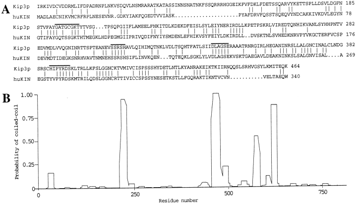
Figure 1
Kip3p sequence alignment and coiled-coil formation probability. ( A ) Alignment of Kip3p residues 86-464 with human kinesin ( huKIN ) ( Navone et al., 1992 ) residues 1u2013340 was performed using the...
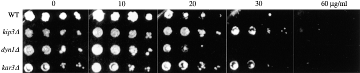
Figure 2
Increased benomyl resistance in the kip3u0394 mutant. Strains wild-type (DS140), kar3u0394102 (DS276), kip3u0394 (DS614), and dyn1u0394 (DS730) were incubated at 23u00b0C for 2 d in liquid YPD medium....
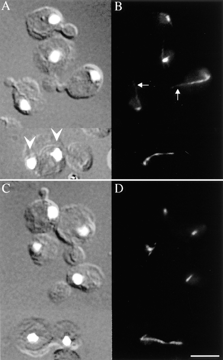
Figure 3
Disruption of KIP3 affects nuclear migration. kip3u0394 strain DS614 ( A and B ) and wild-type strain DS141 ( C and D ) were grown in YPD medium at 30u00b0C to mid-logarithmic phase and prepared for i...
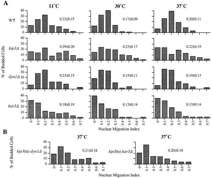
Figure 4
Loss of KIP3 function causes mislocalization of undivided nuclei. ( A ) Histograms of nuclear migration indices of wild-type (DS141), kip3u0394 (DS614), dyn1u0394 (DS730), and kar3u0394 (DS276) strain...
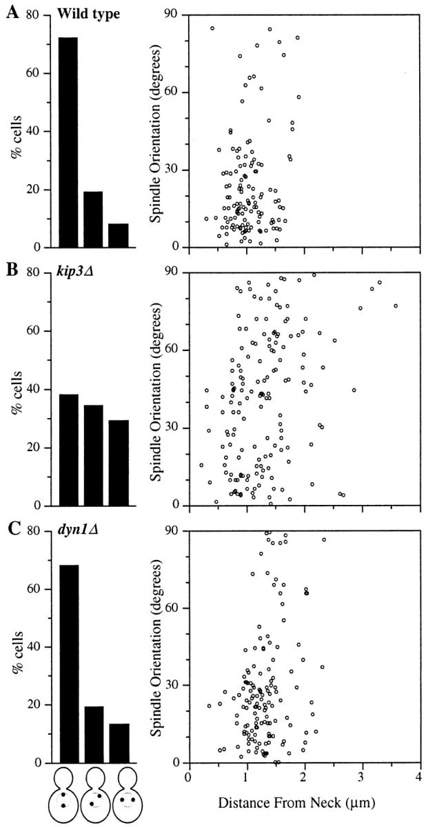
Figure 5
Loss of KIP3 function causes misorientation of preanaphase spindles. The histograms on the left indicate the frequency of cells with bipolar spindles that are oriented from 0u201330 degrees of the mot...
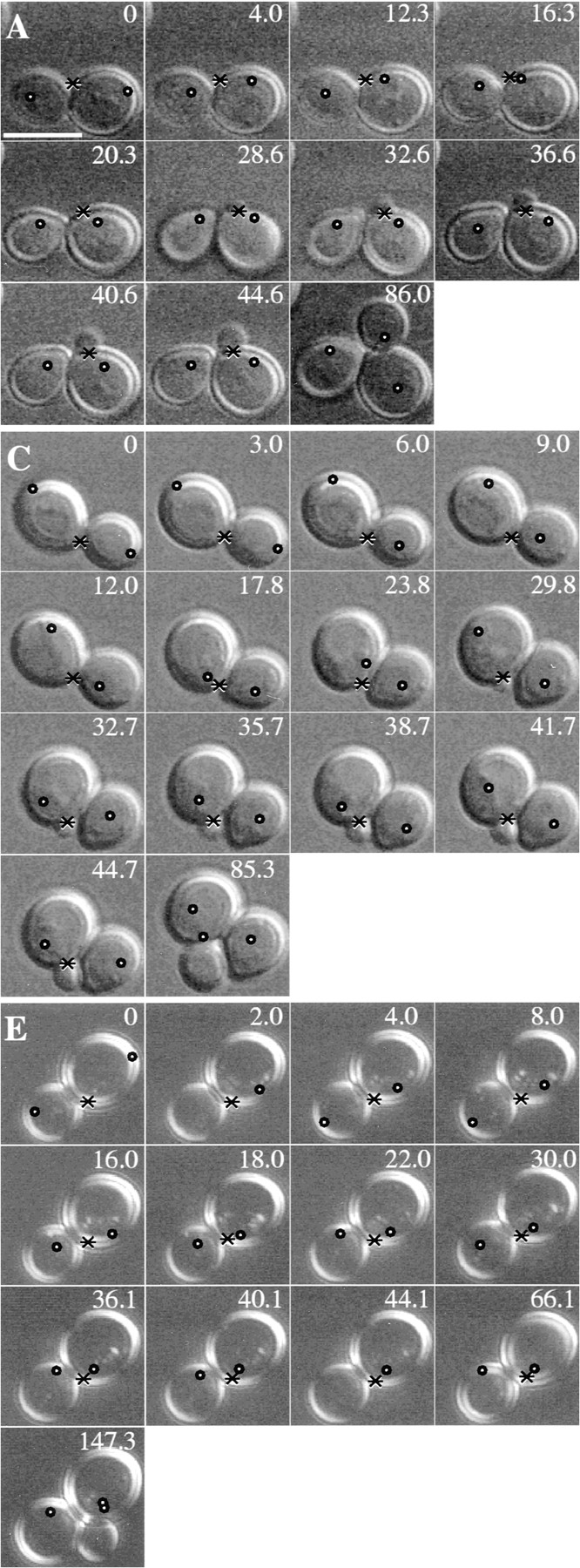
Figure 6
Large oscillations in position of nuclei after spindle disassembly in the kip3u0394 mutant. ( A , C , and E ) A time-lapse series of DIC micrographs of wild-type ( A ), kip3u0394 ( C ), and dynu0394 (...
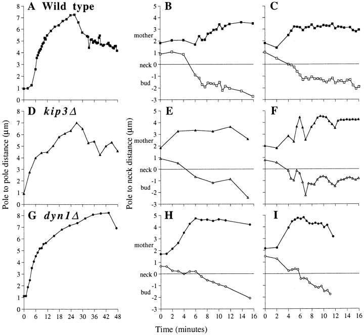
Figure 7
Normal kinetics of anaphase spindle elongation in the kip3u0394 mutant. ( A , D , and G ) Pole-to-pole distance as a function of time in an individual cell of the indicated strain. Each cell exhibited...
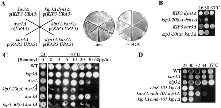
Figure 8
( A ) Synthetic lethality of kip3 , dyn1 , and kar3. Strains kip3 , dyn1 , kar3 , kip3 dyn1 (DS732), kip3 kar3 (DS716), and dyn1 kar3 (DS743) carry a centromere-based plasmid with the markers indicate...
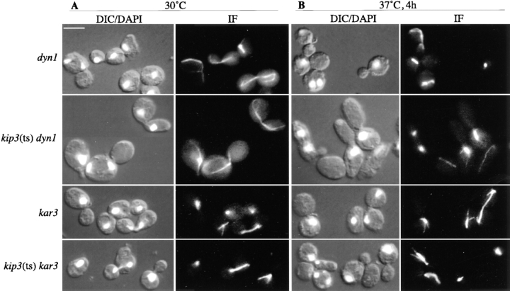
Figure 9
Morphology of kip3 dyn1 and kip3 kar3 double mutants. ( A ) The indicated strains were grown to mid-logarithmic phase at 30u00b0C in YPD medium and fixed for microscopy. ( B ) The same cultures shifte...
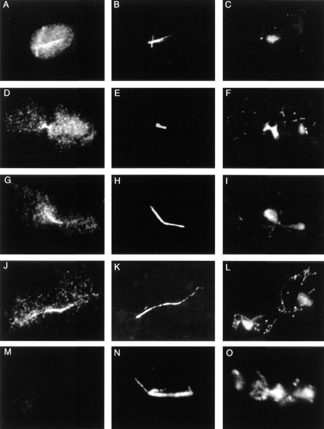
Figure 10
Epitope-tagged Kip3p localizes to astral and spindle microtubules and is present in the cytoplasm and nucleoplasm. The cells are from an asynchronous culture containing epitope-tagged Kip3p expressed ...
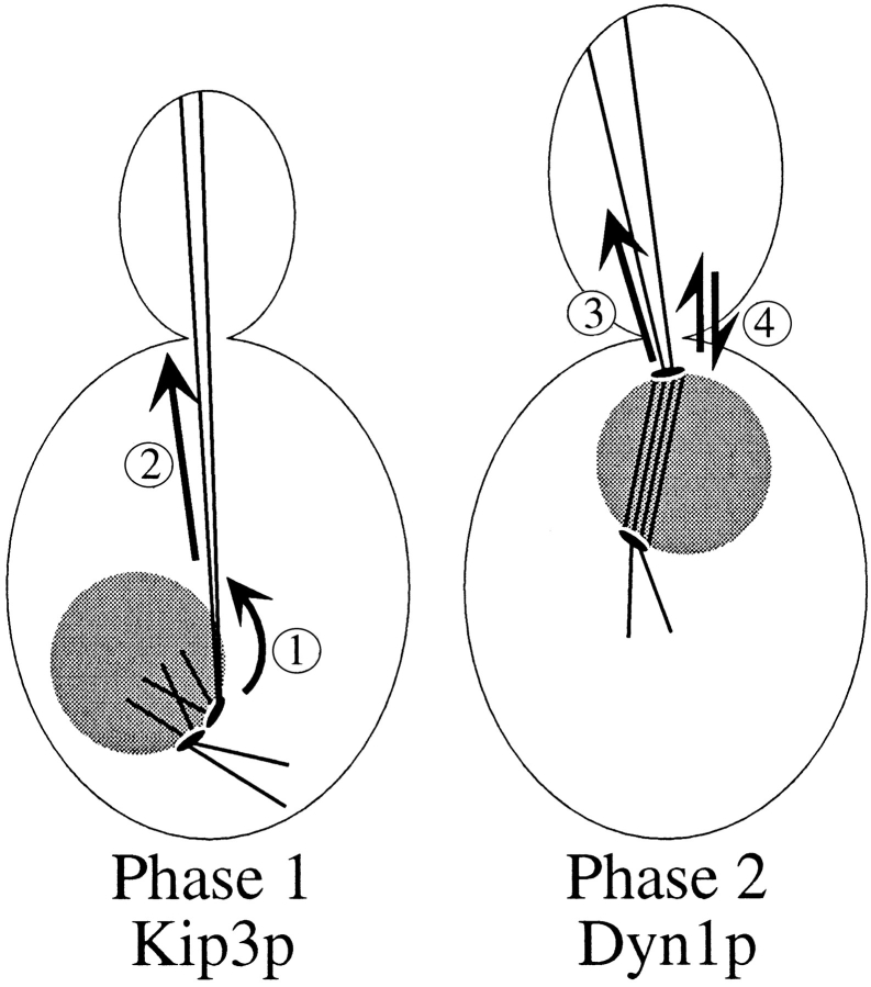
Figure 11
Distinct roles of Kip3p and Dyn1p in the two phases of nuclear migration. Based on the phenotypes of kip3 and dyn1 mutants, Kip3p primarily functions during phase 1 of nuclear migration to ( 1 ) orien...
Figure images are served from the NIH/NLM PubMed Central Open Access Subset or Europe PMC; copyright remains with the publishers and authors.
💬 Discussion
0 commentsNo comments yet. Be the first to start a discussion!
Leave a Comment