Abstract
The maintenance of whole-body glucose homeostasis is critical for survival, and is controlled by the coordination of multiple organs and endocrine systems. Pancreatic islet β cells secrete insulin in response to nutrient stimuli, and insulin then travels through the circulation promoting glucose uptake into insulin-responsive tissues such as liver, skeletal muscle and adipose. Many of the genes identified in human genome-wide association studies of diabetic individuals are directly associated with β cell survival and function, giving credence to the idea that β-cell dysfunction is central to the development of type 2 diabetes. As such, investigations into the mechanisms by which β cells sense glucose and secrete insulin in a regulated manner are a major focus of current diabetes research. In particular, recent discoveries of the detailed role and requirements for reorganization/remodeling of filamentous actin (F-actin) in the regulation of insulin release from the β cell have appeared at the forefront of islet function research, having lapsed in prior years due to technical limitations. Recent advances in live-cell imaging and specialized reagents have revealed localized F-actin remodeling to be a requisite for the normal biphasic pattern of nutrient-stimulated insulin secretion. This review will provide an historical look at the emergent focus on the role of the actin cytoskeleton and its regulation of insulin secretion, leading up to the cutting-edge research in progress in the field today.
🔬 Techniques
✨ Fluorophores
🧪 Sample Preparation
🔬 Cell Lines
🏛️ Research Organizations (ROR)
Affiliated research institutions:
📊 Figures
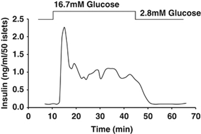
Figure 1
Biphasic glucose-stimulated insulin secretion from perfused mouse islets. In response to a square-wave increase in glucose concentration, islet u03b2 cells dock/fuse u223c50u2013100 granules in the fi...
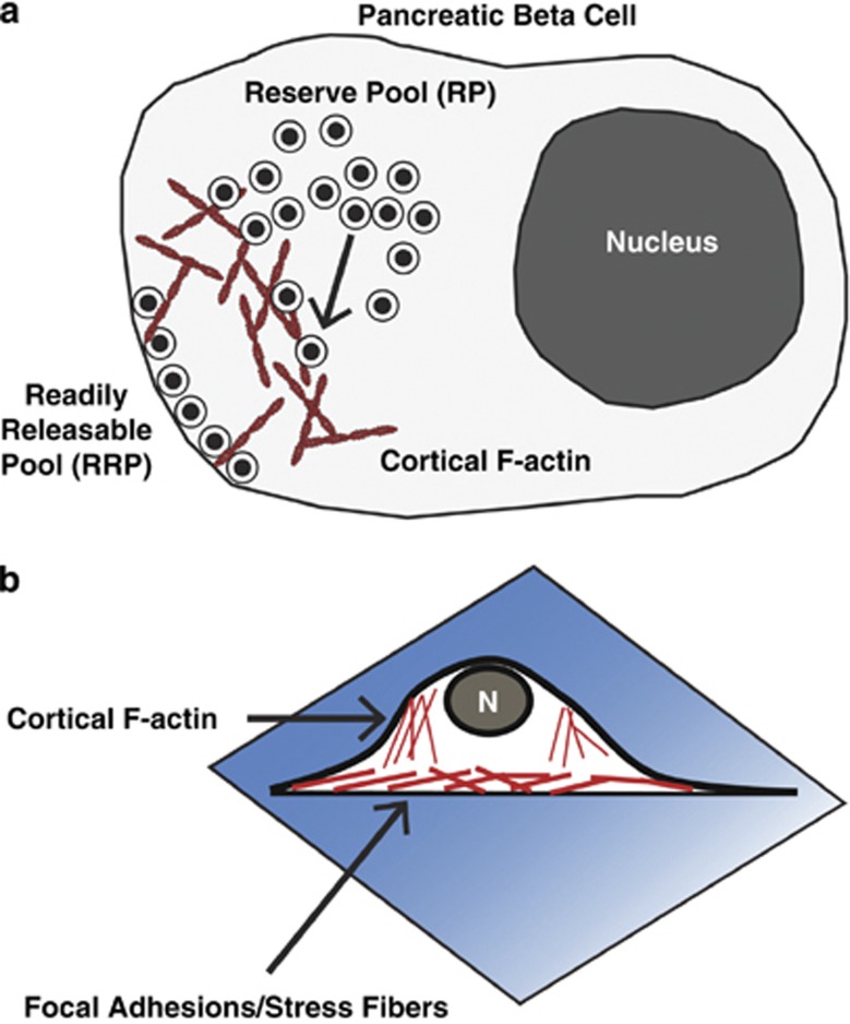
Figure 2
Cortical filamentous actin (F-actin) remodeling in u03b2 cells regulates insulin granule exocytosis. ( a ) Schematic of a pancreatic u03b2 cell depicts how readily releasable pool (RRP) granules are d...
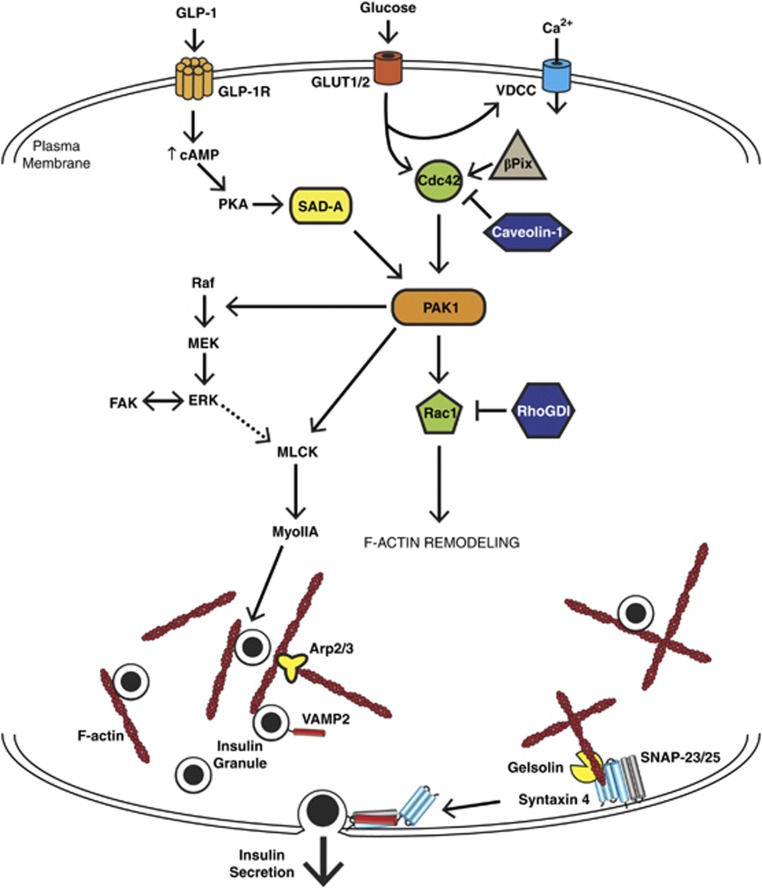
Figure 3
Pathways impinging specifically on glucose-mediated F-actin remodeling in the u03b2 cell. The u03b2 cell responds to certain insulin secretagogues by remodeling its cortical filamentous actin (F-actin...
Figure images are served from the NIH/NLM PubMed Central Open Access Subset or Europe PMC; copyright remains with the publishers and authors.
💬 Discussion
0 commentsNo comments yet. Be the first to start a discussion!
Leave a Comment