Abstract
The bacteriophage T4 capsid is an elongated icosahedron, 120 nm long and 86 nm wide, and is built with three essential proteins; gp23*, which forms the hexagonal capsid lattice, gp24*, which forms pentamers at eleven of the twelve vertices, and gp20, which forms the unique dodecameric portal vertex through which DNA enters during packaging and exits during infection. The past twenty years of research has greatly elevated the understanding of phage T4 head assembly and DNA packaging. The atomic structure of gp24 has been determined. A structural model built for gp23 using its similarity to gp24 showed that the phage T4 major capsid protein has the same fold as that found in phage HK97 and several other icosahedral bacteriophages. Folding of gp23 requires the assistance of two chaperones, the E. coli chaperone GroEL and the phage coded gp23-specific chaperone, gp31. The capsid also contains two non-essential outer capsid proteins, Hoc and Soc, which decorate the capsid surface. The structure of Soc shows two capsid binding sites which, through binding to adjacent gp23 subunits, reinforce the capsid structure. Hoc and Soc have been extensively used in bipartite peptide display libraries and to display pathogen antigens including those from HIV, Neisseria meningitides, Bacillus anthracis, and FMDV. The structure of Ip1*, one of the components of the core, has been determined, which provided insights on how IPs protect T4 genome against the E. coli nucleases that degrade hydroxymethylated and glycosylated T4 DNA. Extensive mutagenesis combined with the atomic structures of the DNA packaging/terminase proteins gp16 and gp17 elucidated the ATPase and nuclease functional motifs involved in DNA translocation and headful DNA cutting. Cryo-EM structure of the T4 packaging machine showed a pentameric motor assembled with gp17 subunits on the portal vertex. Single molecule optical tweezers and fluorescence studies showed that the T4 motor packages DNA at a rate of up to 2000 bp/sec, the fastest reported to date of any packaging motor. FRET-FCS studies indicate that the DNA gets compressed during the translocation process. The current evidence suggests a mechanism in which electrostatic forces generated by ATP hydrolysis drive the DNA translocation by alternating the motor between tensed and relaxed states.
🔬 Techniques
🧬 Organisms
✨ Fluorophores
🧪 Sample Preparation
🏭 Microscope Brands
🧪 Reagent Suppliers
🏛️ Research Organizations (ROR)
Affiliated research institutions:
📊 Figures
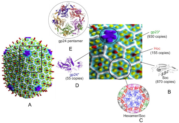
Figure 1
Structure of the bacteriophage T4 head . A) Cryo-EM reconstruction of phage T4 capsid [ 5 ]; the square block shows enlarged view showing gp23 (yellow subunits), gp24 (purple subunits), Hoc (red subun...
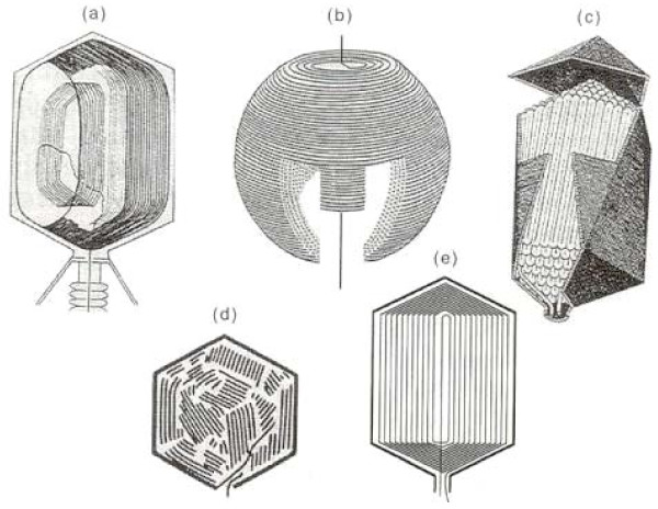
Figure 2
Models of packaged DNA structure . a) T4 DNA is packed longitudinally to the head-tail axis [ 91 ], unlike the transverse packaging in T7 capsids [ 16 ] (b) . Other models shown include spiral fold (c...
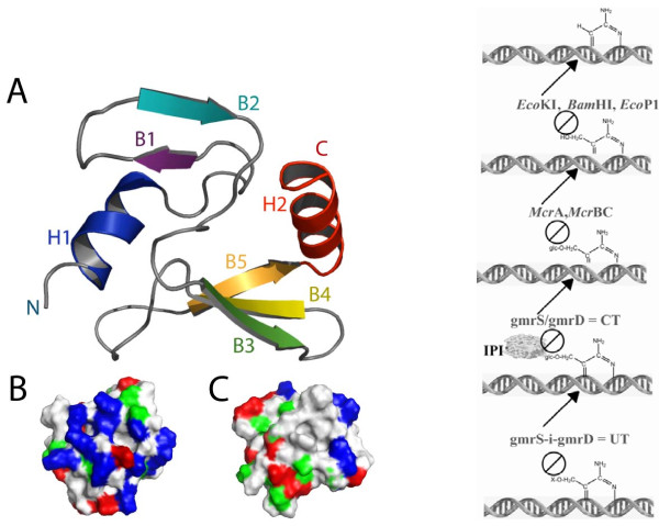
Figure 3
Structure and function of T4 internal protein I* . The NMR structure of IP1*, a highly specific inhibitor of the two-subunit CT (gmrS/gmrD) glucosyl-hmC DNA directed restriction endonuclease (right pa...
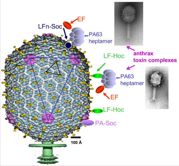
Figure 4
In vitro display of antigens on bacteriophage T4 capsid . Schematic representation of the T4 capsid decorated with large antigens, PA (83 kDa) and LF (89 kDa), or hetero-oligomeric anthrax toxin compl...
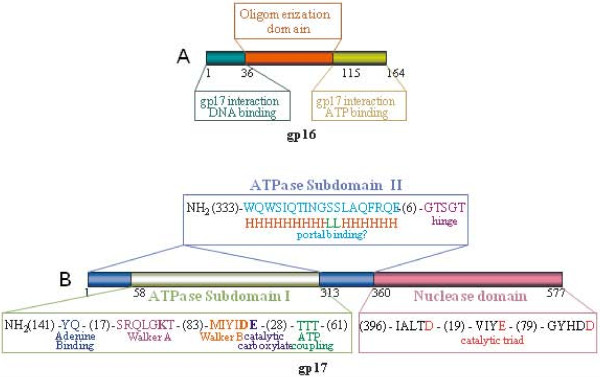
Figure 5
Domains and motifs in phage T4 terminase proteins . Schematic representation of domains and motifs in the small terminase protein gp16. A) and the large terminase protein gp17 (B) . The functionally c...
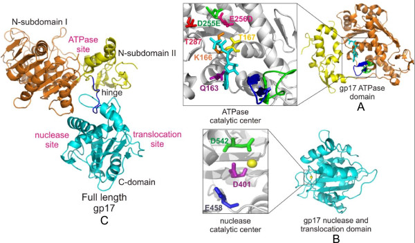
Figure 6
Structures of the T4 packaging motor protein, gp17 . Structures of the ATPase domain: A) nuclease/translocation domain; B) , and full-length gp17; C) . Various functional sites and critical catalytic ...
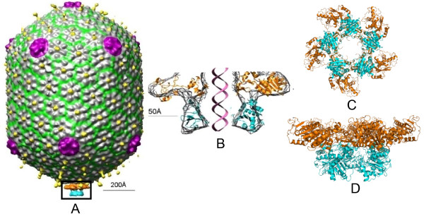
Figure 7
Structure of the T4 DNA packaging machine . A) Cryo-EM reconstruction of the phage T4 DNA packaging machine showing the pentameric motor assembled at the special portal vertex. B-D) Cross section, top...
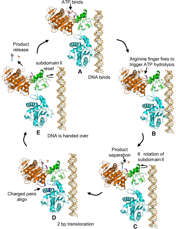
Figure 8
A model for the electrostatic force driven DNA packaging mechanism . Schematic representation showing the sequence of events that occur in a single gp17 molecule to translocate 2 bp of DNA (see the te...
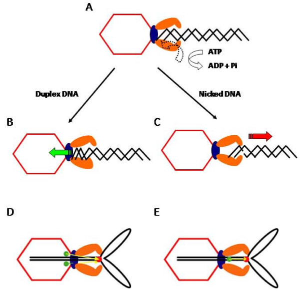
Figure 9
A model for the torsional compression portal-DNA-grip-and-release packaging mechanism . A-C) Short nicked or other abnormal structure containing DNA substrates are released from the motor. D) Leader c...
Figure images are served from the NIH/NLM PubMed Central Open Access Subset or Europe PMC; copyright remains with the publishers and authors.
💬 Discussion
0 commentsNo comments yet. Be the first to start a discussion!
Leave a Comment