Abstract
We have used time-lapse fluorescence microscopy to study the properties of the Cdc25B and Cdc25C phosphatases that have both been implicated as initiators of mitosis in human cells. To differentiate between the functions of the two proteins, we have microinjected expression constructs encoding Cdc25B or Cdc25C or their GFP-chimeras into synchronized tissue culture cells. This assay allows us to express the proteins at defined points in the cell cycle. We have followed the microinjected cells by time-lapse microscopy, in the presence or absence of DNA synthesis inhibitors, and assayed whether they enter mitosis prematurely or at the correct time. We find that overexpressing Cdc25B alone rapidly causes S phase and G2 phase cells to enter mitosis, whether or not DNA replication is complete, whereas overexpressing Cdc25C does not cause premature mitosis. Overexpressing Cdc25C together with cyclin B1 does shorten the G2 phase and can override the unreplicated DNA checkpoint, but much less efficiently than overexpressing Cdc25B. These results suggest that Cdc25B and Cdc25C do not respond identically to the same cell cycle checkpoints. This difference may be related to the differential localization of the proteins; Cdc25C is nuclear throughout interphase, whereas Cdc25B is nuclear in the G1 phase and cytoplasmic in the S and G2 phases. We have found that the change in subcellular localization of Cdc25B is due to nuclear export and that this is dependent on cyclin B1. Our data suggest that although both Cdc25B and Cdc25C can promote mitosis, they are likely to have distinct roles in the controlling the initiation of mitosis.
🔬 Techniques
🧬 Organisms
💻 Software
✨ Fluorophores
🧪 Sample Preparation
🔬 Cell Lines
🏭 Microscope Brands
🧪 Reagent Suppliers
📷 Detectors
💻 Software Details
🏛️ Research Organizations (ROR)
Affiliated research institutions:
📋 Methods
Cell Culture
HeLa cells were grown in DME supplemented with 5% newborn calf serum, 5% FCS, and antibiotics. HeLa cells were synchronized using a thymidine-aphidicolin protocol ( Pines and Hunter 1989 ). Cells were cultured on the microscope stage in a CO 2 -independent medium without phenol red (GIBCO BRL). The medium was overlaid with mineral oil (Sigma Chemical Co.) to prevent evaporation. Hydroxyurea was used at a final concentration of 2.5 mM and was added immediately after release from the aphidicolin block.
Plasmid Constructs and Protein Purification
The cyclin B1-MmGFP, cdc25C(S216G), and Wee1 constructs have been previously described ( Hagting et al. 1998 ). Cyclin B1 R42A -MmGFP was the gift of Paul Clute (Wellcome/CRC Institute). Cdc25B3 and Cdc25C were tagged at the amino terminus with MmGFP ( Zernicka-Goetz et al. 1996 , Zernicka-Goetz et al. 1997 ) by PCR using Taq polymerase and cloned into the pCMX vector ( Umesono et al. 1991 ). The stop codon of GFP was mutated to a Hind III site to link it with the first amino acid of Cdc25B or Cdc25C creating a 3–amino acid linker (Gly-Ile-Pro). Myc-tagged cdc25B2 ( Lammer et al. 1998 ) was cloned into the pCDNA3 vector for expression in HeLa cells. All constructs were sequenced using an Applied Biosystems DNA sequencer. Cyclin B1–cdc2 K33R complexes were expressed in and purified from baculovirus-infected cells as described ( Hagting et al. 1998 ). Cyclin B1 F146A was expressed in Escherichia coli and purified as described ( Hagting et al. 1999 ).
Microinjection and PCC Detection
Constructs expressing cDNAs under the control of the cytomegalovirus promoter were microinjected into cell nuclei using an Eppendorf semi-automatic microinjection apparatus. To assay for condensed chromatin, Hoechst 33342 was added to cells at a concentration of 1 μg/ml at the end of the experiment. Injected cells were identified by green fluorescent protein (GFP) fluorescence and those that had rounded up with abnormally condensed chromatin were scored. At least 50 cells were scored for each injected construct and experiment. Apoptotic cells were assayed using the apoptosis detection kit (R&D Systems, Inc.) and HeLa cells treated with cycloheximide plus tumor necrosis factor α were used as positive controls.
Show full methods section
Cell Culture
HeLa cells were grown in DME supplemented with 5% newborn calf serum, 5% FCS, and antibiotics. HeLa cells were synchronized using a thymidine-aphidicolin protocol ( Pines and Hunter 1989 ). Cells were cultured on the microscope stage in a CO 2 -independent medium without phenol red (GIBCO BRL). The medium was overlaid with mineral oil (Sigma Chemical Co.) to prevent evaporation. Hydroxyurea was used at a final concentration of 2.5 mM and was added immediately after release from the aphidicolin block.
Plasmid Constructs and Protein Purification
The cyclin B1-MmGFP, cdc25C(S216G), and Wee1 constructs have been previously described ( Hagting et al. 1998 ). Cyclin B1 R42A -MmGFP was the gift of Paul Clute (Wellcome/CRC Institute). Cdc25B3 and Cdc25C were tagged at the amino terminus with MmGFP ( Zernicka-Goetz et al. 1996 , Zernicka-Goetz et al. 1997 ) by PCR using Taq polymerase and cloned into the pCMX vector ( Umesono et al. 1991 ). The stop codon of GFP was mutated to a Hind III site to link it with the first amino acid of Cdc25B or Cdc25C creating a 3–amino acid linker (Gly-Ile-Pro). Myc-tagged cdc25B2 ( Lammer et al. 1998 ) was cloned into the pCDNA3 vector for expression in HeLa cells. All constructs were sequenced using an Applied Biosystems DNA sequencer. Cyclin B1–cdc2 K33R complexes were expressed in and purified from baculovirus-infected cells as described ( Hagting et al. 1998 ). Cyclin B1 F146A was expressed in Escherichia coli and purified as described ( Hagting et al. 1999 ).
Microinjection and PCC Detection
Constructs expressing cDNAs under the control of the cytomegalovirus promoter were microinjected into cell nuclei using an Eppendorf semi-automatic microinjection apparatus. To assay for condensed chromatin, Hoechst 33342 was added to cells at a concentration of 1 μg/ml at the end of the experiment. Injected cells were identified by green fluorescent protein (GFP) fluorescence and those that had rounded up with abnormally condensed chromatin were scored. At least 50 cells were scored for each injected construct and experiment. Apoptotic cells were assayed using the apoptosis detection kit (R&D Systems, Inc.) and HeLa cells treated with cycloheximide plus tumor necrosis factor α were used as positive controls.
Time-lapse Differential Interference Contrast
(DIC) and Fluorescence Imaging To visualize GFP-chimeras in living cells, cells were cultured on an inverted Leica DMIRB/E microscope using the ΔTC3 system (Bioptechs) to maintain cells at 37°C. Images were captured with a PentaMax CCD camera (Princeton Instruments) fitted to the lateral photo port. GFP- and yellow fluorescent protein (YFP)–chimeras were detected with custom filter sets JP1 and JP2 (Chroma Technology Corp.) and two Lambda 10-2 filter wheels (Sutter Instrument) controlled by a PowerWave computer (PowerComputing). One filter wheel was used to control the wavelength of the excitation light. The other filter wheel controlled the wavelength of the emission light and also the polarizer for DIC images. To distinguish between GFP and YFP we used the JP3 filter set as described ( Hagting et al. 1999 ). Images were collected and processed using IP Lab Spectrum software (Scanalytics Inc.) and exported to Adobe Photoshop for printing.
Immunofluorescence and Confocal Imaging
For β-tubulin and MPM2 staining, cells were fixed with 3% PFA/Triton X-100 and stained as described ( Pines 1997 ) 3–4 h after microinjection. Tubulin was detected using an anti–β-tubulin mAb (Nycomed Amersham) and mitotic epitopes were detected using the MPM2 mAb (Upstate Biotechnology, Inc.). To detect myc-cdc25B2, pCDNA3/myc-cdc25B2 was microinjected (0.1 μg/μl) and cells were fixed with methanol/acetone (1:1) 3 h after injection and stained with the mAb 9E10 (gift of Erich Nigg, University of Geneva, Geneva). A Cy5 conjugated anti-mouse antibody (Jackson ImmunoResearch Laboratories, Inc.) was used as the secondary antibody. Cells were analyzed by confocal laser scanning microscopy using a Bio-Rad 1024 confocal microscope set on 10% laser power and Kalman averaging. Stacks of images were projected using Lasersharp software (Bio-Rad Laboratories) and exported to Adobe Photoshop for processing and printing. Glutathione-S-transferase (GST) Pulldowns Human cdc25B2, cdc25B3, cdc25C, and cyclin B1 were in vitro translated from pBSK/cdc25B cDNA using the TNT-coupled reticulocyte system (Promega Corp.). GST-cyclin B1, GST-Cdc25B2, GST-Cdc25B3, and GST were expressed in BL21(DE3) cells using the pGEX-4T expression vector and purified on GSH-Sepharose.
📊 Figures
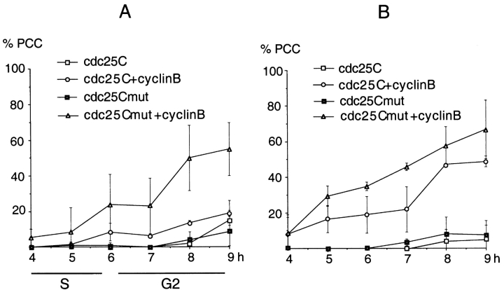
Figure 3
Frequency of PCC induced by overexpressing cdc25C and cdc25C(S216G) in the S phase. Constructs encoding Cdc25C or a Cdc25C(S216G) mutant were microinjected with or without cyclin B1 into S phase HeLa ...
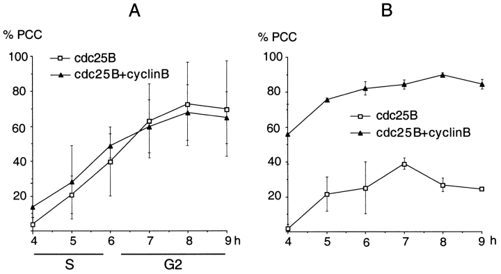
Figure 2
Frequency of PCC induced by overexpressing cdc25B in the S phase. Constructs encoding Cdc25B were microinjected with or without constructs encoding cyclin B1 into S phase HeLa cells, u223c1 h after re...
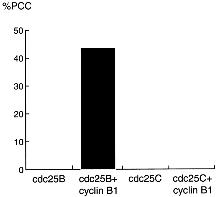
Figure 4
Overexpression of cdc25B together with cyclin B1 can force G1 cells into premature mitosis. Cells in the G1 phase were recognized by virtue of being attached after cytokinesis. Cells were injected wit...
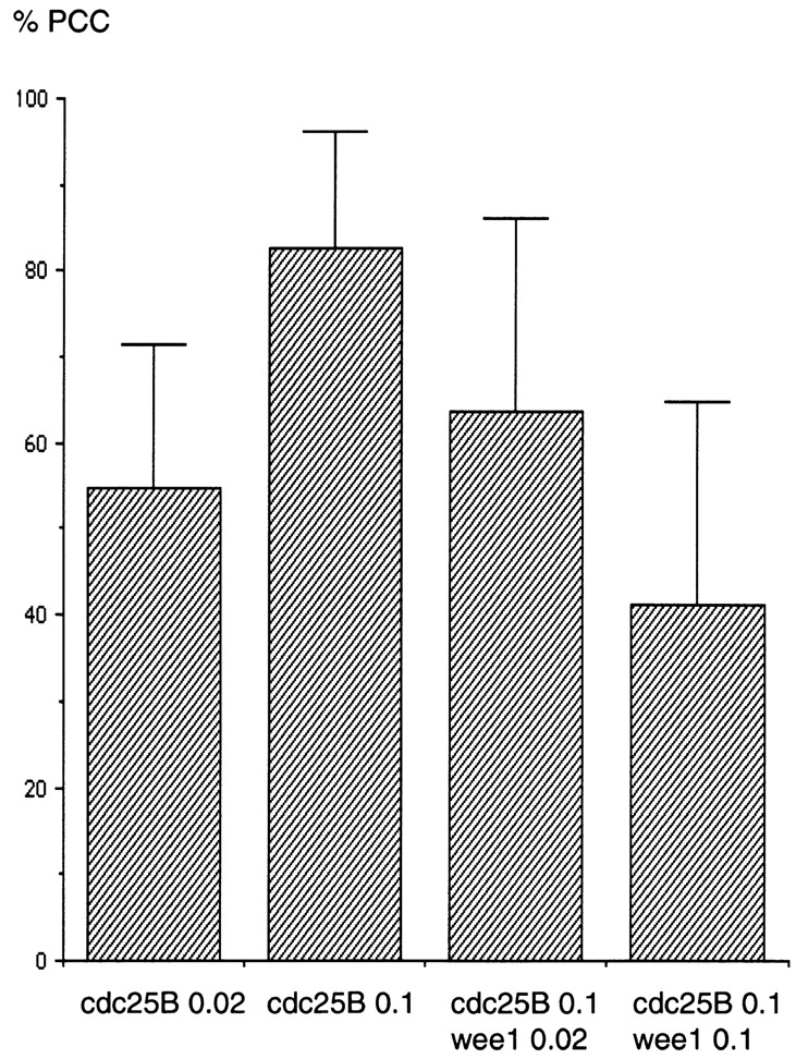
Figure 5
Wee1 can rescue cells overexpressing cdc25B from PCC. Cells were microinjected with expression constructs encoding cdc25B with or without constructs expressing Wee1 at the indicated concentrations (in...
Figure images are served from the NIH/NLM PubMed Central Open Access Subset or Europe PMC; copyright remains with the publishers and authors.
💬 Discussion
0 commentsNo comments yet. Be the first to start a discussion!
Leave a Comment