Abstract
Mitochondrial dysfunction has been implicated in the pathogenesis of acute kidney injury due to ischemia and toxic drugs. Methods for imaging mitochondrial function in cells using confocal microscopy are well established; more recently, it was shown that these techniques can be utilized in ex vivo kidney tissue using multiphoton microscopy. We extended this approach in vivo and found that kidney mitochondrial structure and function can be imaged in anesthetized rodents using multiphoton excitation of endogenous and exogenous fluorophores. Mitochondrial nicotinamide adenine dinucleotide increased markedly in rat kidneys in response to ischemia. Following intravenous injection, the mitochondrial membrane potential-dependent dye TMRM was taken up by proximal tubules; in response to ischemia, the membrane potential dissipated rapidly and mitochondria became shortened and fragmented in proximal tubules. In contrast, the mitochondrial membrane potential and structure were better maintained in distal tubules. Changes in mitochondrial structure, nicotinamide adenine dinucleotide, and membrane potential were found in the proximal, but not distal, tubules after gentamicin exposure. These changes were sporadic, highly variable among animals, and were preceded by changes in non-mitochondrial structures. Thus, real-time changes in mitochondrial structure and function can be imaged in rodent kidneys in vivo using multiphoton excitation of endogenous and exogenous fluorophores in response to ischemia-reperfusion injury or drug toxicity.
🔬 Techniques
🔭 Microscopes
💻 Software
✨ Fluorophores
🧪 Sample Preparation
🔬 Cell Lines
🏭 Microscope Brands
🧪 Reagent Suppliers
💻 Software Details
🏛️ Research Organizations (ROR)
Affiliated research institutions:
📋 Methods
Animals
All experiments were conducted in accordance with NIH Guidelines and were approved by the Institutional Animal Care and Use Committee. Adult male C57 mice (Harlan Labs, Indianapolis, IN) or Sprague–Dawley rats (Harlan Labs) were used (except for experiments in which Munich–Wistar rats were utilized to image superficial glomeruli). Mice weighing 20–20 g were anesthetized with inhaled isoflurane (2%), whereas rats weighing 250–400 g were anesthetized by intraperitoneal injection with thiobutabarbital sodium (Sigma Aldrich, St Louis, MO) at a dose of 160 mg/kg. They were then placed on a warming table to maintain a core temperature of 37°C, which was monitored throughout the experiment using a rectal thermometer. The right internal jugular vein was cannulated to allow the injection of fluorescent dyes. For ischemia–reperfusion experiments, after shaving the abdomen of the rat a midline incision was made through the skin and musculature to expose the abdominal cavity. The left renal artery was isolated through blunt dissection, and a 3.0 silk suture was placed around the vessel and passed through a length of polyethylene tubing that exited the abdominal cavity. The abdomen was then closed around the tubing. To induce ischemia, the tension on the two ends of the thread was increased until there was a visible cessation of kidney perfusion; the tension was subsequently relaxed to allow reperfusion. The left kidney was externalized via a lateral incision and imaged using methods described previously. 73 For gentamicin toxicity experiments, adult male Sprague–Dawley rats weighing 250–350 g were given a daily intraperitoneal injection of gentamicin (Sigma Aldrich) at a dose of 100 mg/kg, as per a previously established protocol. 36 Dye loading and microscope settings Images were acquired using an inverted Fluoview 1000 Olympus microscope adapted for multiphoton microscopy (Olympus Corporation, Tokyo, Japan) and either ×20 or ×60 objectives. Fluorescent dyes were administered via intravenous injection. HEt (200 µg/ml) (Invitrogen Molecular Probes, Eugene, OR), endogenous NADH, and MCB (5 mg/ml) (Invitrogen Molecular Probes) were excited at wavelengths of 720–760nm. TMRM (5 µg/ml) and rhodamine 123 (6 µg/ml) (Invitrogen Molecular Probes) were excited at wavelengths of 800–850nm. Emitted light was collected using three fixed band-pass filters: 420–460nm (blue), 495–540nm (green), and 575–630nm (red). For experiments involving inhibitors of renal transporters, verapamil (Sigma Aldrich) was used at a dose of 1 mg/kg body weight, whereas cimetidine (Sigma Aldrich) was used at a fixed dose of 8 µmoles (0.2 ml of a 40 µM solution) per animal.
Show full methods section
Animals
All experiments were conducted in accordance with NIH Guidelines and were approved by the Institutional Animal Care and Use Committee. Adult male C57 mice (Harlan Labs, Indianapolis, IN) or Sprague–Dawley rats (Harlan Labs) were used (except for experiments in which Munich–Wistar rats were utilized to image superficial glomeruli). Mice weighing 20–20 g were anesthetized with inhaled isoflurane (2%), whereas rats weighing 250–400 g were anesthetized by intraperitoneal injection with thiobutabarbital sodium (Sigma Aldrich, St Louis, MO) at a dose of 160 mg/kg. They were then placed on a warming table to maintain a core temperature of 37°C, which was monitored throughout the experiment using a rectal thermometer. The right internal jugular vein was cannulated to allow the injection of fluorescent dyes. For ischemia–reperfusion experiments, after shaving the abdomen of the rat a midline incision was made through the skin and musculature to expose the abdominal cavity. The left renal artery was isolated through blunt dissection, and a 3.0 silk suture was placed around the vessel and passed through a length of polyethylene tubing that exited the abdominal cavity. The abdomen was then closed around the tubing. To induce ischemia, the tension on the two ends of the thread was increased until there was a visible cessation of kidney perfusion; the tension was subsequently relaxed to allow reperfusion. The left kidney was externalized via a lateral incision and imaged using methods described previously. 73 For gentamicin toxicity experiments, adult male Sprague–Dawley rats weighing 250–350 g were given a daily intraperitoneal injection of gentamicin (Sigma Aldrich) at a dose of 100 mg/kg, as per a previously established protocol. 36 Dye loading and microscope settings Images were acquired using an inverted Fluoview 1000 Olympus microscope adapted for multiphoton microscopy (Olympus Corporation, Tokyo, Japan) and either ×20 or ×60 objectives. Fluorescent dyes were administered via intravenous injection. HEt (200 µg/ml) (Invitrogen Molecular Probes, Eugene, OR), endogenous NADH, and MCB (5 mg/ml) (Invitrogen Molecular Probes) were excited at wavelengths of 720–760nm. TMRM (5 µg/ml) and rhodamine 123 (6 µg/ml) (Invitrogen Molecular Probes) were excited at wavelengths of 800–850nm. Emitted light was collected using three fixed band-pass filters: 420–460nm (blue), 495–540nm (green), and 575–630nm (red). For experiments involving inhibitors of renal transporters, verapamil (Sigma Aldrich) was used at a dose of 1 mg/kg body weight, whereas cimetidine (Sigma Aldrich) was used at a fixed dose of 8 µmoles (0.2 ml of a 40 µM solution) per animal.
Image analysis and statistics
Images were processed using Image J software (National Institutes of Health, MD, http://rsb.info.nih.gov/ij/ ). Regions of interest were drawn around tubules to obtain mean fluorescence signals. Mitochondrial signals were isolated by setting a threshold level to remove the background cytosolic fluorescence. Images were acquired within 5 min of intravenous dye injection, as some signals decayed with time. To account for movement in the z plane during ischemia experiments, images were collected in serial z-stacks and reconstructed to a signal-averaged 2D image using imaging software. For the gentamicin experiments, fields of PTs were chosen randomly using the green autofluorescence channel before obtaining NADH and TMRM signals. The results are presented as means ± s.e.m. Statistical differences among study groups were explored using oneway analysis of variance and groups were compared using the paired or unpaired t -test as appropriate; a P value of < 0.05 was considered significant.
📊 Figures
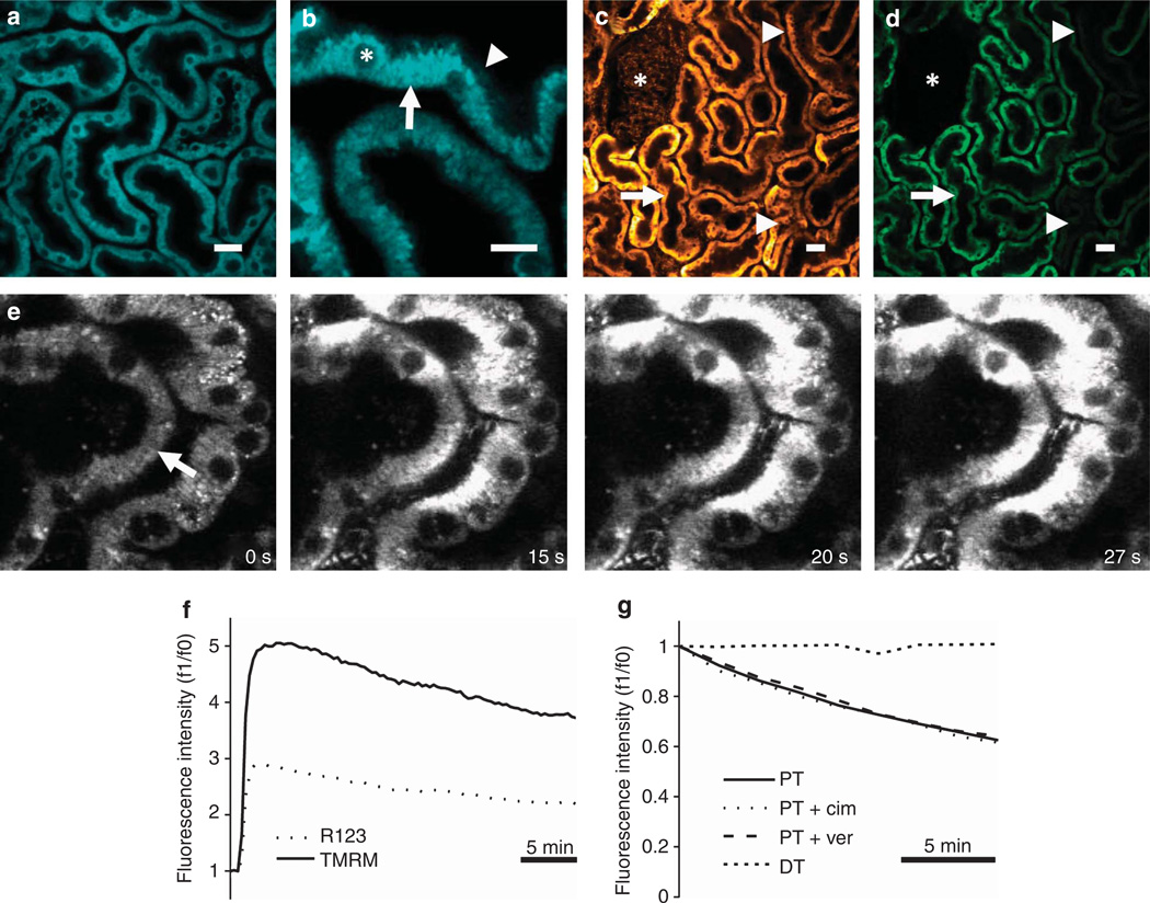
Figure 1
In vivo imaging of mitochondrial nicotinamide adenine dinucleotide (NADH) and membrane potential in the kidney
( a, b ) Mitochondrial NADH was visible at 720 nm excitation in both mouse ( a ) and rat ( b ) kidneys and showed a characteristic basolateral mitochondrial distribution in tubular cells (arrow), with...
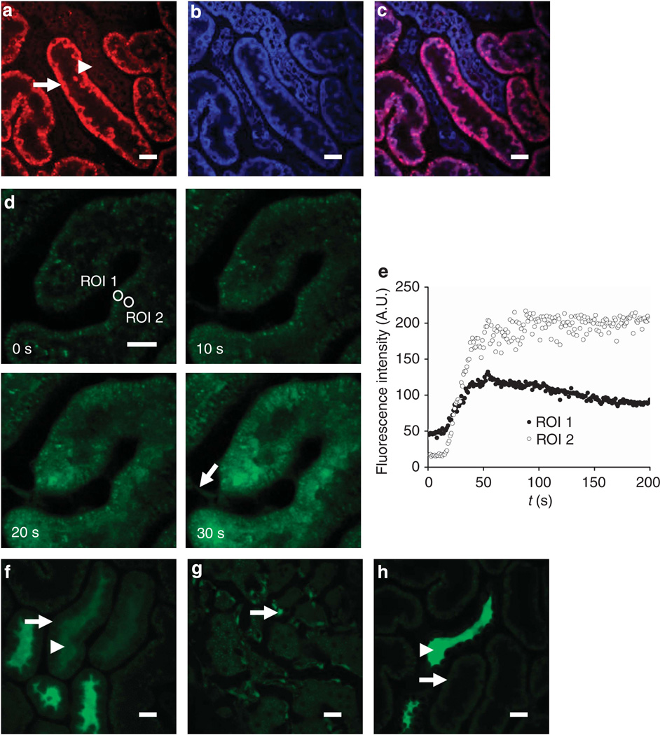
Figure 2
In vivo imaging of reactive oxygen species (ROS) production and glutathione in the kidney
( au2013c ) Imaging of ROS production. Following intravenous injection of the reactive ROS-sensitive dye dihydroethidium (HEt) in rats, the fluorescence signal was higher in proximal tubules (PTsu2014...
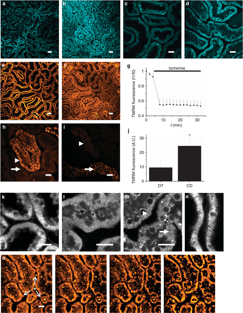
Figure 3
Real-time in vivo imaging of mitochondrial structure and function in the kidney during ischemia reperfusion
( au2013d ) Resting nicotinamide adenine dinucleotide (NADH) signal in rat renal cortical tubules ( a ) increased markedly in response to ischemia ( b ); the image depicted was acquired 2min post occl...
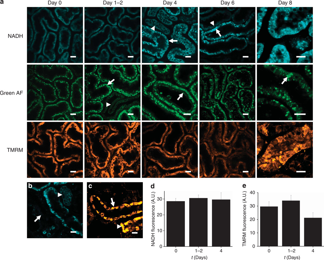
Figure 4
In vivo imaging of gentamicin toxicity in the kidney
(au2013c) Representative images are depicted displaying the time course of intracellular changes in gentamicin toxicity. After 1u20132 days of exposure, bright structures were visible in the proximal ...
Figure images are served from the NIH/NLM PubMed Central Open Access Subset or Europe PMC; copyright remains with the publishers and authors.
💬 Discussion
0 commentsNo comments yet. Be the first to start a discussion!
Leave a Comment