Abstract
Calcium imaging using two-photon scanning microscopy has become an essential tool in neuroscience. However, in its typical implementation, the tradeoffs between fields of view, acquisition speeds, and depth restrictions in scattering brain tissue pose severe limitations. Here, using an integrated systems-wide optimization approach combined with multiple technical innovations, we introduce a new design paradigm for optical microscopy based on maximizing biological information while maintaining the fidelity of obtained neuron signals. Our modular design utilizes hybrid multi-photon acquisition and allows volumetric recording of neuroactivity at single-cell resolution within up to 1 × 1 × 1.22 mm volumes at up to 17 Hz in awake behaving mice. We establish the capabilities and potential of the different configurations of our imaging system at depth and across brain regions by applying it to in vivo recording of up to 12,000 neurons in mouse auditory cortex, posterior parietal cortex, and hippocampus.
🔬 Techniques
💻 Software
✨ Fluorophores
🧪 Sample Preparation
🏭 Microscope Brands
🧪 Reagent Suppliers
📷 Detectors
💻 Software Details
💾 Data Repositories
🏛️ Research Organizations (ROR)
Affiliated research institutions:
📋 Methods
CONTACT FOR REAGENT AND RESOURCE SHARING
Further information and requests for resources and reagents should be directed to the Lead Contact, Alipasha Vaziri ( vaziri@rockefeller.edu ).
EXPERIMENTAL MODEL AND SUBJECT DETAILS Animal subjects
All surgical and experimental procedures were approved by the Institutional Animal Care and Use Committee of The Rockefeller University. Male and female adult C57BL/6J, SST-IRES-Cre and Ai9 were supplied by Jackson Laboratory, and VGlut-IRES-Cre × Ai162 crossed mice were bred in house. All mice were 7–10 weeks of age at the time of the first procedure. Mice were allowed food and water ad libitum .
Virus injection
Mice were anesthetized with isoflurane (1.5% - 2.0% maintenance at a flow rate of 0.7–0.9 l/min) and placed in a stereotaxic frame (RWD Life Science Co., Ltd.). A ~1cm incision was made over the midline of the scalp and the underlying periosteum was cleared from the skull. A grid of four 0.5mm diameter burr holes spaced 400μm apart was drilled in the center of the future window implant. A glass pipette was first back-filled with mineral oil and then front-filled with a genetically expressed Ca 2+ indicator adeno-associated virus (AAV1-syn-GCaMP6f/s). The pipette was subsequently lowered to the injection site and virus was injected (32–64nl each, at 15–25nl/min; titer ~1×10 12 vgs/ml) into the brain parenchyma at 12–28 sites to achieve column-wide cortical (PPC: −2.5 AP, 1.8 ML, −1.2, −1.0, −0.8, −0.6, −0.4 DV or −2.2 AP, −1.75 ML, −1.3, −1.2, −1.1, −1.0, −0.6, −0.4, −0.2 DV) or hippocampal expression (−2.1 AP, 2.0 ML, −1.65 DV). Following injection, the scalp was sutured closed and the animal was allowed to recover 1–2 weeks before undergoing cranial window implantation.
Show full methods section
CONTACT FOR REAGENT AND RESOURCE SHARING
Further information and requests for resources and reagents should be directed to the Lead Contact, Alipasha Vaziri ( vaziri@rockefeller.edu ).
EXPERIMENTAL MODEL AND SUBJECT DETAILS Animal subjects
All surgical and experimental procedures were approved by the Institutional Animal Care and Use Committee of The Rockefeller University. Male and female adult C57BL/6J, SST-IRES-Cre and Ai9 were supplied by Jackson Laboratory, and VGlut-IRES-Cre × Ai162 crossed mice were bred in house. All mice were 7–10 weeks of age at the time of the first procedure. Mice were allowed food and water ad libitum .
Virus injection
Mice were anesthetized with isoflurane (1.5% - 2.0% maintenance at a flow rate of 0.7–0.9 l/min) and placed in a stereotaxic frame (RWD Life Science Co., Ltd.). A ~1cm incision was made over the midline of the scalp and the underlying periosteum was cleared from the skull. A grid of four 0.5mm diameter burr holes spaced 400μm apart was drilled in the center of the future window implant. A glass pipette was first back-filled with mineral oil and then front-filled with a genetically expressed Ca 2+ indicator adeno-associated virus (AAV1-syn-GCaMP6f/s). The pipette was subsequently lowered to the injection site and virus was injected (32–64nl each, at 15–25nl/min; titer ~1×10 12 vgs/ml) into the brain parenchyma at 12–28 sites to achieve column-wide cortical (PPC: −2.5 AP, 1.8 ML, −1.2, −1.0, −0.8, −0.6, −0.4 DV or −2.2 AP, −1.75 ML, −1.3, −1.2, −1.1, −1.0, −0.6, −0.4, −0.2 DV) or hippocampal expression (−2.1 AP, 2.0 ML, −1.65 DV). Following injection, the scalp was sutured closed and the animal was allowed to recover 1–2 weeks before undergoing cranial window implantation.
Craniotomy and window implantation
As previously described, mice were anesthetized with 2% isoflurane and placed in a stereotaxic frame. The scalp was removed, and the underlying connective tissue was cleared from the skull. A custom-made stainless-steel head bar was fixed behind the occipital bone with cyanoacrylate glue (Loctite) or light-curable acrylic resin (Unifast, Henry Schein), and covered with black or pink dental cement (Ortho-Jet, Lang Dental). A circular craniotomy (3, 4, or 5mm diameter) was performed over the desired imaging site (PPC: centered at ~2.5mm caudal and ~1.8mm lateral; AUDp: centered at 2.7mm caudal and 5.35mm lateral, hippocampus: centered at ~2.0mm caudal and ~2.0mm lateral to bregma). A 3, 4, or 5mm circular glass coverslip (#1 thickness, Warner Instruments) was implanted in the craniotomy site and sealed in place with tissue adhesive (Vetbond). The exposed skull surrounding the cranial window was covered with a layer of cyanoacrylate glue and then dental cement. Hippocampal access using an implanted conical cannula was achieved by aspirating cortical tissue prior to implantation of a custom designed metal-sintered conical cannula. Post-operative care consisted of 3 days of subcutaneous delivery of dexamethasone (2mg/kg), antibiotic containing feed (LabDiet #58T7), and meloxicam (0.125mg/tablet) containing food supplements (Bio-Serv #MD275-M). After surgery, animals were returned to their home cages and were given at least one week for recovery and viral gene expression before being subjected to imaging experiments. Mice with damaged dura or unclear windows were euthanized and were not used for imaging experiments. METHOD DETAILS Obtaining sensitivity and precision of neuronal extraction under different acquisition conditions In order to design our HyMS microscope with the optimal performance, we first developed an iterative process between experimental recordings under different acquisition conditions followed by evaluation and analysis of a number of performance metrics. This process allowed us to obtain an experimentally informed model through which different optical and imaging parameters (pulse energy, PSF, sampling) could be optimized. For each microscope configuration, we directly measured the fidelity of neuronal signal extraction by running the generated data stacks through a non-negative matrix factorization algorithm and evaluating the performance against the known ground truth. Performance was quantified using a receiver operating characteristic (ROC) framework comprising “sensitivity S ” (true-positive rate) and “precision P ” (1–false-positive rate) ( Hosmer and Lemeshow, 2005 ). An ideal microscope should provide a high sensitivity without detrimental degradation of precision, for the largest possible volume and volume rate, within the bounds of the tissue heating limit. To generate the above work flow, we conducted experiments recording from awake, head-fixed mice with cytosolic GCaMP6f labeling. Datasets corresponded to 2min recordings from a 300μm FOV in layer 2/3 (depth ~ 200μm) in mouse PPC recorded with high spatial and temporal resolution (0.6 NA, ~ 8μm axial localization, 0.6μm pixel size, 22.9Hz frame rate) using a large-FOV 2p microscope ( Sofroniew et al., 2016 ). The recordings were motion-corrected using NoRMCorre, and neuronal footprints were extracted using the CaImAn software package ( Giovannucci et al., 2019 ; Pnevmatikakis et al., 2016 ). The extraction algorithm was optimized by sweeping the spatial thresholds and comparing the extracted footprints to a visual inspection of the initial dataset. The detected footprints form the ground-truth basis for comparison for all subsequent configurations. The data stack was 2×2 pixel-binned to reduce shot noise, and background subtracted. Finally, the entire data stack was normalized to the average of the baseline values of the detected neurons (estimated by the median value of each neuron’s time series). For each PSF shape, we calculated the expected signal and background counts as a function of the system parameters (collection efficiency, PMT gain, digitizer dynamic range, etc.), the geometry of the laser beam (pulse width, lateral spot size, axial extent, etc.), and the characteristics of the fluorophore (multi-photon interaction cross-section, intracellular concentration, etc.) ( Tsai and Kleinfeld, 2010 ). The denoised data stack was renormalized to the expected signal, offset by the background, and convolved with the current lateral PSF of interest. Finally, shot-noise was added to each image in the stack using the ‘imnoise’ package (Matlab) with Poisson statistics. The resulting stack was spatially and temporally down-sampled to the desired resolution. The prepared stack was then fed into the CaImAn algorithm to extract neuronal footprints. These spatial footprints were compared to the ground-truth in order to compute S and P of signal extraction. For each configuration, we swept CaImAn’s spatial thresholds (as with the initial ground-truth) in order to maximize S and P scores. By evaluating S for each PSF, sampling, and power configuration, we could ensure that a given modality achieves the desired detection fidelity within a tissue-heating-limited power budget. Laser source We used a custom laser system consisting of an Yb-fiber chirped pulse amplifier (FCPA, Active Fiber Systems) and an optical parametric chirped pulse amplifier (OPCPA) (White Dwarf dual, Class 5 Photonics). The OPCPA had two output channels at 960nm and 1,300nm wavelength for 2p and 3p excitation, respectively. The 960nm channel produces >0.8μJ, 1.4μJ, 0.8μJ, 1.4μJ,
📊 Figures
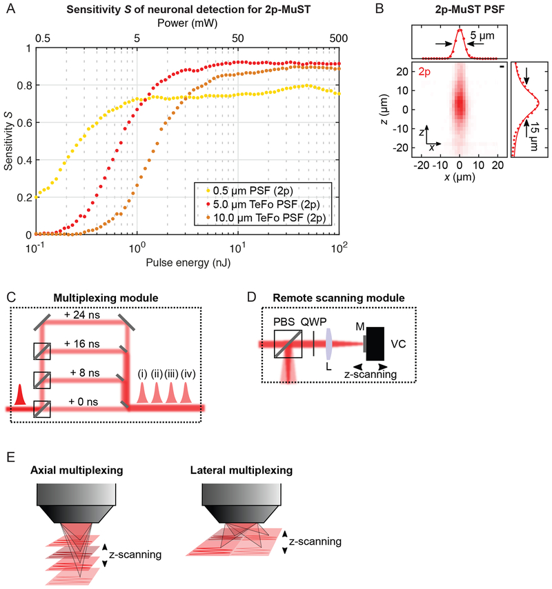
Fig. 1:
2p-MuST microscope with remote scanning.
(A) Sensitivity S to evaluate power penalty in the microscope design for 2p excitation. Analysis for one-pulse-per-voxel acquisition, with DL (0.5u03bcm, light orange), 5u03bcm TeFo (red), and 10u03bc...

Fig. 2:
High-speed volumetric 4u00d7-axial 2p-MuST imaging of mouse PPC.
(A) Mouse brain region under test (PPC). (B) Configuration for 4u00d7-axial 2p-MuST imaging. (C) Mirror holder chuck for 4u00d7 axial multiplexing. Scalebar: 5mm. (D) 3D rendering (MIP) of a 30min vol...

Fig. 3:
High-speed volumetric 4u00d7-lateral 2p-MuST imaging of mouse pAUD.
(A) Configuration for 4u00d7-lateral 2p-MuST imaging. (B) Mouse brain region under test (pAUD). (C) 3D rendering (MIP) of a 20min volumetric 4u00d7-lateral 2p-MuST recording in mouse pAUD, 1u00d71u00d...
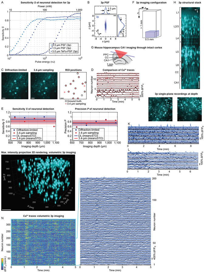
Fig. 4:
High-speed and volumetric 3p imaging at depth of mouse PPC and HPC through intact cortex.
(A) Sensitivity S (at median 3p imaging depth, 1mm) to evaluate power penalty in the microscope design for 3p. Analysis for one-pulse-per-voxel acquisition, and DL (0.5u03bcm, turquoise), 1.5u03bcm (b...

Fig. 5:
Volumetric HyMS microscopy of an entire cortical column in mouse PPC.
(A) HyMS microscope configuration. Red: 2p, blue: 3p excitation volume. (B) 3D rendering (MIP) of a 10min HyMS recording in PPC, 665u00d7730u00d71,000u03bcm FOV, 13.0Hz (4.3Hz: 3p sub-volume), cytosol...

Fig. 6:
Simultaneous volumetric HyMS microscopy of mouse HPC CA1 and DG.
(A) Mouse brain with implanted conical cannula on top of HPC and aspirated cortex. (B) 3D rendering (MIP) of a 15min HyMS recording in HPC, 665u00d7730u00d7800u03bcm FOV, 13.0Hz (4.3Hz: 3p sub-volume)...

Fig. 7:
Simultaneous volumetric imaging of mouse PPC layers 1u20135 and HPC CA1 using HyMS microscopy.
(A) HyMS microscope configuration. Red: 2p, blue: 3p excitation volume. (B) 3D rendering (MIP) of a 10min HyMS recording in PPC and underlying HPC CA1, 720u00d7665u00d71,100u03bcm total volume (record...
Figure images are served from the NIH/NLM PubMed Central Open Access Subset or Europe PMC; copyright remains with the publishers and authors.
💬 Discussion
0 commentsNo comments yet. Be the first to start a discussion!
Leave a Comment