Abstract
Many insects use patterns of polarized light in the sky to orient and navigate. Here, we functionally characterize neural circuitry in the fruit fly, Drosophila melanogaster , that conveys polarized light signals from the eye to the central complex, a brain region essential for the fly’s sense of direction. Neurons tuned to the angle of polarization of ultraviolet light are found throughout the anterior visual pathway, connecting the optic lobes with the central complex via the anterior optic tubercle and bulb, in a homologous organization to the ‘sky compass’ pathways described in other insects. We detail how a consistent, map-like organization of neural tunings in the peripheral visual system is transformed into a reduced representation suited to flexible processing in the central brain. This study identifies computational motifs of the transformation, enabling mechanistic comparisons of multisensory integration and central processing for navigation in the brains of insects.
🔬 Techniques
🔭 Microscopes
🧬 Organisms
💻 Software
✨ Fluorophores
🧪 Sample Preparation
🏭 Microscope Brands
🧪 Reagent Suppliers
🔴 Lasers
💻 Software Details
💾 Data Repositories
🏷️ Research Resource Identifiers (RRIDs)
Verified research resources used in this paper:
🏛️ Research Organizations (ROR)
Affiliated research institutions:
📋 Methods
Key resources table
Reagent type (species) or resource Designation Source or reference Identifiers Additional information Genetic reagent ( D. melanogaster ) Rh3/Rh4-Gal4 Bloomington Drosophila Stock Center FLYB:FBti0058802;RRID: BDSC_8603 Synonym: pan-R7-Gal4 Wernet et al., 2006 Genetic reagent ( D. melanogaster ) DmDRA1 split (R13E04-p65.AD; VT059781-Gal4.DBD) This paper FLYB:FBti0187820;FLYB:FBti0194675 Available upon request. Components: Dionne et al., 2018 ; Tirian and Dickson, 2017 Genetic reagent ( D. melanogaster ) R13E04-LexA Bloomington Drosophila Stock Center FLYB:FBtp0087918;RRID: BDSC_52457 Pfeiffer et al., 2013 Genetic reagent ( D. melanogaster ) R24F06-LexA Bloomington Drosophila Stock Center FLYB:FBti0155501;RRID: BDSC_52695 Pfeiffer et al., 2013 Genetic reagent ( D. melanogaster ) R56F07-Gal4 Bloomington Drosophila Stock Center FLYB:FBti0136983;RRID: BDSC_39160 Jenett et al., 2012 Genetic reagent ( D. melanogaster ) R73C04-Gal4 Bloomington Drosophila Stock Center FLYB:FBti0138074;RRID: BDSC_39815 Jenett et al., 2012 Genetic reagent ( D. melanogaster ) R17F12-Gal4 Bloomington Drosophila Stock Center FLYB:FBti0133538;RRID: BDSC_48779 Jenett et al., 2012 Genetic reagent ( D. melanogaster ) R34H10-Gal4 Bloomington Drosophila Stock Center FLYB:FBti0135093;RRID: BDSC_49808 Jenett et al., 2012 Genetic reagent ( D. melanogaster ) R88A06-Gal4 Bloomington Drosophila Stock Center FLYB:FBti0139280;RRID: BDSC_46847 Jenett et al., 2012 Genetic reagent ( D. melanogaster ) R49E09-Gal4 Bloomington Drosophila Stock Center FLYB:FBti0136346;RRID: BDSC_38692 Jenett et al., 2012 Genetic reagent ( D. melanogaster ) R34D03-Gal4 Bloomington Drosophila Stock Center FLYB:FBti0135043;RRID: BDSC_49784 Jenett et al., 2012 Genetic reagent ( D. melanogaster ) R19C08-Gal4 Bloomington Drosophila Stock Center FLYB:FBti0133678;RRID: BDSC_48845 Jenett et al., 2012 Genetic reagent ( D. melanogaster ) SS00096 (R19G02-p65.AD; R70G12- Gal4.DBD) other FLYB:FBtp0117182;FLYB:FBtp0122173 Kim et al., 2017 Provided by V. Jayaraman and T.Wolff, available via https://www.janelia.org/split-GAL4 Genetic reagent ( D. melanogaster ) R60D05-LexA Bloomington Drosophila Stock Center FLYB:FBti0156166;RRID: BDSC_52867 Pfeiffer et al., 2013 Genetic reagent ( D. melanogaster ) R34D03-LexA Bloomington Drosophila Stock Center FLYB:FBti0135043;RRID: BDSC_49784 Pfeiffer et al., 2013 Genetic reagent ( D. melanogaster ) 20×UAS-IVS-Syn21-opGCaMP6s-p10 other FLYB:FBti0195669 Provided by G. Rubin Genetic reagent ( D. melanogaster ) UAS-mCD4::tdTomato Bloomington Drosophila Stock Center FLYB:FBtp0068010;RRID: BDSC_35837 Han et al., 2011 Genetic reagent ( D. melanogaster ) UAS-sytGCaMP6s Bloomington Drosophila Stock Center FLYB:FBti0181795;RRID: BDSC_64415 Cohn et al., 2015 Genetic reagent ( D. melanogaster ) UAS-tdTomato Bloomington Drosophila Stock Center FLYB:FBti0145104;RRID: BDSC_36328 Schulte, 2011 ; Shaner et al., 2004 Genetic reagent ( D. melanogaster ) 10×UAS-mCD8::GFP Bloomington Drosophila Stock Center FLYB:FBti0131930;RRID: BDSC_32184 Pfeiffer, 2010 Genetic reagent ( D. melanogaster ) 26×LexAop-mCD8::GFP Bloomington Drosophila Stock Center FLYB:FBti0131946;RRID: BDSC_32207 Pfeiffer, 2010 Genetic reagent ( D. melanogaster ) 10×UAS-mCD8::RFP; 13×LexAop-mCD8::GFP Bloomington Drosophila Stock Center FLYB:FBst0032229;RRID: BDSC_32229 Pfeiffer, 2010 Genetic reagent ( D. melanogaster ) UAS-nSyb-spGFP1-10, LexAop-CD4-spGFP11 Bloomington Drosophila Stock Center FLYB:FBtp0111281;RRID: BDSC_64314 Components: Gallio, 2016 ; Macpherson et al., 2015 Genetic reagent ( D. melanogaster ) MCFO-4 Bloomington Drosophila Stock Center FLYB:FBst0064088;RRID: BDSC_64088 Nern et al., 2015 Genetic reagent ( D. melanogaster ) MCFO-5 Bloomington Drosophila Stock Center FLYB:FBst0064089;RRID: BDSC_64089 Nern et al., 2015 Genetic reagent ( D. melanogaster ) MCFO-6 Bloomington Drosophila Stock Center FLYB:FBst0064090;RRID: BDSC_64090 Nern et al., 2015 Genetic reagent ( D. melanogaster ) GRASP Bloomington Drosophila Stock Center FLYB:FBst0064314;RRID: BDSC_64314 Macpherson et al., 2015 Genetic reagent ( D. melanogaster ) trans -Tango Bloomington Drosophila Stock Center FLYB:FBst0077124;RRID: BDSC_77124 Talay et al., 2017 Provided by G. Barnea Antibody Anti-DN-cadherin (rat monoclonal) Developmental Studies Hybridoma Bank Cat#:DN-Ex #8; RRID: AB_528121 (1:20) Antibody Anti-Neuroglian (mouse monoclonal) Developmental Studies Hybridoma Bank Cat#:BP 104; RRID: AB_528402 (1:30) Antibody Anti-GFP (chicken polyclonal) Abcam Cat#:ab13970; RRID: AB_300798 (1:1000) Antibody Anti-DsRed (rabbit polyclonal) Takara Bio Cat#:632496; RRID: AB_10013483 (1:1000) Antibody Anti-HA (rabbit monoclonal) Cell Signaling Technology Cat#: 3724S; RRID: AB_1549585 (1:300) Antibody Anti-V5 (mouse monoclonal) Thermo Fisher Scientific Cat#:37–7500; RRID: AB_2533339 (1:1000) Antibody Anti-Mouse conjugated to Cy5 (goat polyclonal) Jackson ImmunoResearch Cat#:115-175-166; RRID: AB_2338714 (1:300) Antibody Anti-Rat conjugated to Cy3 (goat polyclonal) Jackson ImmunoResearch Cat#:112-165-167;RRID: AB_2338251 (1:300) Antibody Anti-Rat conjugated to Cy5 (goat polyclonal) Jackson ImmunoResearch Cat#:112-175-143;RRID: AB_2338263 (1:300) Antibody Anti-Rabbit conjugated to Cy3 (goat polyclonal) Jackson ImmunoResearch Cat#:111-165-003;RRID: AB_2338000 (1:300) Antibody Anti-GFP conjugated to Alexa488 (rabbit polyclonal) Thermo Fisher Scientific Cat#:A-21311;RRID: AB_221477 (1:1000) Antibody Anti-GFP (mouse monoclonal) Sigma-Aldrich Cat#:G6539; RRID: AB_259941 (1:1000) Antibody Anti-Chicken conjugated to Alexa488 (goat polyclonal) Thermo Fisher Scientific Cat#:A-11039;RRID: AB_2534096 (1:1000) Antibody Anti-Mouse conjugated to Alexa488 (goat polyclonal) Thermo Fisher Scientific Cat#:A-10680;RRID: AB_2534062 (1:1000) Software, algorithm Fiji ImageJ RRID: SCR_002285 Schindelin et al., 2012 Software, algorithm SlideBook 6 Intelligent Imaging Innovations RRID: SCR_014300 Software, algorithm MATLAB R2017a Mathworks RRID: SCR_001622 In vivo calcium imaging Fly preparation Flies were raised at 25°C on a standard cornmeal/molasses diet in 40 ml vials, under a 12:12 hr dark:light cycle. Imaging experiments were performed between ZT0–14, although time of day was not a factor in our experimental design or analysis. We imaged 1–7 day old female flies expressing either UAS-GCaMP6s ( Chen et al., 2013 ) for dendritic regions or UAS-sytGCaMP6s ( Cohn et al., 2015 ) for axon terminals, together with UAS-tdTomato ( Shaner et al., 2004 ) for image registration (for genotypes see Appendix 2—table 1. ). Flies were cold anesthetized and mounted on a custom fly holder, modified from Weir et al., 2016 , with the head pitched forward so that its posterior surface was approximately horizontal ( Figure 1—figure supplement 1A ). Surfaces of the fly holder visible to the fly were covered in matte white paint (Citadel) and roughened to reduce confounding reflected polarized light cues ( Foster et al., 2018 ). We fixed the fly to the holder using UV-curing glue (Fotoplast) around the posterior-dorsal cuticle of the head and at the base of the wings on either side of the thorax. To reduce movement of the brain, we fixed the legs, abdomen, and proboscis with beeswax. We used forceps to remove the cuticle and air-sacs above the optic lobe or central brain, depending on the recording site, and cut muscle 1 ( Demerec, 1950 ) to reduce movement. Physiological saline (103 mM NaCl, 3 mM KCl, 1.5 mM CaCl 2 , 4 mM MgCl 2 , 26 mM NaHCO 3 , 1 mM NaH 2 PO 4 , 10 mM trehalose, 10 mM glucose, 5 mM TES, 2 mM sucrose) was perfused continuously over the brain at 1.5 ml/min via a gravity drip system and the bath was maintained at 22°C for the duration of experiments by an inline solution heater/cooler (SC-20, Warner Instruments) connected to a temperature controller (TC-324, Warner Instruments).
Show full methods section
Key resources table
Reagent type (species) or resource Designation Source or reference Identifiers Additional information Genetic reagent ( D. melanogaster ) Rh3/Rh4-Gal4 Bloomington Drosophila Stock Center FLYB:FBti0058802;RRID: BDSC_8603 Synonym: pan-R7-Gal4 Wernet et al., 2006 Genetic reagent ( D. melanogaster ) DmDRA1 split (R13E04-p65.AD; VT059781-Gal4.DBD) This paper FLYB:FBti0187820;FLYB:FBti0194675 Available upon request. Components: Dionne et al., 2018 ; Tirian and Dickson, 2017 Genetic reagent ( D. melanogaster ) R13E04-LexA Bloomington Drosophila Stock Center FLYB:FBtp0087918;RRID: BDSC_52457 Pfeiffer et al., 2013 Genetic reagent ( D. melanogaster ) R24F06-LexA Bloomington Drosophila Stock Center FLYB:FBti0155501;RRID: BDSC_52695 Pfeiffer et al., 2013 Genetic reagent ( D. melanogaster ) R56F07-Gal4 Bloomington Drosophila Stock Center FLYB:FBti0136983;RRID: BDSC_39160 Jenett et al., 2012 Genetic reagent ( D. melanogaster ) R73C04-Gal4 Bloomington Drosophila Stock Center FLYB:FBti0138074;RRID: BDSC_39815 Jenett et al., 2012 Genetic reagent ( D. melanogaster ) R17F12-Gal4 Bloomington Drosophila Stock Center FLYB:FBti0133538;RRID: BDSC_48779 Jenett et al., 2012 Genetic reagent ( D. melanogaster ) R34H10-Gal4 Bloomington Drosophila Stock Center FLYB:FBti0135093;RRID: BDSC_49808 Jenett et al., 2012 Genetic reagent ( D. melanogaster ) R88A06-Gal4 Bloomington Drosophila Stock Center FLYB:FBti0139280;RRID: BDSC_46847 Jenett et al., 2012 Genetic reagent ( D. melanogaster ) R49E09-Gal4 Bloomington Drosophila Stock Center FLYB:FBti0136346;RRID: BDSC_38692 Jenett et al., 2012 Genetic reagent ( D. melanogaster ) R34D03-Gal4 Bloomington Drosophila Stock Center FLYB:FBti0135043;RRID: BDSC_49784 Jenett et al., 2012 Genetic reagent ( D. melanogaster ) R19C08-Gal4 Bloomington Drosophila Stock Center FLYB:FBti0133678;RRID: BDSC_48845 Jenett et al., 2012 Genetic reagent ( D. melanogaster ) SS00096 (R19G02-p65.AD; R70G12- Gal4.DBD) other FLYB:FBtp0117182;FLYB:FBtp0122173 Kim et al., 2017 Provided by V. Jayaraman and T.Wolff, available via https://www.janelia.org/split-GAL4 Genetic reagent ( D. melanogaster ) R60D05-LexA Bloomington Drosophila Stock Center FLYB:FBti0156166;RRID: BDSC_52867 Pfeiffer et al., 2013 Genetic reagent ( D. melanogaster ) R34D03-LexA Bloomington Drosophila Stock Center FLYB:FBti0135043;RRID: BDSC_49784 Pfeiffer et al., 2013 Genetic reagent ( D. melanogaster ) 20×UAS-IVS-Syn21-opGCaMP6s-p10 other FLYB:FBti0195669 Provided by G. Rubin Genetic reagent ( D. melanogaster ) UAS-mCD4::tdTomato Bloomington Drosophila Stock Center FLYB:FBtp0068010;RRID: BDSC_35837 Han et al., 2011 Genetic reagent ( D. melanogaster ) UAS-sytGCaMP6s Bloomington Drosophila Stock Center FLYB:FBti0181795;RRID: BDSC_64415 Cohn et al., 2015 Genetic reagent ( D. melanogaster ) UAS-tdTomato Bloomington Drosophila Stock Center FLYB:FBti0145104;RRID: BDSC_36328 Schulte, 2011 ; Shaner et al., 2004 Genetic reagent ( D. melanogaster ) 10×UAS-mCD8::GFP Bloomington Drosophila Stock Center FLYB:FBti0131930;RRID: BDSC_32184 Pfeiffer, 2010 Genetic reagent ( D. melanogaster ) 26×LexAop-mCD8::GFP Bloomington Drosophila Stock Center FLYB:FBti0131946;RRID: BDSC_32207 Pfeiffer, 2010 Genetic reagent ( D. melanogaster ) 10×UAS-mCD8::RFP; 13×LexAop-mCD8::GFP Bloomington Drosophila Stock Center FLYB:FBst0032229;RRID: BDSC_32229 Pfeiffer, 2010 Genetic reagent ( D. melanogaster ) UAS-nSyb-spGFP1-10, LexAop-CD4-spGFP11 Bloomington Drosophila Stock Center FLYB:FBtp0111281;RRID: BDSC_64314 Components: Gallio, 2016 ; Macpherson et al., 2015 Genetic reagent ( D. melanogaster ) MCFO-4 Bloomington Drosophila Stock Center FLYB:FBst0064088;RRID: BDSC_64088 Nern et al., 2015 Genetic reagent ( D. melanogaster ) MCFO-5 Bloomington Drosophila Stock Center FLYB:FBst0064089;RRID: BDSC_64089 Nern et al., 2015 Genetic reagent ( D. melanogaster ) MCFO-6 Bloomington Drosophila Stock Center FLYB:FBst0064090;RRID: BDSC_64090 Nern et al., 2015 Genetic reagent ( D. melanogaster ) GRASP Bloomington Drosophila Stock Center FLYB:FBst0064314;RRID: BDSC_64314 Macpherson et al., 2015 Genetic reagent ( D. melanogaster ) trans -Tango Bloomington Drosophila Stock Center FLYB:FBst0077124;RRID: BDSC_77124 Talay et al., 2017 Provided by G. Barnea Antibody Anti-DN-cadherin (rat monoclonal) Developmental Studies Hybridoma Bank Cat#:DN-Ex #8; RRID: AB_528121 (1:20) Antibody Anti-Neuroglian (mouse monoclonal) Developmental Studies Hybridoma Bank Cat#:BP 104; RRID: AB_528402 (1:30) Antibody Anti-GFP (chicken polyclonal) Abcam Cat#:ab13970; RRID: AB_300798 (1:1000) Antibody Anti-DsRed (rabbit polyclonal) Takara Bio Cat#:632496; RRID: AB_10013483 (1:1000) Antibody Anti-HA (rabbit monoclonal) Cell Signaling Technology Cat#: 3724S; RRID: AB_1549585 (1:300) Antibody Anti-V5 (mouse monoclonal) Thermo Fisher Scientific Cat#:37–7500; RRID: AB_2533339 (1:1000) Antibody Anti-Mouse conjugated to Cy5 (goat polyclonal) Jackson ImmunoResearch Cat#:115-175-166; RRID: AB_2338714 (1:300) Antibody Anti-Rat conjugated to Cy3 (goat polyclonal) Jackson ImmunoResearch Cat#:112-165-167;RRID: AB_2338251 (1:300) Antibody Anti-Rat conjugated to Cy5 (goat polyclonal) Jackson ImmunoResearch Cat#:112-175-143;RRID: AB_2338263 (1:300) Antibody Anti-Rabbit conjugated to Cy3 (goat polyclonal) Jackson ImmunoResearch Cat#:111-165-003;RRID: AB_2338000 (1:300) Antibody Anti-GFP conjugated to Alexa488 (rabbit polyclonal) Thermo Fisher Scientific Cat#:A-21311;RRID: AB_221477 (1:1000) Antibody Anti-GFP (mouse monoclonal) Sigma-Aldrich Cat#:G6539; RRID: AB_259941 (1:1000) Antibody Anti-Chicken conjugated to Alexa488 (goat polyclonal) Thermo Fisher Scientific Cat#:A-11039;RRID: AB_2534096 (1:1000) Antibody Anti-Mouse conjugated to Alexa488 (goat polyclonal) Thermo Fisher Scientific Cat#:A-10680;RRID: AB_2534062 (1:1000) Software, algorithm Fiji ImageJ RRID: SCR_002285 Schindelin et al., 2012 Software, algorithm SlideBook 6 Intelligent Imaging Innovations RRID: SCR_014300 Software, algorithm MATLAB R2017a Mathworks RRID: SCR_001622 In vivo calcium imaging Fly preparation Flies were raised at 25°C on a standard cornmeal/molasses diet in 40 ml vials, under a 12:12 hr dark:light cycle. Imaging experiments were performed between ZT0–14, although time of day was not a factor in our experimental design or analysis. We imaged 1–7 day old female flies expressing either UAS-GCaMP6s ( Chen et al., 2013 ) for dendritic regions or UAS-sytGCaMP6s ( Cohn et al., 2015 ) for axon terminals, together with UAS-tdTomato ( Shaner et al., 2004 ) for image registration (for genotypes see Appendix 2—table 1. ). Flies were cold anesthetized and mounted on a custom fly holder, modified from Weir et al., 2016 , with the head pitched forward so that its posterior surface was approximately horizontal ( Figure 1—figure supplement 1A ). Surfaces of the fly holder visible to the fly were covered in matte white paint (Citadel) and roughened to reduce confounding reflected polarized light cues ( Foster et al., 2018 ). We fixed the fly to the holder using UV-curing glue (Fotoplast) around the posterior-dorsal cuticle of the head and at the base of the wings on either side of the thorax. To reduce movement of the brain, we fixed the legs, abdomen, and proboscis with beeswax. We used forceps to remove the cuticle and air-sacs above the optic lobe or central brain, depending on the recording site, and cut muscle 1 ( Demerec, 1950 ) to reduce movement. Physiological saline (103 mM NaCl, 3 mM KCl, 1.5 mM CaCl 2 , 4 mM MgCl 2 , 26 mM NaHCO 3 , 1 mM NaH 2 PO 4 , 10 mM trehalose, 10 mM glucose, 5 mM TES, 2 mM sucrose) was perfused continuously over the brain at 1.5 ml/min via a gravity drip system and the bath was maintained at 22°C for the duration of experiments by an inline solution heater/cooler (SC-20, Warner Instruments) connected to a temperature controller (TC-324, Warner Instruments).
Imaging setup
We used a two-photon excitation scanning microscope controlled by Slidebook (ver. 6, 3i) with a Ti:sapphire laser (Chameleon Vision, Coherent) at 920 nm and a 40× objective (0.8 numerical aperture, NIR Apo, Nikon). For each brain area imaged, we aimed to capture the full extent of the volume of labeled neurons, using a maximum step-size of 4 μm between imaging planes, and maintained a volume-rate of at least 1 Hz. Image resolution varied depending on the number of planes captured but was not less than 100 pixels in the longest dimension. We recorded frame capture markers and stimulus events on a DAQ (6259, NI) sampling at 10 kHz. Polarized light stimulus We used a custom polarized light stimulus device comprising a UV LED (M340D3, Thorlabs), a 7.5 mm diameter aperture, a ground glass diffuser (DGUV10-1500, Thorlabs), a low-pass filter (FGUV11, Thorlabs), and a removable linear polarizer (BVO UV, Bolder Optic). The UV LED was controlled through MATLAB 2017a (Mathworks, MA) via a DAQ (6259, NI) and LED driver (LEDD1B, Thorlabs). The polarizer was rotated with a bipolar stepper motor (ROB-10551, SparkFun) and spur gears (1:1), and a motor driver (ROB-12779, SparkFun) controlled through MATLAB (2017a, Mathworks) via a DAQ (USB1208, MCC), with a minimum step-size of 7.5°. The motor was operated in open-loop and a Hall effect sensor (A1324, Allegro) was used to detect the proximity of a magnet which passed once per revolution, in order to verify correct operation. Angles of polarization and directions of rotation are expressed from an external viewpoint looking toward the fly ( Figure 1—figure supplement 1A ). 0°/180° corresponds to a vertical orientation in the transverse plane and an alignment with the fly’s long-axis in the horizontal plane. We investigated the reproducibility of the polarizer’s angular positions and measured
📊 Figures
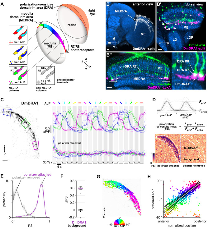
Figure 1.
Polarization processing in the medulla dorsal rim area.
( A ) Schematic of the dorsal rim area (DRA) of the right eye and the projection of DRA R7/R8 photoreceptors to corresponding columns in the medulla dorsal rim area (MEDRA) across the first optic chia...
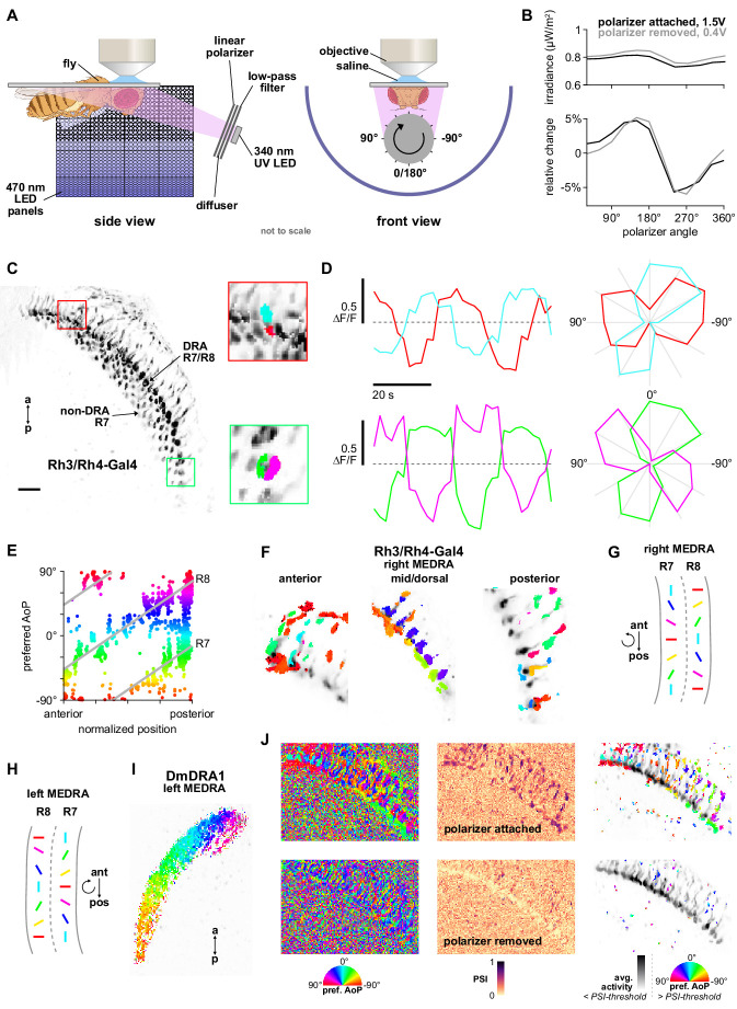
Figure 1u2014figure supplement 1.
Polarizer stimulusu00a0characterization and R7/R8 photoreceptor stimulation.
( A ) Schematic of experimental setup. Volumetric two-photon imaging of the medulla dorsal rim area (MEDRA) was performed while ultraviolet light was presented continuously and a linear polarizing fil...
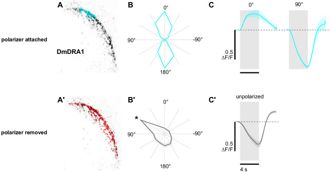
Figure 1u2014figure supplement 2.
Polarization-opponent flash responses in DmDRA1.
( A ) Example time-averaged maximum-intensity projection showing GCaMP activity in DmDRA1 neurons (DmDRA1-split>sytGCaMP6s) and example ROIs automaticallyu00a0generated around areas of DmDRA1 neurons ...
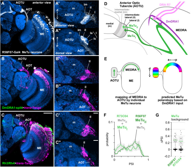
Figure 2.
Medulla projection neurons receive polarization signals from DmDRA1 and DRA photoreceptors.
( A ) Confocal projection (anterior view) of R56F07-Gal4 driving a population of MeTu neurons with dendrites in the dorsal medulla (ME) and projections to theu00a0anterior optic tubercle (AOTU) via th...
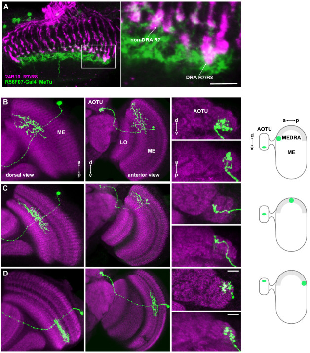
Figure 2u2014figure supplement 1.
Retinotopic mapping of medulla dorsal rim area to AOTU by MeTu neurons.
( A ) Confocal section of the medulla (dorsal view) showing R7/R8 photoreceptors (24B10 antibody staining: green) and their proximity to MeTu neurons (R56F07-Gal4>GFP: magenta). Right: Enlargement of ...
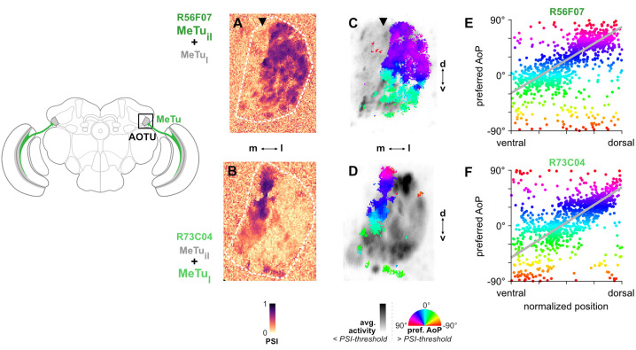
Figure 3.
MeTu neurons convey polarization signals to the AOTU in a polarotopic fashion.
( A ) Example spatial map of polarization-selectivity index (PSI) in MeTu terminals in the AOTU (R56F07-Gal4>sytGCaMP6s; predominantly MeTu il neurons innervating intermediate-lateral (il) domain, wit...
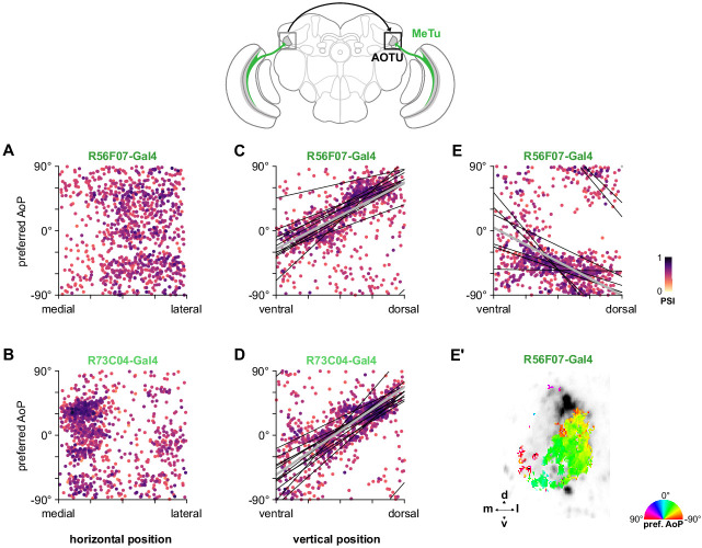
Figure 3u2014figure supplement 1.
Organization of polarization-selective responses in MeTu neurons in the AOTU.
( A ) Scatter plot showing the organization of polarized light responses in R56F07 MeTu neurons (pooled u03c1u00a0=u00a00.04, CI95 [u22120.22 0.22], Nu00a0=u00a017 recordings). Individual points repre...
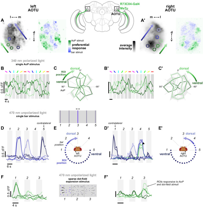
Figure 4.
Retinotopic encoding of multiple visual features in the AOTU.
( A ) Left: Example time-averaged maximum-intensity projection showing GCaMP activity in R73C04 MeTu neuron terminals in the left AOTU and examples of lateral ROIs (green) and medial ROIs (blue) (R73C...
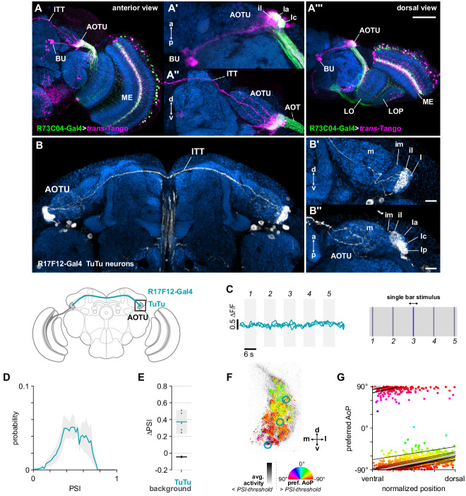
Figure 5.
Bilateral TuTu neurons convey polarization signals to the contralateral AOTU.
( A ) Confocal projection (anterior view) of trans -Tango signal (magenta) labeling putative postsynaptic partners of R73C04-Gal4 MeTu neurons (green). ( Au2019 ) High-magnification dorsal view highli...
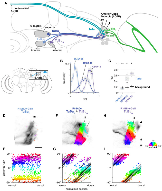
Figure 6.
A population of TuBu neurons receives polarization signals in the AOTU.
( A ) Schematic of TuBu neuron types projecting to the bulb (BU) and connectivity in the AOTU. ( B ) Probability distribution of PSI values in TuBu neurons recorded in the AOTU. Mean u00b1 SEM. Summar...
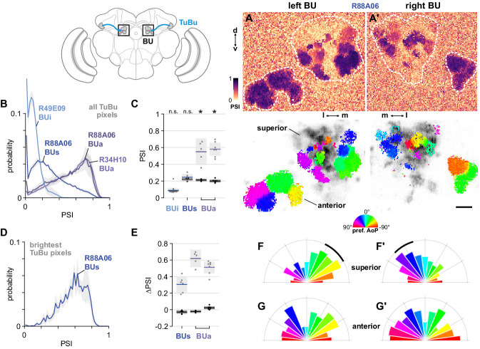
Figure 7.
The anterior bulb is an entry point for polarization signals into the central complex.
( A ) Example spatial maps of polarization-selectivity index (PSI, top) and tuning (bottom) in TuBu neuron output micro-glomeruli in the superior and anterior regions of the left ( A ) and right ( Au2...
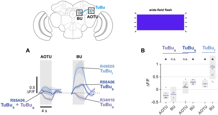
Figure 7u2014figure supplement 1.
Unpolarized flash responses in TuBu neurons.
( A ) Average responses of all TuBu neurons in each population to 4 s blue light flashes, recorded in the anterior optic tubercle (AOTU) (GCaMP6s) and bulb (BU) (sytGCaMP6s). Dashed line indicates 0 u...
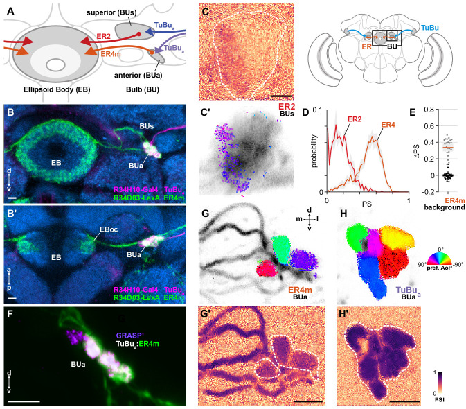
Figure 8.
ER4m ring neurons receive polarization-tuned responses from TuBu neurons.
( A ) Schematic of TuBu and ring neuronu00a0(ER) connectivity in the bulb (BU). ( B ) Confocal projection (anterior view) of dual-labeled TuBu a neurons (R34H10-Gal4>RFP, magenta) and ER4m neurons (R3...
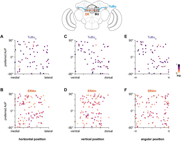
Figure 8u2014figure supplement 1.
Unstructured organization of preferred angle of polarization in the anterior bulb.
( A ) Scatter plot showing the horizontal organization of TuBu a tunings in the anterior bulb (BUa). Individual points represent ROIs drawn on micro-glomeruli, showing their normalized horizontal posi...
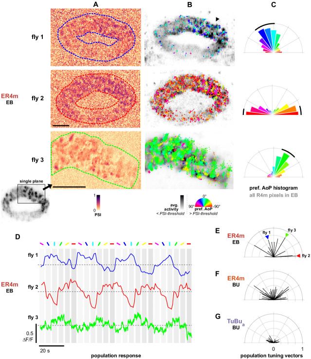
Figure 9.
Populations of ER4m ring neurons exhibit a preferred angle of polarization.
( A ) Example spatial maps of polarization-selectivity index (PSI) in ER4m synapses recorded in the ellipsoid body (EB) (R34D03-Gal4>sytGCaMP6s). Data shown are from maximum-intensity projections thro...
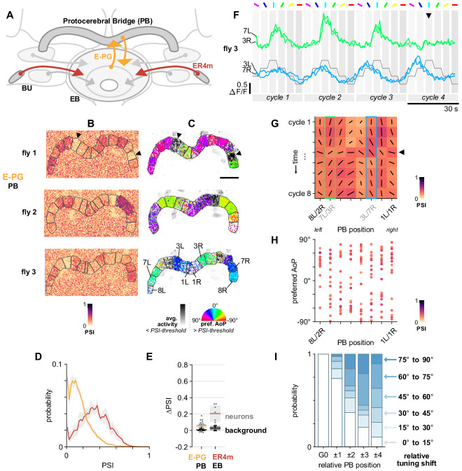
Figure 10.
E-PG neurons respond to polarized light with flexible tuning and no fixed polarotopic map.
( A ) Schematic of E-PG columnar neuron projections and connectivity with tangential ring neurons in the ellipsoid body (EB). See also Figure 10u2014figure supplement 1D . ( B ) Example spatial maps o...
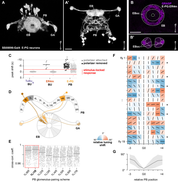
Figure 10u2014figure supplement 1.
E-PG neurons show inconsistent responses and variable tunings.
( A ) Confocal projection (anterior view) of E-PG expression pattern in the ellipsoid body (EB), protocerebral bridge (PB) and gall (GA) (SS00096-Gal4>GFP). ( Au2019 ) Dorsal view. Scale bar denotes 2...
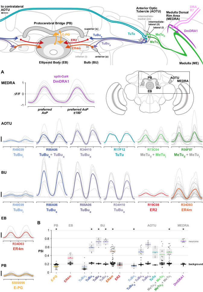
Appendix 1u2014figure 1.
Summary of polarization pathway and response properties.
( A ) Polarization tuning curves for each neuron population recorded. Thin traces show tuning curves for individual animals, thick traces show their mean. All scale bars denote 1 u0394F/F. Dashed hori...
Figure images are served from the NIH/NLM PubMed Central Open Access Subset or Europe PMC; copyright remains with the publishers and authors.
💬 Discussion
0 commentsNo comments yet. Be the first to start a discussion!
Leave a Comment