Abstract
Invadopodia are specialized actin-rich protrusions of metastatic tumor and transformed cells with crucial functions in ECM degradation and invasion. Although early electron microscopy studies described invadopodia as long filament-like protrusions of the cell membrane adherent to the matrix, fluorescence microscopy studies have focused on invadopodia as actin-cortactin aggregates localized to areas of ECM degradation. The absence of a clear conceptual integration of these two descriptions of invadopodial structure has impeded understanding of the regulatory mechanisms that govern invadopodia. To determine the relationship between the membrane filaments identified by electron microscopy and the actin-cortactin aggregates of invadopodia, we applied rapid live-cell high-resolution TIRF microscopy to examine cell membrane dynamics at the cortactin core of the invadopodia of human carcinoma cells. We found that cortactin docking to the cell membrane adherent to 2D fibronectin matrix initiates invadopodium assembly associated with the formation of an invadopodial membrane process that extends from a ventral cell membrane lacuna toward the ECM. The tip of the invadopodial process flattens as it interacts with the 2D matrix, and it undergoes constant rapid ruffling and dynamic formation of filament-like protrusions as the invadopodium matures. To describe this newly discovered dynamic relationship between the actin-cortactin core and invadopodial membranes, we propose a model of the invadopodial complex. Using TIRF microscopy, we also established that - in striking contrast to the invadopodium - membrane at the podosome of a macrophage fails to form any process- or filament-like membrane protrusions. Thus, the undulation and ruffling of the invadopodial membrane together with the formation of dynamic filament-like extensions from the invadopodial cortactin core defines invadopodia as invasive superstructures that are distinct from the podosomes.
🔬 Techniques
🔭 Microscopes
💻 Software
✨ Fluorophores
🧪 Sample Preparation
🔬 Cell Lines
🏭 Microscope Brands
🧪 Reagent Suppliers
📷 Detectors
🔎 Objectives
💻 Software Details
🏛️ Research Organizations (ROR)
Affiliated research institutions:
📋 Methods
Materials Human plasma fibronectin (HPFN) was prepared as described ( Akiyama, 2001 ). Bovine serum albumin (BSA) was purchased from MP Biomedicals (Solon, OH). Phalloidin-AlexaFluor and AlexaFluor protein labeling dyes were from Invitrogen (Carlsbad, CA). Anti-vinculin antibody was from Sigma-Aldrich (St. Louis, MO), and secondary antibodies conjugated to Cy5 were from Jackson ImmunoResearch (West Grove, PA). Rat tail collagen type I was purchased from BD Biosciences (San Jose, CA).
Cell lines and transfections
A stable line of MDA-MB-231 cells transfected with wild-type c-Src (wt c-Src MDA-MB-231) was a generous gift from Dr. Toshiyuki Yoneda ( Myoui et al., 2003 ). Cells were maintained in high glucose-DMEM (HyClone, Logan, UT) supplemented with 10% fetal bovine serum (HyClone), 2 mM L-glutamine, 10 U/ml penicillin, and 10 μg/ml streptomycin. The mouse macrophage IC-21 (TIB-186) cell line was obtained from the American Type Culture Collection (Rockville, MD). This macrophage line was cultured in RPMI-1640 (HyClone, Logan, UT) supplemented with 10% fetal bovine serum, 10 mM HEPES, 1.5 g/l sodium bicarbonate, and 4.5 g/l glucose.
Human foreskin fibroblasts
(HFF) were cultured in high glucose-DMEM supplemented with 10% fetal bovine serum, 10 U/ml penicillin, and 10 μg/ml streptomycin. All cell lines were transiently co-transfected with GFP and mCherry cDNA vectors, and fluorescence of exogenous proteins was imaged at 24 or 48 hours post-transfection. MDA-MB-231 cells were transfected with Lipofectamine 2000 (Invitrogen), IC-21 cells were transfected using AMAXA (Walkersville, MD), and HFF were transfected with PolyJet (SignaGen Laboratories, Ijamsville, MD) according to the manufacturers’ instructions. cDNA vectors Human cortactin cDNA was purchased from Invitrogen. A twelve-amino acid segment missing from the actin-binding domain compared to the original sequence (GenBank) was restored by site-directed mutagenesis (Stratagene, La Jolla, CA). The full-length cortactin cDNA was cloned into Bam H1 and Xba1 sites of pGZ21XdZ (Dr. Shin-ichi Aota, NIDCR, NIH) to produce pGFP-Cortactin. The cortactin sequence was confirmed by sequencing. The plasmid pEIL2RNBC1 was constructed by partially replacing the multiple-cloning site (MCS) of pEGFP-C1 (Clontech, Mountain View, CA) to introduce new MCS sites by inserting the appropriate double-strand oligonucleotides using Bgl11 and Sal1 sites of the parent Clontech pEGFP-C1 plasmid, respectively, after mutating the Bam H1 site in the original MCS using a site-directed mutagenesis kit. The EGFP was then deleted from the construct using Nhe1 and Bgl11 sites and replaced by the extracellular and transmembrane domains of the α subunit of the human IL-2 receptor with deletion of the cytoplasmic domain, which was generated by PCR from pRSVIL2R (Drs. Bruce Howard and Tony Giordano, NICHD) plus addition of a Kozak sequence at the 5‘-end. Fluorescent mCherry flanked by Hind111 and Xba1 sites was produced by PCR from pRSET-B mCherry (a kind gift from Dr. Roger Tsien) and inserted into Hind 111 and Xba1 sites of pEIL2RNBC1 to create pEIL2R-mCherry. The IL-2R and mCherry sequences were confirmed by sequencing. To construct GFP-Vinculin, chicken vinculin cDNA (a kind gift from Dr. Benjamin Geiger) was inserted in Hind111 and Xba1 sites of pGZ21XdZ.
Show full methods section
Materials Human plasma fibronectin (HPFN) was prepared as described ( Akiyama, 2001 ). Bovine serum albumin (BSA) was purchased from MP Biomedicals (Solon, OH). Phalloidin-AlexaFluor and AlexaFluor protein labeling dyes were from Invitrogen (Carlsbad, CA). Anti-vinculin antibody was from Sigma-Aldrich (St. Louis, MO), and secondary antibodies conjugated to Cy5 were from Jackson ImmunoResearch (West Grove, PA). Rat tail collagen type I was purchased from BD Biosciences (San Jose, CA).
Cell lines and transfections
A stable line of MDA-MB-231 cells transfected with wild-type c-Src (wt c-Src MDA-MB-231) was a generous gift from Dr. Toshiyuki Yoneda ( Myoui et al., 2003 ). Cells were maintained in high glucose-DMEM (HyClone, Logan, UT) supplemented with 10% fetal bovine serum (HyClone), 2 mM L-glutamine, 10 U/ml penicillin, and 10 μg/ml streptomycin. The mouse macrophage IC-21 (TIB-186) cell line was obtained from the American Type Culture Collection (Rockville, MD). This macrophage line was cultured in RPMI-1640 (HyClone, Logan, UT) supplemented with 10% fetal bovine serum, 10 mM HEPES, 1.5 g/l sodium bicarbonate, and 4.5 g/l glucose.
Human foreskin fibroblasts
(HFF) were cultured in high glucose-DMEM supplemented with 10% fetal bovine serum, 10 U/ml penicillin, and 10 μg/ml streptomycin. All cell lines were transiently co-transfected with GFP and mCherry cDNA vectors, and fluorescence of exogenous proteins was imaged at 24 or 48 hours post-transfection. MDA-MB-231 cells were transfected with Lipofectamine 2000 (Invitrogen), IC-21 cells were transfected using AMAXA (Walkersville, MD), and HFF were transfected with PolyJet (SignaGen Laboratories, Ijamsville, MD) according to the manufacturers’ instructions. cDNA vectors Human cortactin cDNA was purchased from Invitrogen. A twelve-amino acid segment missing from the actin-binding domain compared to the original sequence (GenBank) was restored by site-directed mutagenesis (Stratagene, La Jolla, CA). The full-length cortactin cDNA was cloned into Bam H1 and Xba1 sites of pGZ21XdZ (Dr. Shin-ichi Aota, NIDCR, NIH) to produce pGFP-Cortactin. The cortactin sequence was confirmed by sequencing. The plasmid pEIL2RNBC1 was constructed by partially replacing the multiple-cloning site (MCS) of pEGFP-C1 (Clontech, Mountain View, CA) to introduce new MCS sites by inserting the appropriate double-strand oligonucleotides using Bgl11 and Sal1 sites of the parent Clontech pEGFP-C1 plasmid, respectively, after mutating the Bam H1 site in the original MCS using a site-directed mutagenesis kit. The EGFP was then deleted from the construct using Nhe1 and Bgl11 sites and replaced by the extracellular and transmembrane domains of the α subunit of the human IL-2 receptor with deletion of the cytoplasmic domain, which was generated by PCR from pRSVIL2R (Drs. Bruce Howard and Tony Giordano, NICHD) plus addition of a Kozak sequence at the 5‘-end. Fluorescent mCherry flanked by Hind111 and Xba1 sites was produced by PCR from pRSET-B mCherry (a kind gift from Dr. Roger Tsien) and inserted into Hind 111 and Xba1 sites of pEIL2RNBC1 to create pEIL2R-mCherry. The IL-2R and mCherry sequences were confirmed by sequencing. To construct GFP-Vinculin, chicken vinculin cDNA (a kind gift from Dr. Benjamin Geiger) was inserted in Hind111 and Xba1 sites of pGZ21XdZ.
Live-cell total internal reflection fluorescence microscopy and image processing
Live cells expressing eGFP and mCherry protein chimeras were imaged with an Olympus TIRF three-channel imaging system: an Olympus CS-71 microscope was equipped with a Photometrics Cascade II:1024 EM-CCD camera (Photometrics, Tucson, AZ) and iFLEX-Mustang 488 nm, 561 nm, and 630 nm lasers (Quoptiq, Hamble, UK), and operated by MetaMorph software (Molecular Devices, Downingtown, PA). Images were acquired with an Olympus 150X/1.4 N.A. oil objective. An environmental chamber mounted on the microscope maintained constant 37°C temperature, CO 2 level, and humidity. For live-cell fluorescence microscopy, cells were seeded in dishes with glass bottoms (MatTek, Ashland, MA) coated with 5 μg/ml HPFN and blocked with 1% heat-denatured BSA. For live-cell time-lapse microscopy, images were acquired with a 2 sec frame delay, i.e., every 2 sec. The acquired raw images were processed using the FFT/Blur filter with sensitivity set to 50 (MetaMorph).
Confocal fluorescence microscopy
Cells transiently expressing eGFP-cortactin and IL2R-mCherry were polymerized between two layers of 3 mg/ml rat tail collagen type I fluorescently labeled with AlexaFluor-647. After overnight incubation, live cells expressing eGFP and mCherry protein chimeras were imaged with a Zeiss Axiovert 200M microscope equipped with a Zeiss Plan-Apochromat 63X/1.4 N.A. oil objective, using a CSU-21 confocal live cell imager (Yokogawa Electric Corporation) and 488nm, 561 nm, and 633 nm lasers lines, all controlled by MetaMorph.
Fluorescent-gelatin degradation assay
Glass coverslips were coated with a thin layer of gelatin fluorescently labeled with AlexaFluor dye as described elsewhere ( Artym et al., 2009 , Artym et al., 2006 ). The fluorescent gelatin layer was coated with 10 μg/ml human plasma FN by incubating for 1 h at room temperature and blocked with 1% heat-denatured bovine serum albumin (BSA) for 1 h at room temperature. To assess the ability of cells to form invadopodia and degrade matrix, cells were plated on coverslips coated with fluorescent gelatin matrix at 4×10 3 cells/ml and incubated at 37°C. Cells were fixed with 4% paraformaldehyde/5% sucrose in PBS for 20 min and permeabilized for 10 min with 0.5% Triton X-100. Cells were immunolabeled for cortactin and actin. Confocal images were collected using a Zeiss510-NLO laser scanning confocal microscope (Carl Zeiss, Thornwood, NY) with a Zeiss Plan-Apochromat 63X/1.4 N.A. oil objective. Images were analyzed with MetaMorph.
Immunocytochemistry
Cells transfected with pEIL2R-mCherry were allowed to adhere to FN-coated glass-bottom dishes. After overnight incubation, cells were fixed with 3.7% formaldehyde for 20 min and then permeabilized with 0.5%Triton X-100 in PBS for 10 min at room temperature. The cells were washed with PBS and labeled with 10 μg/ml primary antibodies for 1 hour followed by labeling with secondary fluorochrome-conjugated antibodies for 30 min. Immuno-stained cells were maintained in PBS and imaged immediately after labeling using TIRF microscopy.
Statistical analysis
The data are presented as the mean value from three pooled independent trials with the corresponding standard deviation.
Materials Human plasma fibronectin (HPFN) was prepared as described ( Akiyama, 2001 ). Bovine serum albumin (BSA) was purchased from MP Biomedicals (Solon, OH). Phalloidin-AlexaFluor and AlexaFluor protein labeling dyes were from Invitrogen (Carlsbad, CA). Anti-vinculin antibody was from Sigma-Aldrich (St. Louis, MO), and secondary antibodies conjugated to Cy5 were from Jackson ImmunoResearch (West Grove, PA). Rat tail collagen type I was purchased from BD Biosciences (San Jose, CA).
Supplementary Material 01 Movie 1. Membrane dynamics at the early invadopodium stage of the wt c-Src MDA-MB-231carcinoma cell. Upper image is cortactin, middle image is the membrane marker, and lower image is an overlay of cortactin in red and membrane marker in green. TIRF images of the cortactin and membrane marker were collected every 2 sec. Total duration of the original data acquisition was 4.6 min, and the movie is played at a speed of 1 frame/30 th of a second. 02 Movie 2. Membrane dynamics at the mature invadopodium of the carcinoma wt c-Src MDA-MB-231 cell. Upper image is cortactin, middle image is membrane marker, and lower image is the overlay of cortactin in red and membrane marker in green. TIRF images of the cortactin and membrane marker were collected every 2 sec. Total acquisition time was 8.5 min, and the movie is played at 30 frames/second. 03 Movie 3. Membrane dynamics at the focal adhesion of HFF. Upper image is vinculin, middle image is membrane marker, and lower image is an overlay of vinculin in red and membrane marker in green. TIRF images of the vinculin and the membrane marker were collected every 2 sec. Total duration was 10 min, and the movie is played at 30 frames/second. 04 Movie 4. Membrane dynamics at podosome of IC-21 macrophage. Upper image is cortactin, middle image is membrane marker, and lower image is an overlay of cortactin in red and membrane marker in green. TIRF images of cortactin and the membrane marker were collected every 2 sec. Total duration was 10.4 min, and the movie is played at 30 frames/second. 05 Supplemental Figure 1. Invadopodia of wt c-Src MDA-MB-231 cells and their matrix-degrading potential. Wt c-Src MDA-MB-231 cells were cultured on 10 μg/ml FN coated on a thin layer of fluorescent gelatin and blocked with 1% heat-denatured BSA. After overnight incubation, cells were fixed and immunostained for actin and cortactin and imaged with confocal microscopy. A, Fluorescent images were analyzed with MetaMorph software to calculate the mean total number ofactin-cortactin aggregates at the ventral cell membrane that form upon cell adhesion to FN (white bar), as well as the mean number of actin-cortactin aggregates localized to areas of matrix degradation (black bar). B, Representative confocal micrograph of actin cores of invadopodia of wt c-Src MDA-MB-231. Mature invadopodia are identified as actin aggregates that localize to areas of degraded matrix. Scale bar indicates 10 μm.
📊 Figures
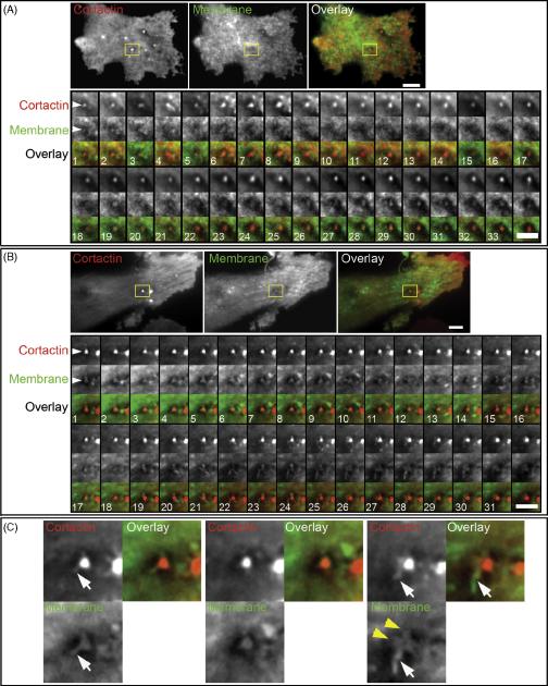
Figure 1
Cell membrane dynamics at invadopodia. Human breast carcinoma wt c-Src MDA-MB-231 cells expressing GFP-Cortactin and membrane marker IL2R-mCherry were cultured on a layer of FN. A. Initiation of invad...
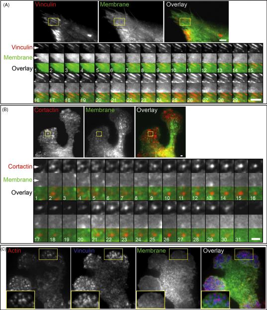
Figure 2
A. Dynamics of the cell membrane at the focal adhesion of HFF transiently expressing GFP-Vinculin and membrane marker IL2R-mCherry. HFF were cultured on a 2D FN matrix. A representative focal adhesion...
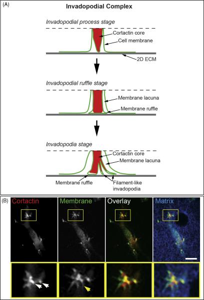
Figure 3
A, Model of the invadopodial complex. Dashed line indicates the upper boundary of the membrane-cytoplasmic zone visualized by TIRF microscopy. B, Invadopodial complex formation in wt c-Src MDA-MB-231 ...
Figure images are served from the NIH/NLM PubMed Central Open Access Subset or Europe PMC; copyright remains with the publishers and authors.
💬 Discussion
0 commentsNo comments yet. Be the first to start a discussion!
Leave a Comment