Abstract
Our capacity to interface with the nervous system remains overwhelmingly reliant on electrical stimulation devices, such as electrode arrays and cuff electrodes that can stimulate both central and peripheral nervous systems. However, electrical stimulation has to deal with multiple challenges, including selectivity, spatial resolution, mechanical stability, implant-induced injury and the subsequent inflammatory response. Optical stimulation techniques may avoid some of these challenges by providing more selective stimulation, higher spatial resolution and reduced invasiveness of the device, while also avoiding the electrical artefacts that complicate recordings of electrically stimulated neuronal activity. This review explores the current status of optical stimulation techniques, including optogenetic methods, photoactive molecule approaches and infrared neural stimulation, together with emerging techniques such as hybrid optical-electrical stimulation, nanoparticle enhanced stimulation and optoelectric methods. Infrared neural stimulation is particularly emphasised, due to the potential for direct activation of neural tissue by infrared light, as opposed to techniques that rely on the introduction of exogenous light responsive materials. However, infrared neural stimulation remains imperfectly understood, and techniques for accurately delivering light are still under development. While the various techniques reviewed here confirm the overall feasibility of optical stimulation, a number of challenges remain to be overcome before they can deliver their full potential.
🔬 Techniques
🧬 Organisms
✨ Fluorophores
🧪 Sample Preparation
🔬 Cell Lines
🏛️ Research Organizations (ROR)
Affiliated research institutions:
📊 Figures
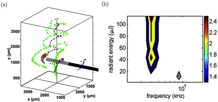
Fig. (1)
(a) 3D reconstruction of a guinea pig cochlea during stimulation. Black cylinder shows the position of the optical fibre, black dotsnindicates the spiral ganglion neurons, green dots are the inner pil...
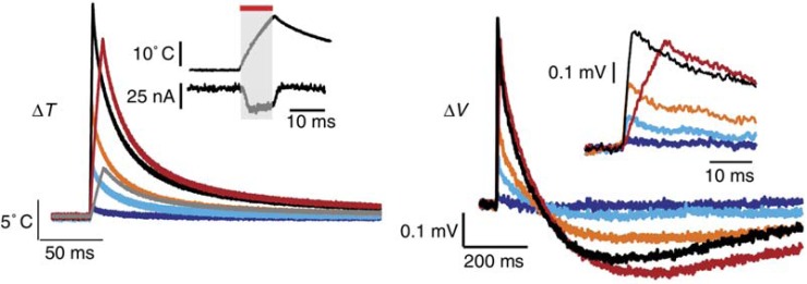
Fig. (2)
Example of temperature measurement (left) and change in cell membrane potential (right) during and after exposure to INS.nAdapted by permission from Macmillan Publishers Ltd: Nature Communications [56...
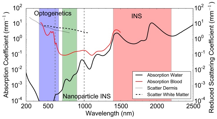
Fig. (3)
Absorption coefficient in water [140] and oxygenated blood (5% haematocrit) [141]. Reduced scattering coefficient in dermis [142]nand white matter [142] over the wavelength range of 200 nm to 2500 nm....
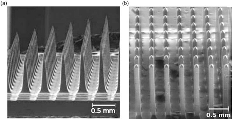
Fig. (4)
SEM image of (a) silicon Utah slant optrode array and (b) glass optrode array reproduced from [153], with permission from SPIE.
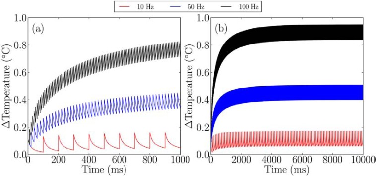
Fig. (5)
Heating from different stimulation rates with INS, when using a fibre with core diameter of 200 u03bcm and NA = 0.22, with anwavelength of 1850 nm and pulse energy of 25 u00b5J. a) Over 1 second b) ov...
Figure images are served from the NIH/NLM PubMed Central Open Access Subset or Europe PMC; copyright remains with the publishers and authors.
💬 Discussion
0 commentsNo comments yet. Be the first to start a discussion!
Leave a Comment