Abstract
Abstract Background The phytocannabinoid cannabidiol (CBD) exhibits antioxidant and antiinflammatory properties. The present study was designed to explore its effects in a mouse model of sepsis-related encephalitis by intravenous administration of lipopolysaccharide (LPS). Methods Vascular responses of pial vessels were analyzed by intravital microscopy and inflammatory parameters measured by qRT-PCR. Results CBD prevented LPS-induced arteriolar and venular vasodilation as well as leukocyte margination. In addition, CBD abolished LPS-induced increases in tumor necrosis factor-alpha and cyclooxygenase-2 expression as measured by quantitative real time PCR. The expression of the inducible-nitric oxide synthase was also reduced by CBD. Finally, preservation of Blood Brain Barrier integrity was also associated to the treatment with CBD. Conclusions These data highlight the antiinflammatory and vascular-stabilizing effects of CBD in endotoxic shock and suggest a possible beneficial effect of this natural cannabinoid.
🔬 Techniques
✨ Fluorophores
🔬 Cell Lines
🏭 Microscope Brands
🧪 Reagent Suppliers
💻 Software Details
🏛️ Research Organizations (ROR)
Affiliated research institutions:
📋 Methods
Vascular responses of pial vessels were analyzed by intravital microscopy and inflammatory parameters measured by qRT-PCR.
Methods
Mice and preparation for intravital microscopy Adult
C57BL/6J mice were maintained in a temperature-controlled specific pathogen-free facility with a strict 12-hour light/dark cycle and with free access to food and water. All experiments were performed in accordance to international and local guidelines as approved by an internal committee (86/609/EEC). Mice were anesthetized with ketamine plus medetomidine (50 mg/kg and 1 mg/kg, respectively). After removing the skin and muscle, a custom-made device was attached to the cranial surface and then fitted to the microscope. Body temperature was continuously monitored by a rectal probe and maintained constant with a thermal blanket. Blood pressure (BP) was monitored by a cuff tail device with a photoelectric sensor (NIPREM 645, Cibertec, Madrid, Spain). A cranial window (2 mm of diameter) was then opened with a high-speed drill, to gain direct access to the brain parenchyma. The tissue was kept humid constantly by subsequent additions of 200 μl-drops of 0.9% saline. Staining of superficial endothelium and microglia was performed by topical administration of Griffonia simplicifolia conjugated with fluorescein (Vector Laboratories, Burlingame, CA, USA), in a 0.9% NaCl solution, for 30 min. At the beginning of the experiment, at 90 and at 180 min, a 50 μL blood sample was obtained by tail puncture to determine blood gases (i-STAT, Abbot Laboratories, NJ, USA). At the end of the experiment mice were killed by decapitation and brains harvested, frozen and conserved at -80°C until use.
Show full methods section
Vascular responses of pial vessels were analyzed by intravital microscopy and inflammatory parameters measured by qRT-PCR.
Methods
Mice and preparation for intravital microscopy Adult
C57BL/6J mice were maintained in a temperature-controlled specific pathogen-free facility with a strict 12-hour light/dark cycle and with free access to food and water. All experiments were performed in accordance to international and local guidelines as approved by an internal committee (86/609/EEC). Mice were anesthetized with ketamine plus medetomidine (50 mg/kg and 1 mg/kg, respectively). After removing the skin and muscle, a custom-made device was attached to the cranial surface and then fitted to the microscope. Body temperature was continuously monitored by a rectal probe and maintained constant with a thermal blanket. Blood pressure (BP) was monitored by a cuff tail device with a photoelectric sensor (NIPREM 645, Cibertec, Madrid, Spain). A cranial window (2 mm of diameter) was then opened with a high-speed drill, to gain direct access to the brain parenchyma. The tissue was kept humid constantly by subsequent additions of 200 μl-drops of 0.9% saline. Staining of superficial endothelium and microglia was performed by topical administration of Griffonia simplicifolia conjugated with fluorescein (Vector Laboratories, Burlingame, CA, USA), in a 0.9% NaCl solution, for 30 min. At the beginning of the experiment, at 90 and at 180 min, a 50 μL blood sample was obtained by tail puncture to determine blood gases (i-STAT, Abbot Laboratories, NJ, USA). At the end of the experiment mice were killed by decapitation and brains harvested, frozen and conserved at -80°C until use.
Drug administration
To clearly observe the cerebrovascular tree throughout the entire experiment, 100 μl of a 70000 MW Texas red-conjugated dextrane solution (Invitrogen, Carlsbad, CA, USA) was administered through the tail vein. This approach stains blood plasma while leaving nucleated cells unstained [ 17 ]. Afterwards, vehicle (Tween/saline, N = 7), LPS (Sigma, St Louis, MO, USA; 1 mg/kg, N = 8), LPS+CBD (1 mg/kg + 3 mg/kg respectively; Tocris Bioscience, Bristol, UK, N = 7) or CBD alone (3 mg/kg, n = 5) were administered i.v. through the tail vein in a total volume of 100 μl. Doses of LPS and CBD were chosen based on previous data [ 5 , 18 ]. A single dose of CBD was chosen because its long half-life time [ 18 ] makes it appropriate for experiments lasting for 3 h as ours.
Image acquisition and analysis
Observations were made using a Nikon 90i upright microscope coupled to a C1 scanhead confocal system with two laser sources (Arg 488 nm and He/Ne 543 nm). Once the area of interest was defined, 60 μm-thick stacks in the Z-axis (3 μm steps) were obtained with the Nikon EZ-C1 software, every 15 min for a total time of 180 min post drug administration. Three-dimensional constructs were analyzed and changes in the diameter of venules (internal diameter 39-112 μm) and third-order arterioles (internal diameter 14-50 μm) (at least 4 of each per animal) measured. Pial vessels of those diameters are considered as optimal for intravital studies on microvessel reactivity [ 19 ]. In addition, the total number of marginated cells (revealed as immobilized black dots inside the vessels) at each time point was counted and the accumulated amount was expressed per area unit (μm 2 ). To that end, the total area corresponding to vessels was estimated in each field of observation by means of ImageJ (NIH) software. BBB integrity 70000 MW dextrane is unable to leave the blood vessels under normal conditions, but diffuses into the brain parenchyma when BBB integrity is compromised. In order to measure this phenomenon, laser-scanning micrographs were analyzed and fluorescence intensity across a cross-section of edematous vessels was measured. This allowed the analysis of fluorochrome distribution inside and outside the affected vessels as an index of BBB damage [ 20 ].
Quantification of markers of oxidative stress Concentrations of 4-hydroxynonenal
(HNE) and of malondialdehyde (MDA) as markers of oxidative stress, were measured in frozen brain tissue by ELISA (OxiSelect HNE-His Adduct and OxiSelect MDA Adduct, Cell Biolabs, San Diego, CA, USA). COX-2, TNF-α and iNOS mRNA levels mRNA levels of cyclooxygenase (COX)-2, tumor necrosis factor-alpha (TNF-α) and inducible nitric oxide synthase (iNOS) were quantified by qRT-PCR from frozen midbrains. Total RNA was extracted using the Tripure Isolation Reagent (Roche Diagnostics, Mannheim, Germany). Single-stranded complementary DNA (cDNA) was synthesized from 1 μg of total RNA using the Transcriptor First Strand cDNA Synthesis Kit (Roche Diagnostics, Mannheim, Germany). PCR primers and TaqMan probes were designed by Tib Molbiol (Berlin, Germany) as shown in table 1 . For normalization, 18S primers and probe number 55 from Universal ProbeLibrary probes 1-165 (Roche) were utilized. Gene expression was quantified using the Quantimix Easy Probes kit (Biotools, Madrid, Spain) in concert with a LightCycler thermocycler (Roche). Standard curves were calculated for quantification purposes using ten-fold serial dilutions of cDNA from brain mouse. The transcript amounts were calculated using the second derivate maximum mode of the LC-sotfware version 4.0. The specific transcript quantities were normalized to the transcript amounts of the reference gene 18S. All further calculations and statistical analyses were carried out with these values referred to as relative expression ratios. Table 1 Sequences for primers and probes employed in this study GENES PRIMER/PROBE SEQUENCES FOR PRIMERS AND PROBES 18S sense AAATCAGTTATGGTTCCTTTGGTC antisense GCTCTAGAATTACCACAGTTATCCAA Probe #55 Use universal ProbeLibrary, Roche Applied Science iNOS sense GCTCCTCCCAGGACCACA antisense GCTGGAAGCCACTGACACTT Probe TaqMan 6FAM-CACCTACCGCACCCGAGATGG--BBQ COX-2 sense TGACCCACTTCAAGGGAGTCT antisense CTGTCAATCAAATATGATCTGGATGTC Probe TaqMan 6FAM-AACAACATCCCCTTCCTGCGAAGTT--BBQ TNF-α sense GCCTATGTCTCAGCCTCTTCTCATT antisense CCACTTGGTGGTTTGCTACGA Probe TaqMan 6FAM-CCATAGAACTGATGAGAGGGAGGCCATTT-BBQ Statistical analysis Results are expressed as mean ± SEM of the indicated number of experiments. Changes in vessel diameter with time were compared between groups by 2-way ANOVA. Differences between groups in enzyme or protein expression were studied by 1-way ANOVA. Newman-Keuls post-hoc test was used for multiple comparisons. A p value of less than 0.05 was considered as statistically significant. Statistical analysis was performed using the 11.0.0 version of SPSS software (SPSS Inc.).
📊 Figures
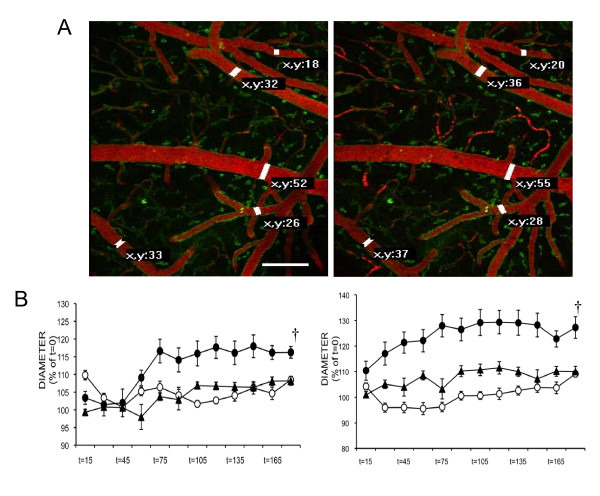
Figure 1
CBD prevented LPS-induced vasodilation . (A) Representative 60 u03bcm Z-stacks of subpial vessels in the mouse brain reflect the increase in blood vessel diameter after LPS administration (t = 0, left...
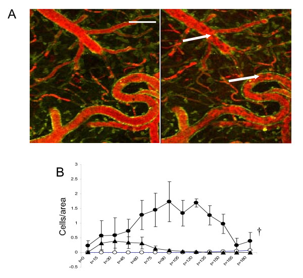
Figure 2
CBD inhibited leukocyte margination . (A) Representative 60 u03bcm Z-stacks of subpial vessels at t = 0 (left) and t = 120 min (right) after LPS administration. Note the intense accumulation of nuclea...
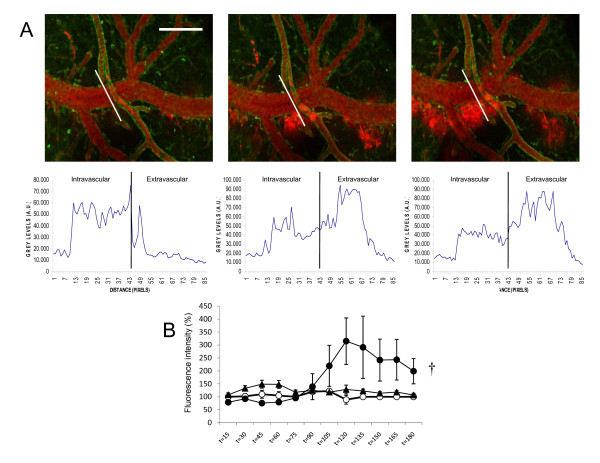
Figure 3
CBD prevented LPS-induced extravasation, as an index of BBB integrity . (A) Representative laser-scanning micrographs of microvasculature at t = 90 (left), t = 105 (center) and t = 120 min (right) aft...
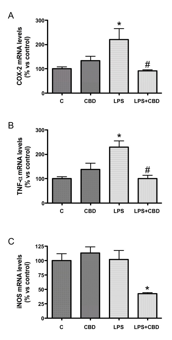
Figure 4
CBD treatment modifies the pattern of induction of COX-2 (A), TNF-u03b1 (B) and iNOS (C) mRNAs, as measured by quantitative real time PCR . Error bars represent mean u00b1 SEM of 7-8 experiments. *p &...
Figure images are served from the NIH/NLM PubMed Central Open Access Subset or Europe PMC; copyright remains with the publishers and authors.
💬 Discussion
0 commentsNo comments yet. Be the first to start a discussion!
Leave a Comment