Abstract
Animal whole-brain functional magnetic resonance imaging (fMRI) provides a non-invasive window into brain activity. A collection of associated methods aims to replicate observations made in humans and to identify the mechanisms underlying the distributed neuronal activity in the healthy and disordered brain. Animal fMRI studies have developed rapidly over the past years, fueled by the development of resting-state fMRI connectivity and genetically encoded neuromodulatory tools. Yet, comparisons between sites remain hampered by lack of standardization. Recently, we highlighted that mouse resting-state functional connectivity converges across centers, although large discrepancies in sensitivity and specificity remained. Here, we explore past and present trends within the animal fMRI community and highlight critical aspects in study design, data acquisition, and post-processing operations, that may affect the results and influence the comparability between studies. We also suggest practices aimed to promote the adoption of standards within the community and improve between-lab reproducibility. The implementation of standardized animal neuroimaging protocols will facilitate animal population imaging efforts as well as meta-analysis and replication studies, the gold standards in evidence-based science.
🔬 Techniques
🧬 Organisms
✨ Fluorophores
🏛️ Research Organizations (ROR)
Affiliated research institutions:
📋 Methods
We searched the Pubmed database 1 on February 11, 2019 for the terms “functional magnetic resonance imaging,” “functional MRI,” or “fMRI” within the abstract or title, excluding studies in human and reviews, from 1990 onward, using the following command. “Search ((fMRI[Title/Abstract]) OR functional MRI[Title/Abstract]) OR functional magnetic resonance imaging[Title/Abstract] Sort by: Best Match Filters: Abstract; Publication date from 1990/01/01 to 2019/12/31; Other Animals.” The query returned 2279 entries. The title and abstract from these were manually screened to exclude studies that did not contain primary research using MRI to assess brain function in animals. In total, 868 research article were considered relevant and could be readily accessed. We recorded the type of study: resting-state or paradigm free RS-FC recordings, pharmacological-evoked, opto-/chemogenetic neuromodulation, deep-brain stimulation (DBS), or stimulus-evoked (including blocks- or events-related designs with sensory stimulation, gas challenge, etc.). We recorded animals species, including strain, gender (male, female, both, N/A), number of animals used, animal preparation (awake, anesthetized free-breathing, anesthetized ventilated), anesthetic used for maintenance during fMRI, field strength, fMRI sequence and contrast, pre-processing softwares, and noted if the datasets were made available by the authors or in online repositories. The resulting table is made available in the Supplementary Material .
Show full methods section
We searched the Pubmed database 1 on February 11, 2019 for the terms “functional magnetic resonance imaging,” “functional MRI,” or “fMRI” within the abstract or title, excluding studies in human and reviews, from 1990 onward, using the following command. “Search ((fMRI[Title/Abstract]) OR functional MRI[Title/Abstract]) OR functional magnetic resonance imaging[Title/Abstract] Sort by: Best Match Filters: Abstract; Publication date from 1990/01/01 to 2019/12/31; Other Animals.” The query returned 2279 entries. The title and abstract from these were manually screened to exclude studies that did not contain primary research using MRI to assess brain function in animals. In total, 868 research article were considered relevant and could be readily accessed. We recorded the type of study: resting-state or paradigm free RS-FC recordings, pharmacological-evoked, opto-/chemogenetic neuromodulation, deep-brain stimulation (DBS), or stimulus-evoked (including blocks- or events-related designs with sensory stimulation, gas challenge, etc.). We recorded animals species, including strain, gender (male, female, both, N/A), number of animals used, animal preparation (awake, anesthetized free-breathing, anesthetized ventilated), anesthetic used for maintenance during fMRI, field strength, fMRI sequence and contrast, pre-processing softwares, and noted if the datasets were made available by the authors or in online repositories. The resulting table is made available in the Supplementary Material .
Experimental Design
Animal fMRI presents the opportunity for new and creative directions in study design, but care must be taken to ensure that experimental changes in the fMRI signal are sufficiently robust for detection and that results are not contaminated by procedural artifacts. Here we highlight evidence supporting standards and reporting strategies to optimize data quality, interpretation, and reproducibility for several common animal fMRI paradigms. Stimulus-Evoked fMRI In animal studies, stimulus-evoked fMRI usually refers to externally applied stimuli during fMRI (e.g., electrical forepaw stimulation), but many principles of study design can be applied to internally delivered stimuli as well, such as with deep-brain stimulation (DBS) and optogenetics. Stimuli can be applied in a block or event-related design. The former alternates between regular stimulation and no-stimulation conditions, while the latter uses brief stimuli presented at varying intervals ( Amaro and Barker, 2006 ). Block designs are best suited to test frequency-related responses and enhance detection power, while event-related designs are best for determining accurate response-time courses and/or frequency-independent functional connectivity ( Amaro and Barker, 2006 ; Van der Linden et al., 2007 ; Maus and van Breukelen, 2013 ; Allen et al., 2015 ; Schlegel et al., 2015 ; Soares et al., 2016 ). Stimulus frequency has a large influence on stimulus-evoked fMRI results. In general, higher frequencies will increase the stimulus input per unit time, thus potentially boosting signal and ability to detect evoked responses ( Amaro and Barker, 2006 ; Kim et al., 2010 ; Maus and van Breukelen, 2013 ), but excessive electrical or optical stimulation can cause tissue damage ( Kiyatkin, 2007 ; Lai et al., 2015 ; Acker et al., 2016 ; Cogan et al., 2016 ), heating and related artifacts ( Zeuthen, 1978 ; Kiyatkin, 2007 ; Cardin et al., 2010 ; Christie et al., 2013 ; Lai et al., 2015 ; Stujenske et al., 2015 ; Acker et al., 2016 ), and non-specific effects ( Tuor et al., 2002 ; Christie et al., 2013 ; Schroeter et al., 2014 ; Shih et al., 2014 ; Schlegel et al., 2015 ; Rungta et al., 2017 ). Stimuli may also change basic physiology and therefore alter the fMRI response ( Tuor et al., 2002 ; Ray et al., 2011 ; Tsubota et al., 2012 ; Li et al., 2013 ; Schroeter et al., 2014 ; Shih et al., 2014 ; Reimann C. et al., 2018 ), thereby occluding signal from the stimulus itself. These findings highlight the importance of carefully monitoring physiology (see below) and establishing frequency-response curves for the stimuli of choice. Functional Connectivity MRI Animal fMRI data acquired in the absence of stimulation or modulation, RS-FC, is commonly used to probe synchronization of spontaneously fluctuating signals between combinations of anatomically, functionally, or procedurally defined brain regions ( Lowe et al., 2000 ; Lu et al., 2007 ; Zhao et al., 2008 ; van Meer et al., 2010 , 2012 ; Lu and Stein, 2014 ; Pan et al., 2015 ; Guadagno et al., 2018 ; Grandjean et al., 2019a ). The use of RS-FC in animal models has rapidly increased over the past decade ( Figure 2 ). To collect the most robust and interpretable RS-FC data, a few principles have been proposed. Recent evidence suggests that brain network components exhibit non-stationary properties ( Hutchison et al., 2013a ; Keilholz et al., 2013 ; Liu and Duyn, 2013 ; Liang et al., 2015a ; Pan et al., 2015 ; Gutierrez-Barragan et al., 2018 ), therefore repetition time should be sufficiently short (e.g., 1 s) to properly sampled the fluctuations and to detect these changes, and scan length should produce enough frames (a minimum of about 300) to account for a large number of temporal clusters ( Majeed et al., 2011 ; Hutchison et al., 2013b ; Jonckers et al., 2015 ). Critical aspects for such analyses are detailed in a later section. Furthermore, if brain modulation/stimulation is included, additional time should be added during the transition periods to and from resting-state to allow for stable connectivity, and subsequent resting periods following each manipulation should be grouped separately to account for potential neuroadaptations ( Pawela et al., 2008 ; Zhao et al., 2008 ; Jonckers et al., 2015 ; Albaugh et al., 2016 ; Chan et al., 2017 ; Decot et al., 2017 ; Chen et al., 2018 ). Importantly, due to the nature of the signal fluctuations on which RS-FC relies, special care must be ensured with regard to physiology and anesthesia to ensure maximal detection. The effects of animal preparations are further discussed below. FIGURE 2 Study design in animal fMRI over time. Stimulus-evoked fMRI (events or blocks related) remain the major component within animal literature. From 2006 and 2010, resting-state fMRI and opto-/chemogenetic fMRI, respectively, have represented an increasing proportion of the animal fMRI studies.
Optogenetics
Many recent stimulus-evoked animal fMRI studies take advantage of the readily MR-compatible optogenetics toolkit ( Figure 2 ; Desai et al., 2011 ; Abe et al., 2012 ; Scott and Murphy, 2012 ; Kahn et al., 2013 ; Iordanova et al., 2015 ; Lemieux et al., 2015 ; Liang et al., 2015b ; Takata et al., 2015 ; Weitz et al., 2015 ; Albaugh et al., 2016 ; Chai et al., 2016 ; Ryali et al., 2016 ; Yu et al., 2016 ; Hinz et al., 2017 ; Lohani et al., 2017 ; Albers et al., 2018 ; Brocka et al., 2018 ; Choe et al., 2018 ; Leong et al., 2018 ; Grandjean et al., 2019b ). Optogenetics allows for robust stimulation of specific cellular and/or anatomical populations ( Zhang et al., 2010 ; Fenno et al., 2011 ; Boyden, 2015 ; Deisseroth, 2015 ; Griessner et al., 2018 ), but despite these advantages this relatively new technique adds layers of complexity over DBS, thereby requiring more rigorous methodology and additional controls. The light-activated channels/pumps expressed in optogenetics, also known as “opsins,” provide a great deal of experimental flexibility ( Fenno et al., 2011 ; Deisseroth, 2015 ; Guru et al., 2015 ). There are several opsins to choose from for optical excitation of cells, including the commonly used ChR2 ( Nagel et al., 2003 ; Boyden et al., 2005 ; Zhang et al., 2006 ; Atasoy et al., 2008 ; Cardin et al., 2010 ) variants activated by penetrating red-shifted light ( Zhang et al., 2008 ; Lin et al., 2013 ; Klapoetke et al., 2014 ) and ultra-fast variants capable of frequencies up to 200 Hz ( Lin et al., 2009 ; Gunaydin et al., 2010 ; Hight et al., 2015 ). If stable excitation over even longer periods is required in fMRI, issues with a continuous light application can be avoided by using step-function opsins which are temporarily activated by a single pulse of light ( Berndt et al., 2009 ; Ferenczi et al., 2016 ). Notably, there are also several opsins for cellular inhibition ( Zhang et al., 2007 ; Berndt et al., 2014 ; Chuong et al., 2014 ), but their application for fMRI is limited as they require longer periods of illumination prone to heat-related artifacts, and anesthetized or sedated animals have low baseline levels of activity ( Lahti et al., 1999 ; Brevard et al., 2003 ; Sicard et al., 2003 ). Injection of viral constructs or expression of foreign genes can potentially change brain function ( Liu et al., 1999 ; Klein et al., 2006 ; Zimmermann et al., 2008 ; Lin, 2011 ; Miyashita et al., 2013 ), and light can induce heating and related MRI artifacts, tissue damage, and non-specific effects ( Elias et al., 1987 ; Christie et al., 2013 ; Stujenske et al., 2015 ; Schmid et al., 2016 ; Rungta et al., 2017 ) thus it is critical to characterize opsin expression and activation of the light source with light delivery to empty-vector (e.g., EYFP) controls. It follows that histological confirmation of fiber placement and construct co-localization with targeted promoters is required ( Bernstein and Boyden, 2011 ; Witten et al., 2011 ; Madisen et al., 2012 ; Zeng and Madisen, 2012 ; Allen et al., 2015 ; Gompf et al., 2015 ; Lin et al., 2016 ; Decot et al., 2017 ). In addition, given the spatial nature of fMRI, the reporting of single-point measurements of light power should be avoided in favor of irradiance (mW/mm 2 ; Aravanis et al., 2007 ; Huber et al., 2008 ; Kahn et al., 2011 ; Yizhar et al., 2011 ; Schmid et al., 2017 ). Finally, light stimulation at frequencies at or below 20 Hz can produce a visual response by activating the visual-related network, requiring light masking or careful control comparison to view experimental effects ( Ferenczi et al., 2016 ; Lin et al., 2016 ; Decot et al., 2017 ; Schmid et al., 2017 ). Chemogenetics Chemogenetics, initially termed “pharmacogenetics,” utilizes pharmacologically inert ligands to stimulate genetically encoded designer receptors, with the aim to produce drug-like sustained activation or inhibition of specific neuronal populations. Initial attempts to combine this approach with fMRI have involved the regional re-expression of pharmacologically targetable endogenous G-coupled protein receptors (e.g., Htr1a, Gozzi et al., 2012 ). The recent development of a modular set of evolved G protein-coupled receptors, termed Designer Receptors Exclusively Activated by Designer Drugs (DREADDs) has greatly expanded the capabilities of this approach ( Armbruster et al., 2007 ; Alexander et al., 2009 ; Lee et al., 2014 ; English and Roth, 2015 ; Roth, 2016 ; Sciolino et al., 2016 ; Smith et al., 2016 ; Zhu et al., 2016 ; Aldrin-Kirk et al., 2018 ). Like optogenetics, chemogenetics is readily MRI compatible ( Giorgi et al., 2017 ; Roelofs et al., 2017 ; Chen et al., 2018 ; Griessner et al., 2018 ; Markicevic et al., 2018 ). Despite its potential, there is, however, an ongoing debate about the specificity of chemogenetics ligands both in neurobehavioral studies ( MacLaren et al., 2016 ; Gomez et al., 2017 ; Mahler and Aston-Jones, 2018 ; Manvich et al., 2018 ) and in chemo-fMRI applications ( Giorgi et al., 2017 ), thereby requiring rigorous methodology to control for potential off-target effects. Both hM3Dq and hM4Di DREADDs are classically activated with infusion of the effector clozapine-N-oxide (CNO) ( Armbruster et al., 2007 ; Alexander et al., 2009 ; Roth, 2016 ; Smith et al., 2016 ; Giorgi et al., 2017 ; Markicevic et al., 2018 ), but new evidence suggests that CNO does not cross the blood-brain barrier and instead is back-metabolized in vivo into its precursor, clozapine ( Gomez et al., 2017 ; Mahler and Aston-Jones, 2018 ; Manvich et al., 2018 ). Importantly, unlike CNO, clozapine is a psychoactive drug, that possesses an affinity for many endogenous receptors. As a result, the use of high CNO doses may result in a plethora of undesirable off-target effects ( Ashby and Wang, 1996 ; Selent et al., 2008 ; MacLaren et al., 2016 ; Roth, 2016 ), including unspecific fMRI response ( Giorgi et al., 2017 ). Overall, it is apparent that chemogenetics effects cannot be interpreted without proper non-DREADD expressing controls. Specifically, the effect of effector administration should be compared between DREADD expressing, and non-DREADD expressing animals and/or hemispheres. Finally, as with optogenetics, validation of DREADD expression and co-localization with target promoters is essential for data interpretation ( Farrell et al., 2013 ; Smith et al., 2016 ; Giorgi et al., 2017 ; Gomez et al., 2017 ; Roelofs et al., 2017 ; Aldrin-Kirk et al., 2018 ; Chen et al., 2018 ; Markicevic et al., 2018 ). Pharmacological fMRI Modulating the brain with pharmacological agents during animal fMRI has a wide variety of traditional applications such as studying the global effects of compounds and their target neurotransmitter systems ( Mueggler et al., 2001 ; Shah et al., 2004 ; Ferrari et al., 2012 ; Razoux et al., 2013 ; van der Marel et al., 2013 ; Jonckers et al., 2015 ). This approach does not require surgical methods, and is apt for identifying global or regional changes in function associated with new or existing drug therapies for neurotransmitter-related brain disorders ( Leslie and James, 2000 ; Martin and Sibson, 2008 ; Canese et al., 2011 ; Bifone and Gozzi, 2012 ; Klomp et al., 2012 ; Minzenberg, 2012 ; Medhi et al., 2014 ), or to map the effect of exogenously administered neuromodulators. In addition, pharmacological challenges can be used to probe how targets and neurotransmitter systems modulate BOLD responses evoked by other stimuli or pharmacological agents ( Marota et al., 2000 ; Hess et al., 2007 ; Schwarz et al., 2007 ; Knabl et al., 2008 ; Rauch et al., 2008 ; Shih et al., 2012a ; Squillace et al., 2014 ; Shah et al., 2016 ; Decot et al., 2017 ; Bruinsma et al., 2018 ; Griessner et al., 2018 ). However, functional imaging with pharmacological agents may not be ideal for dynamic or repetitive studies as effects are dependent on diffusion and receptor kinetics ( Steward et al., 2005 ; Ferris et al., 2006 ; Mandeville et al., 2013 ; Bruinsma et al., 2018 ), and subject to receptor desensitization and downregulation ( Chen et al., 1999 ; Arey, 2014 ; Berg and Clarke, 2018 ); which in some instances may be species-specific ( Knabl et al., 2008 ). It is important to consider dose-response effects and the pharmacokinetics of each drug used in the experimental design. Ideally several doses of drug, and sufficiently long time series should be included in order to interpret the results according to dose-response and absorption/elimination functions ( Leslie and James, 2000 ; Marota et al., 2000 ; Mueggler et al., 2001 ; Steward et al., 2005 ; Ferris et al., 2006 ; Rauch et al., 2008 ; Jenkins, 2012 ; Minzenberg, 2012 ; Jonckers et al., 2015 ; Shah et al., 2015 ; Bruinsma et al., 2018 ). Indeed, many pharmacological agents have known systemic effects which can influence animal physiology and the BOLD signal ( Shah et al., 2004 ; Wang et al., 2006 ; Martin and Sibson, 2008 ; Ferrari et al., 2012 ; Klomp et al., 2012 ), and some drugs have direct effects on the vascular endothelium in the brain, which could alter properties of the hemodynamic response ( Luo et al., 2003 ; Gozzi et al., 2007 ; Shih et al., 2012b ). It is imperative to closely control and monitor animal physiology, and use appropriate doses in order to control for unwanted side effects. Importantly, vehicle controls are necessary for any pharmacological fMRI study, as increased blood flow/volume and increased blood pressure from systemic infusions can alter the MRI signal ( Kalisch et al., 2001 ; Tuor et al., 2002 ; Gozzi et al., 2007 ; Reimann H. M. et al., 2018 ).
Supplementary Material The Supplementary Material for this article can be found online at: https://www.frontiersin.org/articles/10.3389/fninf.2019.00078/full#supplementary-material Click here for additional data file.
📊 Figures
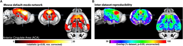
FIGURE 1
(A) A seed-based analysis of the anterior cingulate area in 98 resting-state fMRI scans reveals the topological distribution of the mouse default-mode network. The regions co-activating with the seed ...
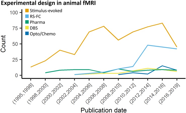
FIGURE 2
Study design in animal fMRI over time. Stimulus-evoked fMRI (events or blocks related) remain the major component within animal literature. From 2006 and 2010, resting-state fMRI and opto-/chemogeneti...
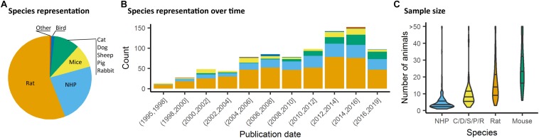
FIGURE 3
Species distribution and sample size. (A) Animal representation in the documented studies. (B) Animal species occurrence in the literature over time. Rats and non-human primate (NHP) represent the maj...
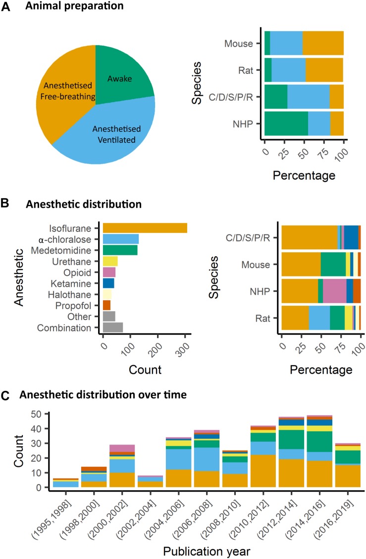
FIGURE 4
Animal preparation and anesthesia trends. (A) Animal fMRI relies mainly on anesthesia to help restrain animals. NHP remain the major species acclimated to awake fMRI. (B) Isoflurane is the principal a...
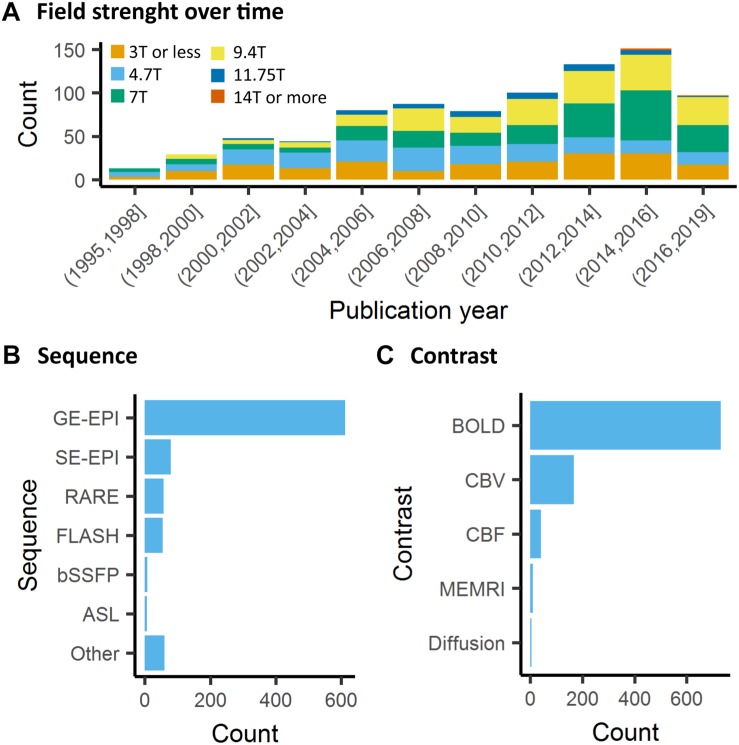
FIGURE 5
Data acquisition. (A) There is a general trend toward higher strength of the main magnetic field in animal fMRI over time. In the past decade, the majority of studies were performed on 7T and 9.4T sys...
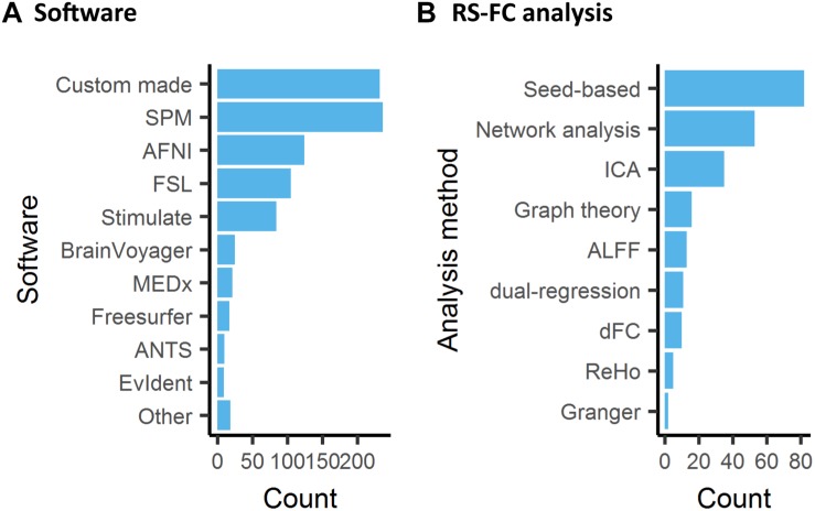
FIGURE 6
Software used for data analysis of animal fMRI and functional connectivity analysis. (A) Custom made software or combination of existing software pipelines remained the most common approach to animal ...
Figure images are served from the NIH/NLM PubMed Central Open Access Subset or Europe PMC; copyright remains with the publishers and authors.
💬 Discussion
0 commentsNo comments yet. Be the first to start a discussion!
Leave a Comment