Abstract
Abstract Introduction Liver microcirculation disturbances are a cause of hepatic failure in sepsis. Increased leukocyte-endothelial interaction, platelet adherence and impaired microperfusion cause hepatocellular damage. The time course and reciprocal influences of ongoing microcirculatory events during endotoxemia have not been clarified. Methods Male Wistar rats (232 ± 17 g) underwent cecal ligation and puncture (CLP). Intravital microscopy (IVM) was performed 0, 1, 3, 5, 10 and 20 hours after CLP. Mean erythrocyte velocity, leukocyte and platelet rolling in postsinusoidal venules and sticking of leukocytes and platelets in postsinusoidal venules and hepatic sinusoids were determined. Heart rate (HR), mean arterial pressure (MAP) and portal venous blood flow (PBF) were measured. Blood count and investigation of hepatic enzyme release was performed after each IVM time point. Results Hepatic platelet-endothelial adherence in liver sinusoids and postsinusoidal venules occurred one hour after the induction of endotoxemia. Leukocyte-endothelial interaction started three to five hours after CLP. A decrease of hepatic microperfusion could be observed at three hours in sinusoids and ten hours in postsinusoidal venules after CLP, although PBF was reduced one hour after CLP. HR remained stable and MAP decreased ten hours after CLP. Hepatic enzymes in blood were significantly elevated ten hours after CLP. Conclusion Hepatic platelet-endothelial interaction is an early event during endotoxemia. Leukocyte adherence occurs later, which underlines the probable involvement of platelets in leukocyte recruitment. Although PBF is reduced immediately after CLP, the later onset of hepatic microperfusion decrease makes the existence of autoregulatory liver mechanisms likely.
🔬 Techniques
✨ Fluorophores
🏭 Microscope Brands
🧪 Reagent Suppliers
🏛️ Research Organizations (ROR)
Affiliated research institutions:
📋 Methods
Male Wistar rats (232 ± 17 g) underwent cecal ligation and puncture (CLP).
Intravital microscopy
(IVM) was performed 0, 1, 3, 5, 10 and 20 hours after CLP. Mean erythrocyte velocity, leukocyte and platelet rolling in postsinusoidal venules and sticking of leukocytes and platelets in postsinusoidal venules and hepatic sinusoids were determined. Heart rate (HR), mean arterial pressure (MAP) and portal venous blood flow (PBF) were measured. Blood count and investigation of hepatic enzyme release was performed after each IVM time point.
Materials and methods
Animals and protocols
All experimental procedures and protocols used in this investigation were approved by the Governmental Animal Protection Committee (Karlsruhe, Germany). Male Wistar rats (232 ± 17 g) were anaesthetized by intraperitoneal injection of 20 mg/kg body weight sodium pentobarbital (Nembutal; Sanofi, Düsseldorf, Germany) and 30 mg/kg body weight intramuscular injection of Ketamin. The right jugular vein was cannulated for the infusion of reagents. Sepsis was induced by cecal ligation and puncture (CLP) [ 21 , 22 ]. Laparotomy of 2 cm in the lower abdomen was performed and the cecum was exteriorised. After non-obstructive ligation of the cecum, two stitches with an 18G needle were performed. The right carotid artery was cannulated for the measurement of heart rate and mean arterial pressure (MAP). To maintain anaesthesia during the observation period, the left femoral vein was cannulated for continuous sodium pentobarbital (8 mg/h/kg body weight) and Ketamin (4 mg/h/kg body weight) infusion. Rectal temperature was measured and maintained at 37°C using a heating pad. IVM was performed in eight animals of each group immediately (0 h) and 1 h, 3 h, 5 h, 10 h and 20 h after CLP. After the IVM blood count in venous blood was performed, hepatocellular enzyme release (AST, ALT), albumin and bilirubin levels in blood, heart rate and MAP were measured. The blood flow of the portal vein (PBF) was determined using the flow probe of a small animal ultrasonic flowmeter (Transonic Systems, New York, USA [ 16 ].
Show full methods section
Male Wistar rats (232 ± 17 g) underwent cecal ligation and puncture (CLP).
Intravital microscopy
(IVM) was performed 0, 1, 3, 5, 10 and 20 hours after CLP. Mean erythrocyte velocity, leukocyte and platelet rolling in postsinusoidal venules and sticking of leukocytes and platelets in postsinusoidal venules and hepatic sinusoids were determined. Heart rate (HR), mean arterial pressure (MAP) and portal venous blood flow (PBF) were measured. Blood count and investigation of hepatic enzyme release was performed after each IVM time point.
Materials and methods
Animals and protocols
All experimental procedures and protocols used in this investigation were approved by the Governmental Animal Protection Committee (Karlsruhe, Germany). Male Wistar rats (232 ± 17 g) were anaesthetized by intraperitoneal injection of 20 mg/kg body weight sodium pentobarbital (Nembutal; Sanofi, Düsseldorf, Germany) and 30 mg/kg body weight intramuscular injection of Ketamin. The right jugular vein was cannulated for the infusion of reagents. Sepsis was induced by cecal ligation and puncture (CLP) [ 21 , 22 ]. Laparotomy of 2 cm in the lower abdomen was performed and the cecum was exteriorised. After non-obstructive ligation of the cecum, two stitches with an 18G needle were performed. The right carotid artery was cannulated for the measurement of heart rate and mean arterial pressure (MAP). To maintain anaesthesia during the observation period, the left femoral vein was cannulated for continuous sodium pentobarbital (8 mg/h/kg body weight) and Ketamin (4 mg/h/kg body weight) infusion. Rectal temperature was measured and maintained at 37°C using a heating pad. IVM was performed in eight animals of each group immediately (0 h) and 1 h, 3 h, 5 h, 10 h and 20 h after CLP. After the IVM blood count in venous blood was performed, hepatocellular enzyme release (AST, ALT), albumin and bilirubin levels in blood, heart rate and MAP were measured. The blood flow of the portal vein (PBF) was determined using the flow probe of a small animal ultrasonic flowmeter (Transonic Systems, New York, USA [ 16 ].
Intravital microscopy
After placing the animal beneath the microscope, a 30 minute stabilisation period followed. The upper surface of the left liver lobe was exteriorised on a specially designed mechanical stage. To maintain body temperature, the liver lobe was continuously superfused by thermostat-controlled (37.0°C) Ringer solution. Hepatic microcirculation was oserved using a specially designed microscope for epi-illumination (Orthoplan; Leica, Wetzlar, Germany; lens with 40-fold magnification, Archoplan 40/0.75 W; Zeiss, Jena, Germany). To protect the liver lobe from heat, a heat protection filter (KG 1; Leica) was located in the body of the microscope. Microscopic images were transferred to a monitor (PVM 1444QM; Sony Corp., Tokyo, Japan) by a low light camera (Kappa CF 8/1; Kappa Messtechnik, Gleichen, Germany) and recorded on a video tape for later evaluation using a computer assisted system for microcirculation analysis (Cap image; Zeintl, Heidelberg, Germany).
Platelet preparation
Whole heparine-blood (1 ml) from donor rats was collected and platelets were stained with rhodamine 6G (Sigma Chemical, St. Louis, USA) as described elsewhere [ 23 ]. The collected blood was diluted with Alserver's buffer after addition of prostaglandin E 1 . Following a four-cycle washing procedure in phosphate-buffered saline, platelets were separated and injected in septic animals prior to IVM.
Analysis of leukocyte-endothelial and platelet-endothelial interactions
Leukocytes were visualized by staining them with rhodamin 6G (0.1 μg/kg body weight). The leukocyte-endothelial and platelet-endothelial interactions were investigated in separate animal groups and analysed in each animal within a minimum of 10 hepatic lobuli and 10 postsinusoidal venules. Adherent leukocytes and platelets that did move or detach from the endothelium prior to a period of 30 s were defined as 'rollers'. Those that adhered to the endothelial wall for longer were classified as 'stickers'. The number of rollers and stickers in postsinusoidal venules were calculated per mm 2 of endothelial surface (length of observed vessel segment × diameter × π = rollers or stickers per mm 2 ). Sticking leukocytes and platelets in sinusoids were quantified as stickers per mm 2 liver surface. Thrombotic sinusoids were calculated as not perfused sinusoids/sinusoids in hepatic lobuli (%).
Analysis of blood flow in liver sinusoids and postsinusoidal venules
Erythrocytes from seperate donor rats were labelled with fluorescein isothiocyanate (FITC, Isomer I, No. F-7250; Sigma Chemical, Deisenhofen, Germany). Blood was washed three times with Alserver's buffer solution and one time with bicine-saline buffer solution to remove plasma. The washed erythrocytes were diluted 1:2 with bicine-saline buffer solution and incubated with FITC (9 mg/ml erythrocytes) for 180 minutes at 25°C. Labelled erythrocytes were further washed five times in bicine-saline buffer solution. Then the erythrocytes were diluted with saline until the hematocrit was 50% in citrate-phosphate-dextrose solution (No.C-7165, Sigma Chemical, Germany). Thirty minutes prior to IVM, the animals received 1.0 ml/kg bodyweight FITC-labelled erythrocytes. For the measurement of sinusoidal perfusion, the velocity of 50 erythrocytes in 10 acini was measured and calculated as a mean of erythrocyte velocyte per mm 2 liver surface. The velocity of 10 erythrocytes in 10 postsinusoidal venules was measured and calculated as a mean of erythrocyte velocity (MEV) per mm 2 of endothelial surface.
Statistical analysis
All data are presented as mean ± standard error of the mean (SEM). Differences were considered significant for p < 0.05. Comparisons between groups were performed by one-way ANOVA followed by LSD test after Shapiro-Wilk's analysis for normal distribution.
Animals and protocols
All experimental procedures and protocols used in this investigation were approved by the Governmental Animal Protection Committee (Karlsruhe, Germany). Male Wistar rats (232 ± 17 g) were anaesthetized by intraperitoneal injection of 20 mg/kg body weight sodium pentobarbital (Nembutal; Sanofi, Düsseldorf, Germany) and 30 mg/kg body weight intramuscular injection of Ketamin. The right jugular vein was cannulated for the infusion of reagents. Sepsis was induced by cecal ligation and puncture (CLP) [ 21 , 22 ]. Laparotomy of 2 cm in the lower abdomen was performed and the cecum was exteriorised. After non-obstructive ligation of the cecum, two stitches with an 18G needle were performed. The right carotid artery was cannulated for the measurement of heart rate and mean arterial pressure (MAP). To maintain anaesthesia during the observation period, the left femoral vein was cannulated for continuous sodium pentobarbital (8 mg/h/kg body weight) and Ketamin (4 mg/h/kg body weight) infusion. Rectal temperature was measured and maintained at 37°C using a heating pad. IVM was performed in eight animals of each group immediately (0 h) and 1 h, 3 h, 5 h, 10 h and 20 h after CLP. After the IVM blood count in venous blood was performed, hepatocellular enzyme release (AST, ALT), albumin and bilirubin levels in blood, heart rate and MAP were measured. The blood flow of the portal vein (PBF) was determined using the flow probe of a small animal ultrasonic flowmeter (Transonic Systems, New York, USA [ 16 ].
📊 Figures
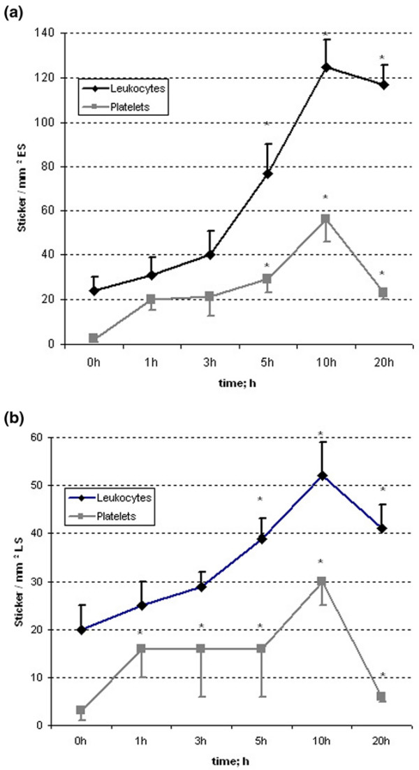
Figure 1
Sticking leukocytes and platelets in (a) postsinusoidal venules and (b) hepatic sinusoids. Asterisks indicate p < 0.05 versus 0 h. ES, endothelial surface; LS, liver surface.
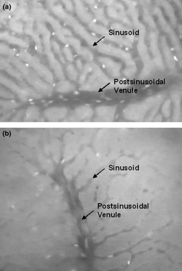
Figure 2
Hepatic sinusoids and postsinusoidal venules during intravital microscopy (a) 0 h and (b) 20 h after CLP demonstrate the decrease of perfused sinusoids and sinusoidal diameter during endotoxemia.
Figure images are served from the NIH/NLM PubMed Central Open Access Subset or Europe PMC; copyright remains with the publishers and authors.
💬 Discussion
0 commentsNo comments yet. Be the first to start a discussion!
Leave a Comment