Abstract
AbstractClean surgical margins in breast‐conserving surgery (BCS) are essential for preventing recurrence. Intraoperative pathologic diagnostic methods, such as frozen section analysis and imprint cytology, have been recognized as crucial tools in BCS. However, the complexity and time‐consuming nature of these pathologic procedures still inhibit their broader applicability worldwide. To address this situation, two issues should be considered: 1) the development of nonpathologic intraoperative diagnosis methods that have better sensitivity, specificity, speed, and cost; and 2) the promotion of new imaging algorithms to standardize data for analyzing positive margins, as represented by artificial intelligence (AI), without the need for judgment by well‐trained pathologists. Researchers have attempted to develop new methods or techniques; several have recently emerged for real‐time intraoperative management of breast margins in live tissues. These methods include conventional imaging, spectroscopy, tomography, magnetic resonance imaging, microscopy, fluorescent probes, and multimodal imaging techniques. This work summarizes the traditional pathologic and newly developed techniques and discusses the advantages and disadvantages of each method. Taking into consideration the recent advances in analyzing pathologic data from breast cancer tissue with AI, the combined use of new technologies with AI algorithms is proposed, and future directions for real‐time intraoperative margin assessment in BCS are discussed.
🔬 Techniques
✨ Fluorophores
🧪 Sample Preparation
🔬 Cell Lines
🏭 Microscope Brands
🏛️ Research Organizations (ROR)
Affiliated research institutions:
📋 Methods
2 Pathologic Methods Frozen section analysis and imprint cytology are the traditional pathologic methods for real‐time intraoperative margin assessment in BCS. [ 5 ] Many intraoperative examples have been reported for pathologic analysis. These methods have the highest diagnostic accuracy in terms of both sensitivity and selectivity and are currently recognized as the most promising methods for lowering the rates of positive margins during BCS. [ 6 ] The reoperation rate for patients who undergo imprint cytology or intraoperative frozen section margin assessment is lower than that of patients who do not receive any intraoperative margin status assessment. [ 5 , 19 ] In addition, the routine use of intraoperative frozen section analysis can be cost‐effective for both the patient and the hospital. [ 20 ] 2.1 Frozen Section Analysis In rapid frozen section analysis, breast tissue samples, i.e., stumps specimens, from the surgical resection are embedded in the optimal cutting temperature compound, frozen, and cut into slices. The slices are then put on a glass slide and fixed with paraformaldehyde for immunohistochemical staining. Next, light microscopy of sections stained with hematoxylin and eosin (H&E) are used to perform the pathologic evaluation. A significant advantage of H&E staining analysis of the frozen section is that this method can determine the presence of cancer in the surgical samples of interest as well as diagnose the type of various cancers. Thus, further surgery could be guided by whether morphological analysis shows that the sample consists of DCIS, invasive ductal carcinoma (IDC), ductal hyperplasia (DH), or normal breast gland (NBG). Nevertheless, currently these methods are not used for BCS in hospitals worldwide. Reasons include complicated sample preparation, which requires an additional skilled technician to cut the specimens for frozen section interpretation and tedious pathologic analysis that requires ≈30 min for a single assessment. [ 5b,c,e ] If additional examination is needed, tremendous efforts and amounts of time are necessary. A worldwide shortage of trained pathologists, which is more severe than the shortage of physicians in general, is another limiting factor for routine frozen section analysis as part of intraoperative margin assessment in BCS.
Show full methods section
2 Pathologic Methods Frozen section analysis and imprint cytology are the traditional pathologic methods for real‐time intraoperative margin assessment in BCS. [ 5 ] Many intraoperative examples have been reported for pathologic analysis. These methods have the highest diagnostic accuracy in terms of both sensitivity and selectivity and are currently recognized as the most promising methods for lowering the rates of positive margins during BCS. [ 6 ] The reoperation rate for patients who undergo imprint cytology or intraoperative frozen section margin assessment is lower than that of patients who do not receive any intraoperative margin status assessment. [ 5 , 19 ] In addition, the routine use of intraoperative frozen section analysis can be cost‐effective for both the patient and the hospital. [ 20 ] 2.1 Frozen Section Analysis In rapid frozen section analysis, breast tissue samples, i.e., stumps specimens, from the surgical resection are embedded in the optimal cutting temperature compound, frozen, and cut into slices. The slices are then put on a glass slide and fixed with paraformaldehyde for immunohistochemical staining. Next, light microscopy of sections stained with hematoxylin and eosin (H&E) are used to perform the pathologic evaluation. A significant advantage of H&E staining analysis of the frozen section is that this method can determine the presence of cancer in the surgical samples of interest as well as diagnose the type of various cancers. Thus, further surgery could be guided by whether morphological analysis shows that the sample consists of DCIS, invasive ductal carcinoma (IDC), ductal hyperplasia (DH), or normal breast gland (NBG). Nevertheless, currently these methods are not used for BCS in hospitals worldwide. Reasons include complicated sample preparation, which requires an additional skilled technician to cut the specimens for frozen section interpretation and tedious pathologic analysis that requires ≈30 min for a single assessment. [ 5b,c,e ] If additional examination is needed, tremendous efforts and amounts of time are necessary. A worldwide shortage of trained pathologists, which is more severe than the shortage of physicians in general, is another limiting factor for routine frozen section analysis as part of intraoperative margin assessment in BCS.
Imprint Cytology
Imprint cytology analysis, on the other hand, is a simpler method used during BCS in some hospitals. Live tissue samples are rubbed onto a glass slide. The attached cells are immediately fixed with ethanol. Next, H&E or Papanicolaou staining is performed on the ethanol‐fixed cells. Imprint cytology analysis is based on the idea that malignant cells will adhere to the slides, whereas adipose cells will not. Imprint cytology analysis examines the entire surface of the resected tissue, unlike the spot checks that occur with frozen section analysis. Many reports on BCS show that imprint cytology can achieve similarly high diagnostic accuracy as frozen section analysis. [ 5a,d,e ] However, this method can only determine the presence of cancer. It cannot analyze morphology. Rapid and accurate interpretation requires a professional trained in cytopathology in the operating room in addition to the regular surgical team. [ 21 ] The increase in operative time resulting from the pathologic procedure and the increased workload for pathologists inhibit the broader applicability of this method as an established global standard for intraoperative diagnosis. Thus, although frozen section analysis and imprint cytology are the most reliable methods currently available, only a limited number of hospitals in some developed countries actually use them for rapid intraoperative diagnosis during BCS.
H&E Pathologic Assessment by Deep Learning and AI Algorithms
To circumvent the shortage of and workload burden for pathologists and improve the diagnostic accuracy of intraoperative margin assessment in BCS, significant attention has been focused on automated deep learning algorithms. An exciting international competition (CAMELYON16) to assess the effectiveness of automated deep learning algorithms in diagnosing the H&E sections of axillary lymph node metastasis was conducted during November 2015 to November 2016. [ 18a ] Thirty‐two algorithms and 12 pathologists (with and without time constraints) were tested with 129 whole‐slide images (49 with and 80 without metastasis); the task was to classify images as definitely normal tissue, probably normal tissue, equivocal, probably tumor, or definitely tumor. The algorithms developed by Harvard Medical School and Massachusette Institute of Technology achieved the highest score (true‐positive fraction, 72.4%), which was comparable to scores from pathologists without time constraints. It is noteworthy to mention that the best algorithms, i.e., those with an area under the receiver operating characteristic curve (AUC) of 0.994, performed significantly better than pathologists when there is time constraint for diagnosis (AUC, 0.810). The top five algorithms had a mean AUC of 0.960, which was comparable with pathologists without time constraints (AUC, 0.966). These data showed that the deep learning algorithms exhibited better diagnostic performance than 12 pathologists taking part in a simulation exercise designed to mimic the routine pathology workflow for axillary lymph nodes. Alternatively, the performance of the algorithms was comparable with an expert pathologist interpreting images with H&E staining under no time constraints. Thus, deep learning algorithms have significant potential to circumvent the problems associated with pathologic methods. However, other than for axillary lymph nodes in breast cancer, there have been no reports to date on using new algorithms to interpret pathologic information for margin assessment in BCS. Alternatively, we could combine these AI algorithms with other newly emerging rapid and convenient techniques that could give the same pathologic information for diagnosis during BCS in the future. Recently, Tsirigos and co‐workers developed a deep learning model using publicly available whole‐slide images in the Cancer Genome Atlas to accurately and automatically classify histopathologic images of non‐small cell lung cancer from different cohorts collected at their institution. [ 18b ] They demonstrated that a convolutional neural network, such as Google's inception v3, can be used to assist in the diagnosis of lung cancer from histopathologic slides, reaching sensitivity and specificity comparable to that of a pathologist. Furthermore, by analyzing only the pathology images, the network was also able to predict the most commonly mutated genes in lung adenocarcinoma. These findings suggest that deep learning and AI algorithms have the potential to predict gene mutations in various kinds of cancers.
3 Conventional Imaging Methods Although BCS has been used as the primary treatment for early‐stage breast cancer, more accurate techniques are needed to assess resection margins during surgery to avoid the need for re‐excision and reoperation. Intraoperative specimen imaging methods such as conventional SR and IOUSG provide timely information on whether re‐excision of a cavity shave margin is indicated during routine BCS. Conventional Specimen Radiography (SR) SR is used for immediate assessment of tissue samples following biopsy or surgical excision. Conventional SR involves X‐ray imaging of excised tissue using mammography or a specimen radiography system ( Figure 2 A ). SR is performed on a nonpalpable lesion to exploit the X‐ray projection of the imaged tissue and produce contrast based on beam attenuation through the tissue. The standard of care involves using X‐ray projections to localize the center of the visible tumor and verify that the specimen contains the observed lesion. Three methods commonly used for preoperative localization of nonpalpable tumors are radioactive seed localization, wire‐guided localization, and radio‐guided occult lesion localization. [ 7d,e ] Figure 2 A‐i) Digital specimen mammography device for identifying lumpectomy targets during breast‐conserving surgery (BCS). A‐ii) Mammography images obtained using the intraoperative digital specimen mammography (IDSM) method. A‐iii) Nonpalpable breast lesions were wire‐localized. Adapted with permission. [ 7c ] Copyright 2007, Springer Nature. B) Ultrasound images of breast specimens with B‐i) transverse and B‐ii) longitudinal scans. The images show a hypoechoic mass close to the lateral margin, which was considered positive based on ultrasonographic assessment. Adapted with permission. [ 8b ] Copyright 2019, Elsevier. Compared to conventional SR, which needs significant time for transporting the surgical specimen from the operating room to the diagnostic imaging room for specimen radiography, intraoperative digital specimen mammography (IDSM), which can occur in the operating room, enables immediate radiography of the specimen. However, one study that compared tumor localization and margin estimation determined using conventional SR and IDSM reported that IDSM did not reduce overall operative times significantly, but it leads to a significant reduction in the positive margin rate. [ 7c,f ] However, clear margin width for specimen radiography has not yet been defined, and low sensitivity and specificity associated with conventional SR methods remain problematic. Intraoperative Ultrasonography (IOUSG) IOUSG imaging of specimen margins allows for visualization of structural features and associated heterogeneity (Figure 2B ). Surgeons locate the tumor in the breast using ultrasound and compare findings with preoperative digital images. After excision, the surgeon can use ultrasound to examine the specimen ex vivo to confirm that it resembles the candidate lesion targeted preoperatively. In a multicenter, randomized controlled trial, IOUSG‐guided surgery significantly lowered the proportion of tumor‐involved resection margins compared with palpation‐guided surgery, thus reducing the need for re‐excision, mastectomy, and boost radiotherapy. [ 7 , 8 ] Since mammography has difficulty imaging through dense breast tissue, IOUSG may be a better alternative than specimen radiography. Moreover, IOUSG is much faster and more cost‐efficient than more commonly used radiography techniques. [ 8d ] However, the necessity of larger margins, low sensitivity, and a requirement to be scanned make IOUSG unlikely to be a full solution to the margin status problem.
4 Newly Emerging Diagnosis Methods Given the disadvantages of pathologic analysis, newly emerging technologies have been developed that are advantageous in terms of speed, cost, and reliability, in addition to diagnostic accuracy. We describe methods based on computed tomography, MRI, spectroscopy, and chemical approaches. We discuss the advantages and disadvantages of these techniques in improving future real‐time intraoperative margin assessment in BCS.
Optical Coherence Tomography
Optical coherence tomography
(OCT) is the optical version of ultrasound imaging, which applies a light wave instead of a sound wave in an entire live BCS specimen. The application of near‐infrared light leads to a high‐resolution, real‐time, multidimensional image of a cancer tissue sample up to 2 mm beneath the tissue surface ( Figure 3 A ). [ 12a ] The OCT light penetrates the entire live specimen, which is scattered back to the detector. Since cancer typically has a higher nuclear‐to‐cytoplasm ratio, higher cellular density, and higher nuclear density than fibrous and fatty tissue of normal mammary regions, cancer tissue has higher scattering properties. Adipocytes are imaged with depths of up to 2 mm, but tumors are imaged to depths of 200 to 1000 µm. Thus, normal gland tissue and cancer tissue are differentiated with OCT imaging. Figure 3 A‐i) Schematic of the spectral‐domain optical coherence tomography (OCT) system. Light from a superluminescent diode (SLD) is directed into an optical circulator (OC) and to a fiber coupler (FC), which splits 5% of the light to a reference arm mirror (RM) and 95% of the light to a sample arm containing focusing optics and an automated x – y translation stage (TS). Light is collimated through fiber collimators (Col). Reflected light from each arm is coupled through polarization paddles (PP), interfered within the fiber coupler, and spectrally dispersed onto a line camera. A‐ii,iii) OCT images of positive tumor margins show a distinct region with heterogeneous scattering. The corresponding histologic images with hematoxylin and eosin (H&E) staining confirmed the presence of positive margins. Adapted with permission. [ 12a ] Copyright 2009, American Association for Cancer Research. B) Micro‐computed tomography (Micro‐CT) images of a breast lumpectomy specimen with the largest dimension in the sagittal, transverse, and coronal planes. Arrow: a close margin (
📊 Figures
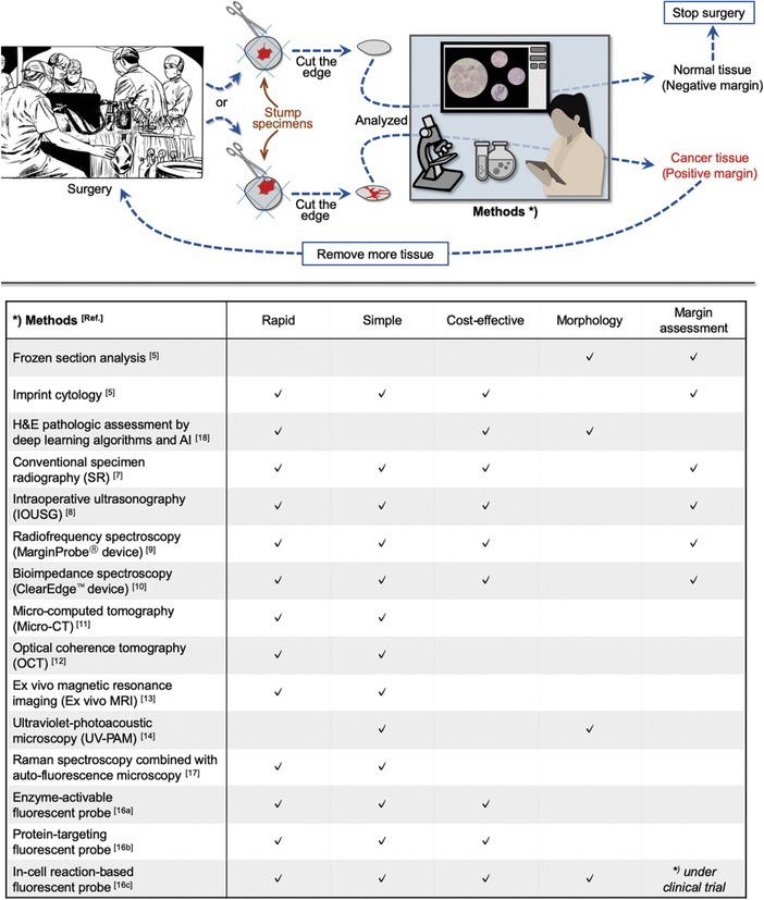
Figure 1
Schematic of intraoperative diagnosis methods. Frozen section analysis has been used for decades as the gold standard during breastu2010conserving surgery (BCS). Despite its reliability, this traditio...
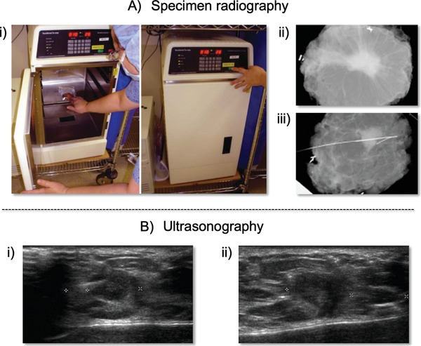
Figure 2
Au2010i) Digital specimen mammography device for identifying lumpectomy targets during breastu2010conserving surgery (BCS). Au2010ii) Mammography images obtained using the intraoperative digital speci...
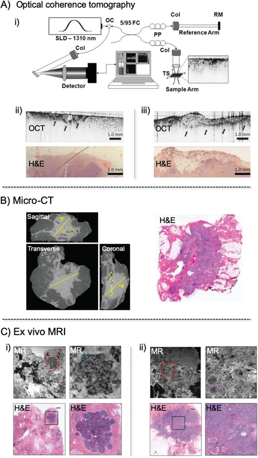
Figure 3
Au2010i) Schematic of the spectralu2010domain optical coherence tomography (OCT) system. Light from a superluminescent diode (SLD) is directed into an optical circulator (OC) and to a fiber coupler (F...
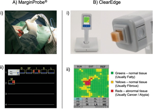
Figure 4
Au2010i) Intraoperative use of the MarginProbe device with a breast lumpectomy specimen. Measurement is performed by applying the tip of the probe to a point on the resected lumpectomy specimen. Durin...
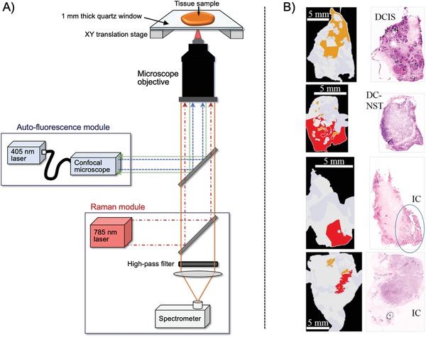
Figure 5
A) Multimodal spectral histopathology (MSH) instrument consist of an inverted optical microscope with an integrated Raman spectrometer (excitation 785 nm, detection Raman shift range, 600u20131800 cm ...
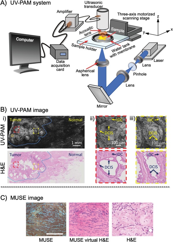
Figure 6
A) Schematic of the ultravioletu2010photoacoustic microscopy (UVu2010PAM) system for surgical margin imaging. A pair of lenses and a pinhole filter and expand the UV laser beam. The aspherical lens fo...
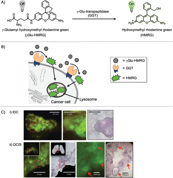
Figure 7
A) Explicit activiation of the u03b3u2010glutamyl hydroxymethyl rhodamine green (u03b3Gluu2010HMRG) probe by u03b3u2010glutamyltranspeptidase (GGT). B) Mechanism of u03b3Gluu2010HMRG activation. u03b3...
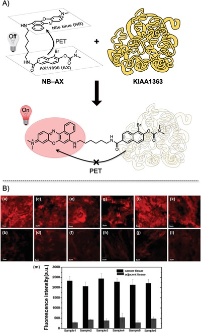
Figure 8
A) The NBu2010AX probe is composed of Nile blue (NB) dye and AX11890, a selective inhibitor of enzyme KIAA1363, linked by a flexible linear hexanediamine chain. Free NBu2010AX (top) is in a folded str...
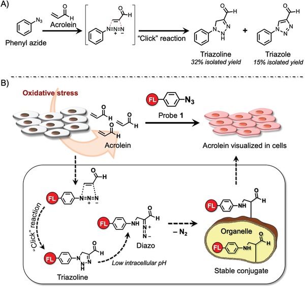
Figure 9
A) Phenyl azide reaction with acrolein. B) u201cClicku2010tou2010senseu201d method and mechanism. Fluorescenceu2010labeled phenyl azide smoothly reacts with acrolein generated by cancer cells through ...
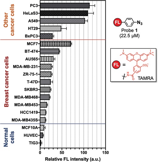
Figure 10
Intensity of fluorescence observed for cell lines over 22.5 u00d7 10 u22126 m of probe 1 . Three normal cell lines were tested, from bottom to top: TIG3 (normal human diploid cells), HUVEC (human umbi...
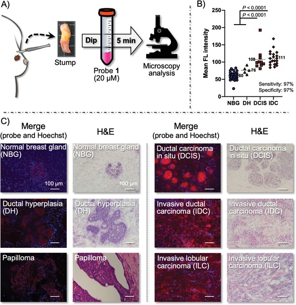
Figure 11
A) Schematic of the procedure for labeling of breast cancer stumps with probe 1 . B) Morphology of normal breast gland (NBG), ductal hyperplasia (DH), papilloma, ductal carcinoma in situ (DCIS), invas...
Figure images are served from the NIH/NLM PubMed Central Open Access Subset or Europe PMC; copyright remains with the publishers and authors.
💬 Discussion
0 commentsNo comments yet. Be the first to start a discussion!
Leave a Comment