Abstract
Abstract Oxytocin is an important regulator of the social brain. In some animal models of autism, notably in Magel2 tm1.1Mus -deficient mice, peripheral administration of oxytocin in infancy improves social behaviors until adulthood. However, neither the mechanisms responsible for social deficits nor the mechanisms by which such oxytocin administration has long-term effects are known. Here, we aimed to clarify these oxytocin-dependent mechanisms, focusing on social memory performance. Using in situ hybridization (RNAscope), we have established that Magel2 and oxytocin receptor are co-expressed in the dentate gyrus and CA2/CA3 hippocampal regions involved in the circuitry underlying social memory. Then, we have shown that Magel2 tm1.1Mus - deficient mice, evaluated in a three-chamber test, present a deficit in social memory. Next, in hippocampus, we conducted neuroanatomical and functional studies using immunostaining, oxytocin-binding experiments, ex vivo electrophysiological recordings, calcium imaging and biochemical studies. We demonstrated: an increase of the GABAergic activity of CA3-pyramidal cells associated with an increase in the quantity of oxytocin receptors and of somatostatin interneurons in both DG and CA2/CA3 regions. We also revealed a delay in the GABAergic development sequence in Magel2 tm1.1Mus -deficient pups, linked to phosphorylation modifications of KCC2. Above all, we demonstrated the positive effects of subcutaneous administration of oxytocin in the mutant neonates, restoring hippocampal alterations and social memory at adulthood. Although clinical trials are debated, this study highlights the mechanisms by which peripheral oxytocin administration in neonates impacts the brain and demonstrates the therapeutic value of oxytocin to treat infants with autism spectrum disorders.
🔬 Techniques
🧪 Sample Preparation
🧪 Reagent Suppliers
💻 Software Details
💾 Data Repositories
🏛️ Research Organizations (ROR)
Affiliated research institutions:
📋 Methods
Animals and primary hippocampal cultures
Magel2 tm1.1Mus +/+ (WT) and Magel2 tm1.1Mus −/− ( Magel2- KO) mice were maintained on a C57BL/6J genetic background. Experimental protocols were approved by the institutional Ethical Committee guidelines for animal research with the accreditation no. B13-055-19 from the French Ministry of Agriculture. Magel2 -deficient mice were generated as previously published [ 18 ]. Embryonic day 18 dissociated hippocampal neurons were obtained from timed pregnant mice. See Supplementary Information for details.
Oxytocin treatment
WT and Magel2- KO pups were removed from their mother, placed on a heating pad, given a subcutaneous (s.c.) injection and quickly returned to the mother. The solutions injected were isotonic saline (10 µl) for control mice and 2 µg of OT (Phoenix Pharmaceuticals Inc., cat #051-01) diluted in isotonic saline (10 µl) for treated mice.
Behavior
The effects of Magel2 deletion and OT-treatment were evaluated on social behavior, locomotor and vertical activity, anxiety-like behavior and nonsocial memory. For detailed procedures, see Supplementary Information .
Calcium imaging recordings
Calcium imaging experiments were carried out as previously reported [ 36 ] and are described in Supplementary Information section.
Electrophysiological recordings and morphological analysis Whole-cell patch clamp
Spontaneous and miniature synaptic activity was recorded in voltage-clamp mode on P20–P25 CA3-pyramidal neurons. The morphology of recorded aCA3d neurons was defined by adding biocytin in the recording solution and performing Neurolucida reconstruction followed by a Sholl analysis. See Supplementary Information , for details. Single GABA A channel recordings were performed on hippocampal CA3-pyramidal neurons at P1, P7, and P15 in cell-attached configuration, as described in Supplementary Information .
Show full methods section
Animals and primary hippocampal cultures
Magel2 tm1.1Mus +/+ (WT) and Magel2 tm1.1Mus −/− ( Magel2- KO) mice were maintained on a C57BL/6J genetic background. Experimental protocols were approved by the institutional Ethical Committee guidelines for animal research with the accreditation no. B13-055-19 from the French Ministry of Agriculture. Magel2 -deficient mice were generated as previously published [ 18 ]. Embryonic day 18 dissociated hippocampal neurons were obtained from timed pregnant mice. See Supplementary Information for details.
Oxytocin treatment
WT and Magel2- KO pups were removed from their mother, placed on a heating pad, given a subcutaneous (s.c.) injection and quickly returned to the mother. The solutions injected were isotonic saline (10 µl) for control mice and 2 µg of OT (Phoenix Pharmaceuticals Inc., cat #051-01) diluted in isotonic saline (10 µl) for treated mice.
Behavior
The effects of Magel2 deletion and OT-treatment were evaluated on social behavior, locomotor and vertical activity, anxiety-like behavior and nonsocial memory. For detailed procedures, see Supplementary Information .
Calcium imaging recordings
Calcium imaging experiments were carried out as previously reported [ 36 ] and are described in Supplementary Information section.
Electrophysiological recordings and morphological analysis Whole-cell patch clamp
Spontaneous and miniature synaptic activity was recorded in voltage-clamp mode on P20–P25 CA3-pyramidal neurons. The morphology of recorded aCA3d neurons was defined by adding biocytin in the recording solution and performing Neurolucida reconstruction followed by a Sholl analysis. See Supplementary Information , for details. Single GABA A channel recordings were performed on hippocampal CA3-pyramidal neurons at P1, P7, and P15 in cell-attached configuration, as described in Supplementary Information .
Immunohistochemistry and quantification
Immunostaining was carried out on 50 μm-thick coronal sections following standard procedure, as described in Supplementary Information . OT-binding assay Adult WT and mutant mice were killed and non-perfused mouse brain were frozen in −25 °C isopentane and stored at −80 °C until cut. 14 µm-thick brain slices were cut using a cryostat (Frigocut-2700, Reichert-Jung) and collected on chromallume-coated slides and stored at −80 °C until use. Localization of OT-binding sites was performed by autoradiography as detailed in Supplementary Information .
Chromogenic in situ hybridization
Fresh-frozen brains from WT mice at P7 and P28 were sectioned in a cryostat in the coronal plane at 20 μm thickness and mounted on Superfrost Plus slides and stored at −80 °C. RNA detection was performed on tissue sections using RNAscope 2.5HD Duplex Assay (Cat #322430, Advanced Cell Diagnostics (ACD), Hayward, CA) as detailed in Supplementary Information .
Western blot
Western blotting experiments were performed on hippocampal tissue and specific bands were visualized with secondary HRP-conjugated antibodies using ChemiDoc™ Imaging Systems (Bio-Rad). The relative intensities of immunoblot bands were determined by densitometry with ImageJ software. See Supplementary Information for details.
Statistical analysis
Statistical analyses were performed using GraphPad Prism (GraphPad Software, Prism 8.0 software, Inc, La Jolla, CA, USA). All the statistical analyses are reported in a specific file. For details, see Supplementary Information .
Supplementary information Supplemental Information - Materials and Methods Supplemental Information - Statistical Tables Supplementary Figure 1 Supplementary Figure 2 Supplementary Figure 3 Supplementary Figure 4 Supplementary Figure 5 Supplementary Figure 6 Supplementary Figure 7
📊 Figures
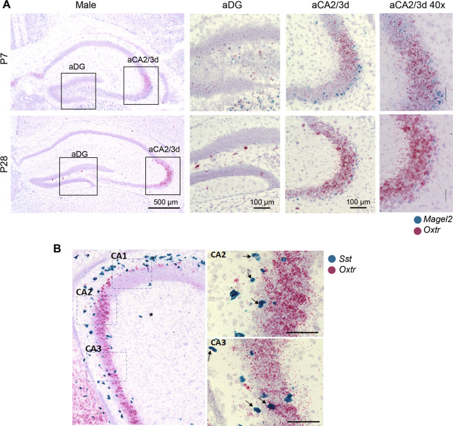
Fig. 1
Expression of Magel2 and Oxytocin receptor ( Oxtr ) transcripts in hippocampus of wild-type male mice at P7 and P28.
A Representative image obtained by RNAscope technology showing the respective localization of Magel2 (blue) and Oxtr (pink) transcripts in dentate gyrus (DG) and aCA2/CA3d region of hippocampus. B Rep...
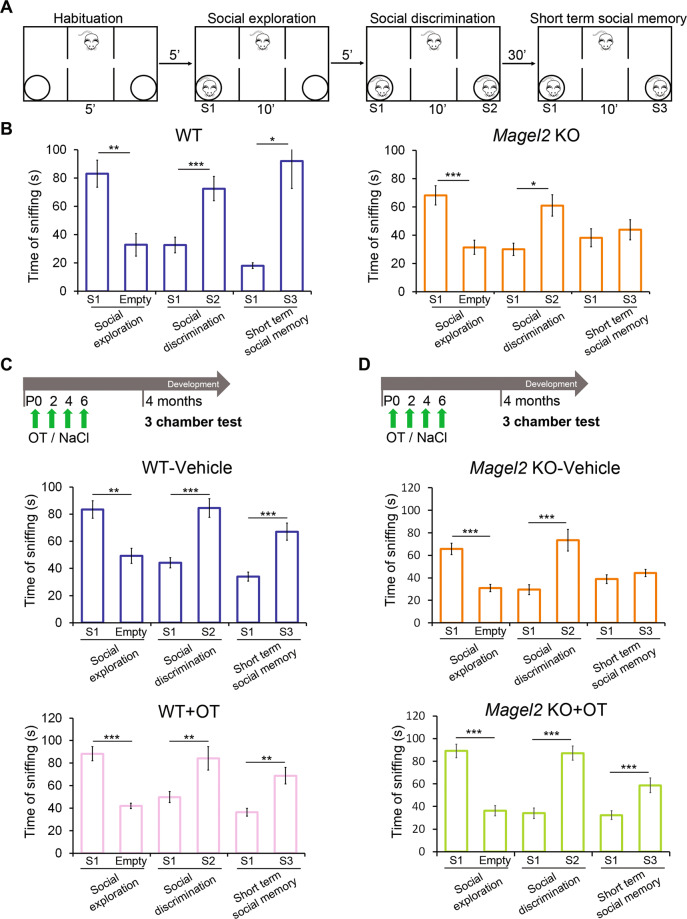
Fig. 2
Social behavior in the three-chamber test of adult male WT versus Magel2 KO and adult male WT or Magel2 KO vehicle-treated or OT-treated as neonates.
A Paradigm of the three-chamber test. Sniffing time between mice is measured in each test. B WT males ( N =u200912) show normal behavior in all the steps of the test; Magel2 KO males ( N =u20099) show...
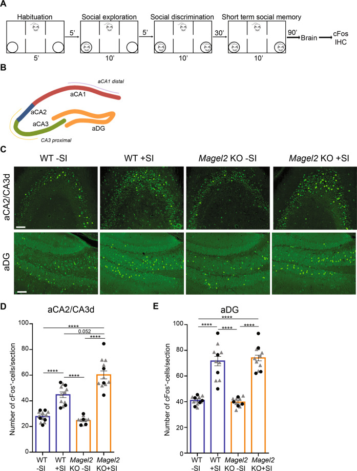
Fig. 3
cFos activity in aCA2/CA3d and aDG regions of Magel2 KO and WT adult male mice following the social memory task in the three-chamber test.
A Paradigm of the three-chamber test (+SI) followed 90u2009min later by dissection of the brains and immunohistochemistry experiments. Control mice (u2212SI) were not tested in the three-chamber test....
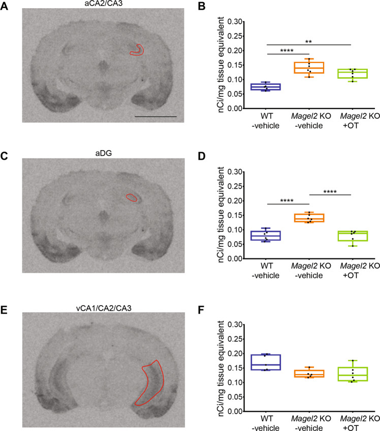
Fig. 4
Quantification of OT-binding sites by brain autoradiography in the adult hippocampus of Magel2 KO male mice treated with OT or vehicle versus WT-vehicle male mice.
A , C , E Representative sections of autoradiographic labeling of OT-binding sites displayed in grayscale, showing the regions of interest (ROI) selected for analysis: ( A ) anterior CA2/CA3 (aCA2/CA3...
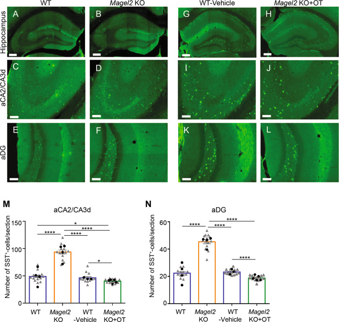
Fig. 5
Quantification of somatostatin (SST) immunopositive cells in the anterior hippocampus region of adult Magel2 KO, WT, Magel2 KO treated with OT and WT-vehicle male mice.
Au2013L Immunolabeling on coronal hippocampal sections at adulthood in WT ( A , C , E ) and Magel2 KO ( B , D , F ), and in WT-vehicle ( G , I , K ) and Magel2 KOu2009+u2009OT ( H , J , L ) with a mag...
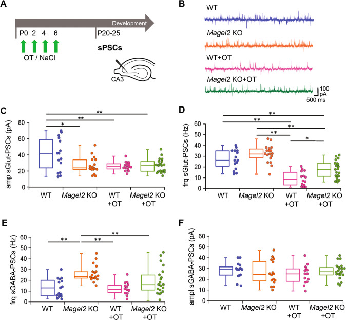
Fig. 6
Spontaneous Glutamatergic and GABAergic synaptic activity of CA3-pyramidal neurons in the anterior hippocampus region of Magel2 KO mice versus WT juvenile mice with or without OT-treatment as neonates.
A Paradigm of the test. WT or Magel2 KO mice were either not injected or were injected with OT in the first week of life. At P20u201325, neurons were recorded in brain slices. B Examples of whole-cell...
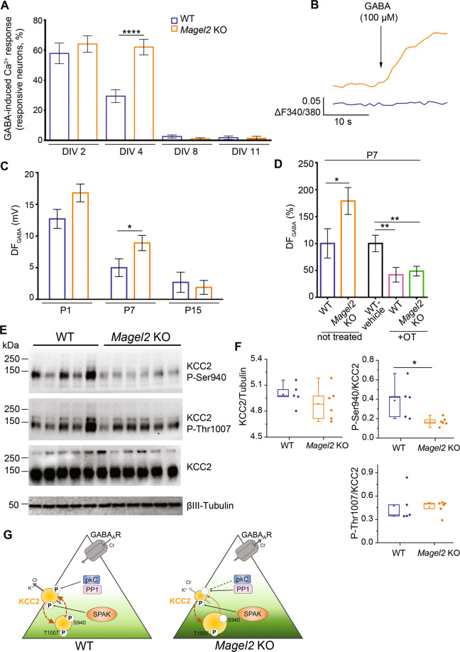
Fig. 7
The excitatory-to-inhibitory developmental GABA shift in Magel2 KO versus WT hippocampi and the effect on an OT-treatment.u00a0Abundance and phosphorylation state of KCC2 in WT and Magel2 KO pups in relation with this GABA shift.
A , B GABA-induced Ca 2+ responses in Magel2 KO developing hippocampal neuronal cultures versus WT. A Percentage of WT and Magel2 KO E18 hippocampal neurons showing GABA-induced Ca 2+ responses at sel...
Figure images are served from the NIH/NLM PubMed Central Open Access Subset or Europe PMC; copyright remains with the publishers and authors.
💬 Discussion
0 commentsNo comments yet. Be the first to start a discussion!
Leave a Comment