Abstract
Cell replacement therapies are often enhanced by utilizing polymer scaffolds to improve retention or direct cell orientation and migration. Obstacles to refinement of such polymer scaffolds often include challenges in controlling the microstructure of biocompatible molecules in three dimensions at cellular scales. Two-photon polymerization of acrylated poly(caprolactone) (PCL) could offer a means of achieving precise microstructural control of a material in a biocompatible platform. In this work, we studied the effect of various formulation and two-photon polymerization parameters on minimum laser power needed to achieve polymerization, resolution, and fidelity to a target 3D model designed to be used for retinal cell replacement. Overall, we found that increasing the concentration of crosslink-able groups decreased polymerization threshold and the size of resolvable features while increasing fidelity of the scaffold to the 3D model. In general, this improvement was achieved by increasing the number of acrylate groups per prepolymer molecule, increasing the acrylated PCL concentration, or decreasing its molecular weight. Resulting two-photon polymerized PCL scaffolds successfully supported human iPSC derived retinal progenitor cells in vitro. Sub-retinal implantation of cell free scaffolds in a porcine model of retinitis pigmentosa did not cause inflammation, infection or local or systemic toxicity after one month. In addition, comprehensive ISO 10993 testing of photopolymerized scaffolds revealed a favorable biocompatibility profile. These results represent an important step towards understanding how two-photon polymerization can be applied to a wide range of biologically compatible chemistries for various biomedical applications. STATEMENT OF SIGNIFICANCE: Inherited retinal degenerative blindness results from the death of light sensing photoreceptor cells. To restore high-acuity vision a photoreceptor cell replacement strategy will likely be necessary. Unfortunately, single cell injection typically results in poor cell survival and integration post-transplantation. Polymeric biomaterial cell delivery scaffolds can be used to promote donor cell viability, control cellular polarity and increase packing density. A challenge faced in this endeavor has been developing methods suitable for generating scaffolds that can be used to deliver stem cell derived photoreceptors in an ordered columnar orientation (i.e., similar to that of the native retina). In this study we combined the biomaterial poly(caprolactone) with two-photon lithography to generate a biocompatible, clinically relevant scaffold suitable for retina cell delivery.
🔬 Techniques
✨ Fluorophores
🧪 Sample Preparation
🔬 Cell Lines
🏭 Microscope Brands
🧪 Reagent Suppliers
🎨 Filters
💻 Software Details
🏛️ Research Organizations (ROR)
Affiliated research institutions:
📋 Methods
Formulation
To prepare samples for photopolymerization, functionalized PCL prepolymers, which were synthesized by reacting PCL diol and triol with acryloyl chloride (Sigma-Aldrich, St. Louis, MO; see Supplemental Materials and Methods ), were dissolved in dioxane (Sigma-Aldrich) to form PCL:dioxane solutions (Ratios of 3:1, 1:1, 1:3). A photoinitiator, 2-Benzyl-2-dimethylamino-1-(4-morpholinophenyl)-butanone-1 (commonly known as Irgacure 369 or I-369, Ciba Specialty Chemicals, BASF, Ludwigshafen, Germany) was added to the solution (at 1, 3, and 5 wt%). Two Photon Polymerization 3D models were created in AutoCAD 2015 (Autodesk Inc., San Rafael, CA). Describe version 2.2.1 (Nanoscribe GmbH; Eggenstein-Leopoldshafen, Germany) was used to slice and hatch the models. “Slicing” refers to the process of dividing a 3D solid model into a series of horizontal layers, where the distance between each of these layers is defined as the slicing distance. On the other hand, “hatching” refers to dividing each horizontal layer into a series of 2D line commands that are separated by a defined distance known as the hatching distance. After slicing and hatching, the resulting code file was used to vary laser power and scanning speed. Sample preparation is described in greater detail in the Supplemental Information . Each specimen was printed using a Nanoscribe Photonic Professional GT two-photon lithography system (Nanoscribe GmbH) via regular 3D direct-laser-writing with a 25X objective (NA = 0.8).
Scanning Electron Microscopy
Two-photon polymerized samples were coated with a gold-palladium mixture using an argon beam K550 sputter coater (Emitech Ltd.; Kent, England). Images were collected with a 1.0kV accelerating voltage at suitable magnifications for the model size using a Hitachi S-4800 scanning electron microscope (Hitachi High-Technologies; Ontario, Canada). For scaffolds, images were also collected with a 30° tilt applied to the stage. Threshold and Resolution A 10 μm wide (point-to-point) by 1 μm tall star was designed with a 0.1 μm slicing distance and 0.1 μm hatching distance. Based on previously published optimizations, hatching type was fixed as parallel lines with the direction alternating by 90° between each layer.[ 44 ] For each formulation, attempts to form this star were performed using laser power ranging from 2% to 100% in increments of 2% and scanning speeds ranging from 6 mm/s to 40 mm/s in increments of 2 and 60 mm/s to 160 mm/s in increments of 20 mm/s. The presence of the stars was determined qualitatively. The minimum laser power at which a star was present was deemed the polymerization threshold. At the fastest scanning speed that did not result in undesirable artifacts, the width (point-to-point) of each star near the polymerization threshold was measured in triplicate using image analysis (ImageJ 1.48v) of scanning electron micrographs.
Show full methods section
Formulation
To prepare samples for photopolymerization, functionalized PCL prepolymers, which were synthesized by reacting PCL diol and triol with acryloyl chloride (Sigma-Aldrich, St. Louis, MO; see Supplemental Materials and Methods ), were dissolved in dioxane (Sigma-Aldrich) to form PCL:dioxane solutions (Ratios of 3:1, 1:1, 1:3). A photoinitiator, 2-Benzyl-2-dimethylamino-1-(4-morpholinophenyl)-butanone-1 (commonly known as Irgacure 369 or I-369, Ciba Specialty Chemicals, BASF, Ludwigshafen, Germany) was added to the solution (at 1, 3, and 5 wt%). Two Photon Polymerization 3D models were created in AutoCAD 2015 (Autodesk Inc., San Rafael, CA). Describe version 2.2.1 (Nanoscribe GmbH; Eggenstein-Leopoldshafen, Germany) was used to slice and hatch the models. “Slicing” refers to the process of dividing a 3D solid model into a series of horizontal layers, where the distance between each of these layers is defined as the slicing distance. On the other hand, “hatching” refers to dividing each horizontal layer into a series of 2D line commands that are separated by a defined distance known as the hatching distance. After slicing and hatching, the resulting code file was used to vary laser power and scanning speed. Sample preparation is described in greater detail in the Supplemental Information . Each specimen was printed using a Nanoscribe Photonic Professional GT two-photon lithography system (Nanoscribe GmbH) via regular 3D direct-laser-writing with a 25X objective (NA = 0.8).
Scanning Electron Microscopy
Two-photon polymerized samples were coated with a gold-palladium mixture using an argon beam K550 sputter coater (Emitech Ltd.; Kent, England). Images were collected with a 1.0kV accelerating voltage at suitable magnifications for the model size using a Hitachi S-4800 scanning electron microscope (Hitachi High-Technologies; Ontario, Canada). For scaffolds, images were also collected with a 30° tilt applied to the stage. Threshold and Resolution A 10 μm wide (point-to-point) by 1 μm tall star was designed with a 0.1 μm slicing distance and 0.1 μm hatching distance. Based on previously published optimizations, hatching type was fixed as parallel lines with the direction alternating by 90° between each layer.[ 44 ] For each formulation, attempts to form this star were performed using laser power ranging from 2% to 100% in increments of 2% and scanning speeds ranging from 6 mm/s to 40 mm/s in increments of 2 and 60 mm/s to 160 mm/s in increments of 20 mm/s. The presence of the stars was determined qualitatively. The minimum laser power at which a star was present was deemed the polymerization threshold. At the fastest scanning speed that did not result in undesirable artifacts, the width (point-to-point) of each star near the polymerization threshold was measured in triplicate using image analysis (ImageJ 1.48v) of scanning electron micrographs.
Scaffold Fidelity to Model
Generic scaffolds were designed as previously described.[ 44 ] Briefly, each scaffold had 20 μm diameter vertical pores intended to house cells and 7 μm diameter horizontal pores intended to allow fluid and nutrient transport. For each formulation, the optimum scanning speed and laser power were selected based on the conditions that resulted in stars with widths closest to the model (see experimental design above). The slicing distance was held constant at 0.5 μm while the hatching distance was manipulated: 0.1, 0.5, 1.0, and 1.5 μm. Likewise, the hatching distance was held constant at 0.5 μm while the slicing distance was manipulated: 0.1, 0.5, 1.0, and 1.5 μm. The diameter and roundness of the vertical pores, as well as the scaffold width and height, were evaluated using SEM and ImageJ, as described in detail in the Supplemental Information . The quality of the horizontal pores was determined using a 4-point scale (0 – 3) with zero representing a complete absence of pores and three representing pores that appeared to traverse the full width of the scaffold, see Figure S3 for visual representation of the scale. For each scaffold, a single value was assigned to each layer of horizontal pores for a total of three observations per scaffold. To better understand the reproducibility of the fabrication process, triplicate sets of scaffolds with varying slicing and hatching were created using 900 g/mol PCLTA at 50 wt% with 3 wt% photoinitiator. The scaffold dimensions were measured as described above and variability between sets at various slicing and hatching distances was reported as the standard error of the mean.
Scale-Up Optimization and Cell Loading Validation
Once the optimal printing parameters, concentration of monomer, concentration of photoinitiator and molecular weight were identified, a prototype of a graft to be used for sub-retinal transplantation in a small animal model was created. This circular scaffold had pore dimensions identical to those described above with an overall diameter of 1 mm and height of 0.1 mm. Samples were prepared, printed and processed as described above, and images were collected using bright field, scanning electron, and confocal microscopy and analyzed as described above. To demonstrate cellular compatibility (i.e., the ability of cells to adhere to and survive within two-photon polymerized PCL scaffolds), clinical-grade human retinal progenitor cells (OTX2+) were generated and seeded onto two-photon polymerized scaffolds as previously described. [ 44 , 45 ] Cell localization within the scaffolds nine days after seeding was evaluated using nuclear staining, the inherent autofluorescence of PCL, and confocal microscopy (Leica DM 2500 SPE confocal microscope, Leica Microsystems; Wetzlar, Germany).[ 44 , 45 ] Retinal Biocompatibility Prior to transplantation, six two-photon polymerized PCL scaffolds were sterilized by submersion in 100% ethanol for 24 hours, followed by three 30-minute rinses with sterile 1xHBSS. All animal procedures were performed with permission of the University of Iowa IACUC and complied with the ARVO Statement for the Use of Animals in Ophthalmic and Vision Research ( https://www.arvo.org/About/policies/statement-for-the-use-of-animals-in-ophthalmic-and-vision-research/ ). Four-month-old transgenic pigs carrying the human Pro23His mutation in the gene rhodopsin were used as transplant recipients in order to model the ocular dimensions and sub-retinal conditions expected in human retinal degeneration.[ 46 , 47 ] Each of six animals (two males and four females) underwent sub-retinal transplantation of a 1 x 3 x 0.1 mm two-photon polymerized PCL scaffold (50 wt% PCLTA 900 created using 3 wt% photoinitiator) with vertical and horizontal pores as described above. In the contralateral eye, four animals (two males and two females) received the same surgical treatment with no polymer and two animals (both female) received no treatment. Animals were sacrificed by barbiturate overdose at one-month post-surgery, at which time indirect ophthalmoscopy and spectral-domain OCT (SD-OCT) were performed (Bioptigen Envisu R2200, Bioptogen, Inc., Morrisville, NC). Immediately post-ophthalmoscopic evaluation, eyes were enucleated and fixed in 4% paraformaldehyde overnight, then processed for morphologic analysis. Each animal subsequently underwent a complete necropsy to identify evidence of systemic toxicity and tumorigenicity. Retinal sections were assessed by using paraffin sections stained using hematoxylin and eosin (H&E) and immunohistochemistry and imaged using light and confocal microscopy, respectively. For immunohistochemistry, tissues were treated with anti-recoverin antibody (Millipore, 1:250) to detect photoreceptor cells and DAPI for cell nuclei. Comprehensive Biocompatibility To rigorously determine the safety and biocompatibility of photopolymerized PCL, we performed exhaustive testing according to ISO 10993 standards. Due to the high volume of samples required for this testing, we created samples for these assays using UV photopolymerization (see Supplemental Information ). As per the samples used in the retinal biocompatability study described above, scaffolds were extensively rinsed with 1,4-dioxane and balanced salt solution prior to sterilization (see Supplemental Information ). Assays were performed according to ISO 10993 standards by an independent testing service (WuXi AppTec, St. Paul, MN). Briefly, for cytotoxicity analysis PCL scaffold extracts (obtained over 72hrs in E-MEM with 5% FBS) were feed to L-929 mouse fibroblast cells, which were evaluated at 24, 48, and 72 hours post-feeding. To evaluate genotoxicity, two independent assays, 1) an in vitro mouse lymphoma assay and 2) a bacterial mutagenicity assay, were performed. To test pryogenicity, PCL scaffold extracts were injected into rabbits and febrile responses were measured. To determine if photopolymerized PCL scaffolds have the ability to activate the immune system and stimulate an allergic response, a guinea pig sensitization assay was performed. To determine if photopolymerized PCL elicits any subacute toxicity or acute systemic toxicity, three independent mouse injection studies were performed. In Study 1, animals received IV extract injections and were evaluated over a 14 day time period (subacute toxicity). In Study 2, animals received intraperitoneal (IP) extract injections and were evaluated over a 14 day time period (subacute toxicity). Finally, Study 3 consisted of animals that received a single IP or intravenous (IV) injection of PCL extract and were evaluated at 72 hours (acute toxicity). Extracts from photopolymerized PCL samples were also analyzed by gas chromatography-mass spectrometry to identify volatile to semi-volatile compounds, liquid chromatography-mass spectrometry to identify semi-volatile to non-volatile compounds, inductively coupled plasma mass spectrometry to identify elemental (metal and other) components and headspace gas chromatography-mass spectrometry to identify residual solvents and other volatile components. All compounds identified were subsequently evaluated for potential toxicity via worst-case exposure (i.e., effect of 100% daily bioavailability), based on their concentrations identified in the extract (HDE). Levels of each compound that could be tolerated (TI) were derived from existing regulatory values, toxicity data, and in silico prediction, and a margin of safety value was subsequently calculated. Margin of safety (MOS) values of greater or equal to 1 (where MOS = TI / HDE), were considered safe.
Statistical Analyses Three-way analysis of variance Three-way
ANOVA was used to assess the relative contributions of three variables (polymer concentration, polymer molecular weight and slicing or hatching distance) on scaffold width and height as well as pore roundness and diameter. The limitations of the test required reducing the number of levels for each variable to two. For molecular weight, no more than two levels (300 g/mol and 900 g/mol) had been selected for the experiment, so all levels were included. For slicing and hatching, only the upper and lower bounds of the experimental range (0.1 μm and 0.5 μm) were selected for three-way ANOVA. Since the use of 900 g/mol PCLTA at 75 wt% resulted in poor or no printing and thus data could not be collected from this group, 25 wt% and 50 wt% were selected as the levels of interest for polymer concentration. Three-way ANOVA also demands an equal number of replicates for each sample, yet some scaffolds were incomplete and did not have as many pores as designed. Thus, for pore diameter and roundness, we only included the first three measurements for each sample in our analysis. A few scaffolds were void of pores altogether, in which case zeros were used as the measurement of pore diameter and roundness. Data were assumed to be normally distributed and were assessed at a confidence interval of 95%. For each measured outcome, the percentage of variation attributed to each variable or interaction was reported as a pie chart. Two-way analysis of variance For each measured outcome, the two variables that contributed most to measurement variation (either alone or in interactions) were further analyzed via two-way ANOVA. For molecular weight, the levels were 300 g/mol and 900 g/mol. For slicing and hatching, levels were 0.1 μm, 0.5 μm, 1.0 μm and 1.5 μm. For polymer concentration, the experimental levels used for two-way ANOVA were 25 wt% and 50 wt% (as no data could be collected for 900 g/mol PCLTA at 75 wt%). Data were assumed to be normally distributed and were assessed at a confidence level of 95%. Where relevant, post-hoc Tukey’s multiple comparisons tests were performed for slicing or hatching distance, while Sidak’s multiple comparisons tests were performed for molecular weight and polymer concentration, each at a confidence level of 95%. One-way analysis of variance In the instance that polymer concentration was identified by three-way ANOVA as contributing strongly to variance, one-way ANOVA and Tukey’s multiple comparisons tests were performed for 300 g/mol PCLTA only with 25 wt%, 50 wt%, and 75 wt% in addition to the two-way ANOVA described above that excluded 75 wt%. Data were assumed to be normally distributed and were assessed at a confidence interval of 95%.
Supplementary Material 1
📊 Figures
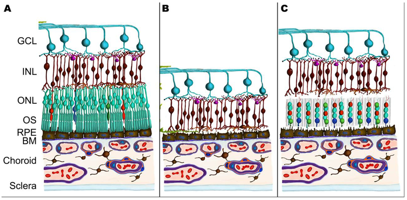
Figure 1.
Schematic of scaffold-assisted retinal regeneration. Compared to a healthy retina (A), a retina affected by late-stage inherited or age-related degeneration encounters a loss of photoreceptor cells (B...
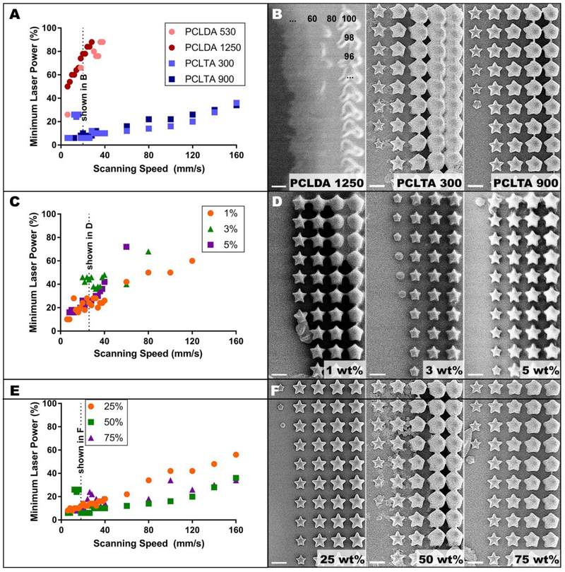
Figure 2.
Two-photon polymerization threshold. A) Threshold profiles for four acrylated PCL prepolymers at fixed composition (50 wt% monomer, 3 wt% photoinitiator) with B) corresponding images of stars fabricat...
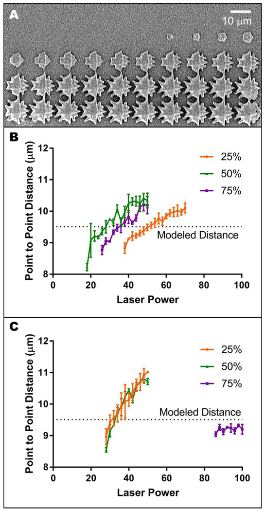
Figure 3.
Determining optimal laser power and scanning speed. A) Representative image of undesirable, delaminated two-photon polymerized structures. B-C) Width of stars created using the fastest scanning speed ...
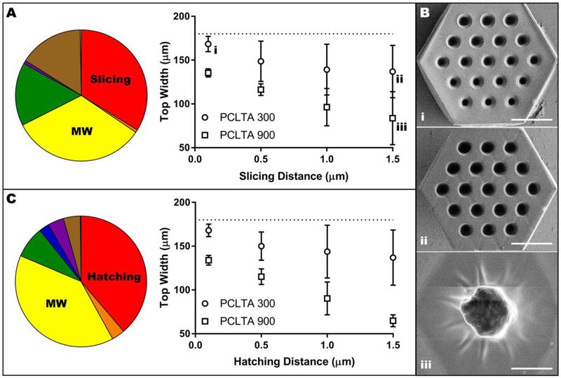
Figure 4.
Influence of slicing distance, hatching distance and molecular weight on scaffold width. A) Pie chart representing the contribution of experimental variables on scaffold width with constant hatching o...
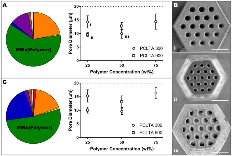
Figure 5.
Influence of molecular weight and prepolymer concentration on the vertical pore diameter of PCLTA scaffolds. A) Pie chart representing the contribution of experimental variables on pore diameter with ...
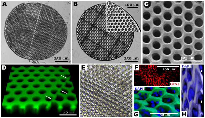
Figure 6.
Scale-up and validation of optimized two-photon polymerization to transplantation relevant size. Light micrographs of scaffolds created using 75 wt% 300 g/mol PCLTA with slicing and hatching distances...
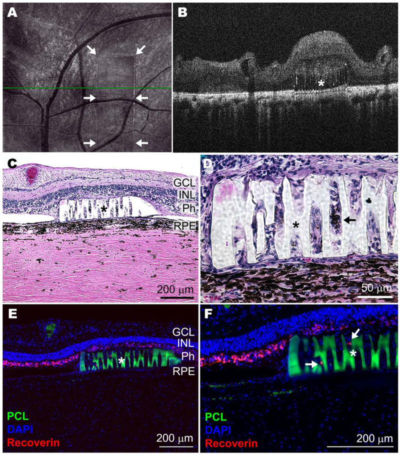
Figure 7.
Scaled-up prototype of crosslinked PCLTA 900 in the degenerating pig sub-retinal space 30 days after transplantation. Representative en face (A) and horizontal B-scan (B) optical coherence tomographs ...
Figure images are served from the NIH/NLM PubMed Central Open Access Subset or Europe PMC; copyright remains with the publishers and authors.
💬 Discussion
0 commentsNo comments yet. Be the first to start a discussion!
Leave a Comment