Abstract
OBJECTIVE: Monocyte (MN) recruitment is an essential inflammatory component of many autoimmune diseases, including rheumatoid arthritis (RA). In this study we investigated the ability of 2-fucosyllactose (H-2g), a glucose analog of blood group H antigen to induce MN migration in vivo and determined if H-2g-induced interleukin-8 (IL-8/CXCL8) plays a role in MN ingress in RA. METHODS: Sponge granuloma and intravital microscopy assays were performed to examine H-2g-induced in vivo MN migration and rolling, respectively. MNs were stimulated with H-2g, and the production of IL-8/CXCL8 was assessed by enzyme-linked immunosorbent assay and quantitative polymerase chain reaction. Lastly, in vitro MN migration assays and an in vivo RA synovial tissue severe combined immunodeficiency mouse model were used to determine the role of IL-8/CXCL8 in H-2g-induced MN migration. RESULTS: In vivo, H-2g induced significantly greater MN migration compared to phosphate buffered saline. Intravital microscopy revealed that H-2g mediates MN migration in vivo by inducing MN rolling. In addition, H-2g induced MN production of IL-8/CXCL8, a process that was dependent on Src kinase. Moreover, we found that H-2g mediated MN migration in vitro, and in vivo migration was inhibited by a neutralizing anti-IL-8/CXCL8 antibody. CONCLUSION: These findings suggest that H-2g mediates MN recruitment in vitro and in vivo (in part) via IL-8/CXCL8.
🔬 Techniques
🧪 Sample Preparation
🔬 Cell Lines
🧪 Reagent Suppliers
🏛️ Research Organizations (ROR)
Affiliated research institutions:
📋 Methods
Sponge granuloma and intravital microscopy assays were performed to examine H-2g-induced in vivo MN migration and rolling, respectively. MNs were stimulated with H-2g, and the production of IL-8/CXCL8 was assessed by enzyme-linked immunosorbent assay and quantitative polymerase chain reaction. Lastly, in vitro MN migration assays and an in vivo RA synovial tissue severe combined immunodeficiency mouse model were used to determine the role of IL-8/CXCL8 in H-2g-induced MN migration.
Materials and methods Patients and controls
Peripheral blood (PB) samples were collected from healthy volunteers. RA synovial tissue (ST) samples were obtained from patients undergoing synovectomy or total joint replacement. The study was approved by the University of Michigan Institutional Review Board.
Animals
All experiments were performed with approval from the University of Michigan Committee on Use and Care of Animals.
Sponge granuloma model
We performed a mouse sponge model to evaluate MN recruitment in vivo as previously described. 9 Briefly, phosphate-buffered saline (PBS) or H-2g-treated polyvinyl sponges were inserted subcutaneously into C57BL/6 mice (National Cancer Institute at the National Institutes of Health, Bethesda, MD). Simultaneously, human PB MNs, dye-tagged with PKH26 (Sigma-Aldrich, St Louis, MO), were injected intravenously. After 48 hours, mice were euthanized, sponges removed, and MNs isolated. Cytospins were performed and dye-tagged cells were counted by a blinded observer.
Intravital microscopy
Intravital microscopy was performed as previously described using C57BL/6 mice (National Cancer Institute at the National Institutes of Health, Bethesda, MD). 10 H-2g (100 μM) or PBS was injected intravenously 2 hours before executing intravital microscopy (three mice per group). 11 Three to five videos were taken per mouse. Rolling leukocytes were defined as leukocytes that rolled at a velocity slower than red blood cells.
Show full methods section
Sponge granuloma and intravital microscopy assays were performed to examine H-2g-induced in vivo MN migration and rolling, respectively. MNs were stimulated with H-2g, and the production of IL-8/CXCL8 was assessed by enzyme-linked immunosorbent assay and quantitative polymerase chain reaction. Lastly, in vitro MN migration assays and an in vivo RA synovial tissue severe combined immunodeficiency mouse model were used to determine the role of IL-8/CXCL8 in H-2g-induced MN migration.
Materials and methods Patients and controls
Peripheral blood (PB) samples were collected from healthy volunteers. RA synovial tissue (ST) samples were obtained from patients undergoing synovectomy or total joint replacement. The study was approved by the University of Michigan Institutional Review Board.
Animals
All experiments were performed with approval from the University of Michigan Committee on Use and Care of Animals.
Sponge granuloma model
We performed a mouse sponge model to evaluate MN recruitment in vivo as previously described. 9 Briefly, phosphate-buffered saline (PBS) or H-2g-treated polyvinyl sponges were inserted subcutaneously into C57BL/6 mice (National Cancer Institute at the National Institutes of Health, Bethesda, MD). Simultaneously, human PB MNs, dye-tagged with PKH26 (Sigma-Aldrich, St Louis, MO), were injected intravenously. After 48 hours, mice were euthanized, sponges removed, and MNs isolated. Cytospins were performed and dye-tagged cells were counted by a blinded observer.
Intravital microscopy
Intravital microscopy was performed as previously described using C57BL/6 mice (National Cancer Institute at the National Institutes of Health, Bethesda, MD). 10 H-2g (100 μM) or PBS was injected intravenously 2 hours before executing intravital microscopy (three mice per group). 11 Three to five videos were taken per mouse. Rolling leukocytes were defined as leukocytes that rolled at a velocity slower than red blood cells.
Isolation of human MNs
MNs were isolated from the PB of normal healthy volunteers as previously described. 8 The viability of MNs determined by trypan blue exclusion was found to be >98%, and the purity was >90%. Enzyme-linked immunosorbent assay (ELISA) Isolated human MNs were serum-starved for 2 hours and incubated with PP2 529573 (Src inhibitor), LY294002 (phosphatidylinositol 3-kinase [PI3K] inhibitor), PDTC (nuclear factor kappa-light-chain-enhancer of activated B cells [NFκB] inhibitor), Ro31 (protein kinase C [PKC] inhibitor), or dimethyl sulfoxide (vehicle control) for 1 hour. All inhibitors were purchased from Calbiochem (Calbiochem Novabiochem Corp, San Diego, CA) and were used at 10 μM. The MNs were then stimulated with H-2g (500 nM) in the presence of each inhibitor, based on our previous study. 8 Cell culture supernatants were collected after 24 hours and assayed using a human IL-8/CXCL8 ELISA kit (R&D Systems, Inc, Minneapolis, MN). 12 RNA extraction and quantitative polymerase chain reaction (qPCR) MNs were cultured as described above, RNA was extracted, and qPCR was performed as previously described. 12 , 13 Primers for human IL-8/CXCL8 (5′-GAGGGTTGTGGAGAAGTTTTTG-3′, 5′-CTGGCATCTTCACTGATTCTTG-3′) and human β-actin (5′-GTCAGGCAGCTCGTAGCTCT-3′, 5′-GCCATGTACGTTGCTATCCA-3′) were used. All samples were run in duplicate and the relative abundance of each gene was normalized to β-actin. MN chemotaxis MN chemotaxis was performed using 48-well modified Boyden chambers (Neuro Probe, Inc, Cabinjohn, MD) as previously described. 8 H-2g (500 nM) in the presence of mouse anti-human IL-8/CXCL8 antibody (R&D Systems, Inc) or an isotype-matched control antibody (R&D Systems, Inc) was added to the bottom wells of the chambers. Formyl-met-leu-phe and Hank’s balanced salt solution served as positive and negative controls, respectively. Each test group was assayed in quadruplicate. Three high-power fields (hpfs; ×400) were counted in each replicate well.
Human RA ST-SCID chimeras Severe combined immunodeficiency
(SCID) mice (National Cancer Institute at the National Institutes of Health) were anesthetized, and each mouse received one RA ST graft. After 4 weeks of engraftment, human PB MNs were dye-tagged with PKH26 fluorescent dye (Sigma-Aldrich), and were injected intravenously. Simultaneously, H-2g (10 μM) with an anti-IL-8/CXCL8 antibody or an isotype control antibody (R&D Systems, Inc) was injected into the RA ST grafts. Mice were euthanized after 48 hours and grafts were removed and snap frozen in liquid nitrogen. Cryosections were examined for cell homing using a fluorescence microscope. Migrated MNs were quantified by counting three slides per mouse, with two sections per slide, and at 6 hpfs per section.
Statistical analysis
Student’s t -tests were performed, and P -values less than 0.05 were considered significant. All values presented were the mean ± standard error of the mean (SEM).
Statistical analysis
Student’s t -tests were performed, and P -values less than 0.05 were considered significant. All values presented were the mean ± standard error of the mean (SEM).
📊 Figures
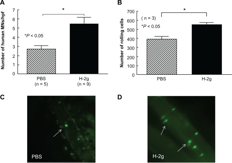
Figure 1
H-2g induces MN migration and leukocyte rolling in vivo. ( A ) H-2g or PBS was applied to sponges, which were then inserted into C57BL/6 mice. The sponges were removed, sectioned, and MN were counted....
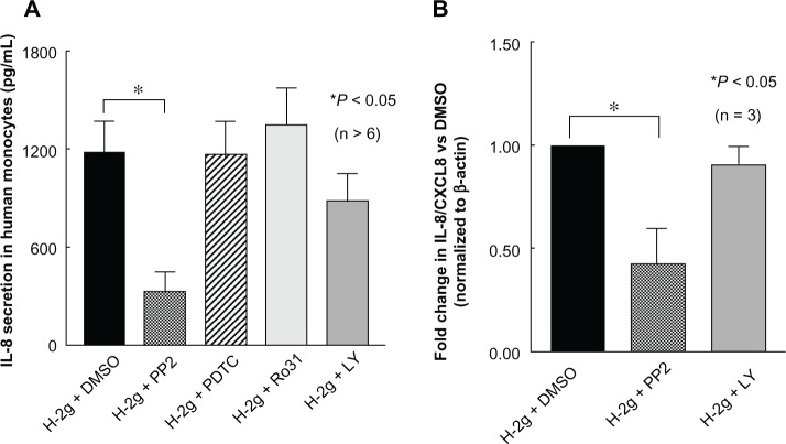
Figure 2
Src is required for H-2g induced MN production of IL-8/CXCL8. ( A ) Isolated MNs were cultured in the presence of H-2g with DMSO (vehicle control), PP2 529573 (Src inhibitor), PDTC (NF-u03baB inhibito...
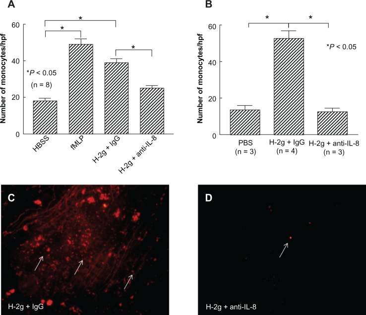
Figure 3
H-2g mediates in vitro and in vivo MN chemotaxis via IL-8/CXCL8. ( A ) MN chemotaxis assays were performed using a modified Boyden chamber. MN migration was determined in response to H-2g (500 nM) wit...
Figure images are served from the NIH/NLM PubMed Central Open Access Subset or Europe PMC; copyright remains with the publishers and authors.
💬 Discussion
0 commentsNo comments yet. Be the first to start a discussion!
Leave a Comment