Abstract
AbstractActin filament assembly and disassembly are vital for cell functions. MICAL Redox enzymes are important post-translational effectors of actin that stereo-specifically oxidize actin’s M44 and M47 residues to induce cellular F-actin disassembly. Here we show that Mical-oxidized (Mox) actin can undergo extremely fast (84 subunits/s) disassembly, which depends on F-actin’s nucleotide-bound state. Using near-atomic resolution cryoEM reconstruction and single filament TIRF microscopy we identify two dynamic and structural states of Mox-actin. Modeling actin’s D-loop region based on our 3.9 Å cryoEM reconstruction suggests that oxidation by Mical reorients the side chain of M44 and induces a new intermolecular interaction of actin residue M47 (M47-O-T351). Site-directed mutagenesis reveals that this interaction promotes Mox-actin instability. Moreover, we find that Mical oxidation of actin allows for cofilin-mediated severing even in the presence of inorganic phosphate. Thus, in conjunction with cofilin, Mical oxidation of actin promotes F-actin disassembly independent of the nucleotide-bound state.
🔬 Techniques
🔭 Microscopes
🧬 Organisms
💻 Software
✨ Fluorophores
🧪 Sample Preparation
🏭 Microscope Brands
🧪 Reagent Suppliers
💻 Software Details
💾 Data Repositories
🏛️ Research Organizations (ROR)
Affiliated research institutions:
📋 Methods
Protein purification
Rabbit skeletal actin (RSA) was purified from acetone powder 27 . Recombinant Drosophila 5C actins (WT and M47L mutant) were purified from baculovirus-infected Sf9 cells 12 . Recombinant human profilin-1 was expressed in E. coli and purified using poly-L-proline affinity column 28 . Recombinant gelsolin segment-1 (6His-GS1) was expressed in E. coli and purified using metal chelate chromatography (Ni-NTA, Qiagen) 26 . Human cofilin-1 29 was expressed in E. coli BL21(DE3)pLysS cells and purified using SP Sepharose FF (GE Healthcare) followed by gel filtration on HiLoad 16/60 Superdex 75 (Amersham Biosciences). Drosophila Mical redoxCH construct (referred to as Mical in this study) was expressed in E. coli ArcticExpress cells (Stratagene) and purified by two rounds of Ni 2+ affinity chromatography followed by anion exchange on HiTrap Q-FF (GE Healthcare) 30 . Acanthamoeba castellanii actin (AA) and mouse heterodimeric capping protein (CP) were a kind gift from Dr. Margot Quinlan (UCLA).
Preparation of actin forms
RSA was labeled with Alexa488-succinimidyl ester (SE) using standard approach 13 that included: (1) actin polymerization with 100 mM KCl; (2) high speed pelleting (TLA110 rotor at 60,000 rpm for 30 min at 4 °C); (3) dialysis (3 h) against labeling buffer (50 mM PIPES, pH 6.8, 50 mM KCl, 0.2 mM CaCl 2 , 0.2 mM ATP); (4) overnight incubation at 4 °C with three-fold molar excess of Alexa488SE dye. The labeling reaction was stopped by addition of 1–2 mM DTT followed by high speed pelleting, actin depolymerization on dialysis (GB 2 : 2 mM Tris, pH 8, 0.2 mM CaCl 2 , 0.2 mM ATP, 0.5–1 mM DTT), and gel-filtration (Superdex S200 10/300 GL). RSA labeling with pyrene maleimide was carried out in thiol-free GB 2 supplemented with 2 mM MgCl 2 and 100 mM KCl at 1:2.5 (actin:dye) molar ratio for 1 h on ice. The resulting pyrene-labeled F-actin was pelleted, depolymerized (GB 2 ), and gel-filtered on Superdex S200 16/60 column. Mical-oxidized RSA (Mox-actin) was prepared and purified according to the published protocol 13 with minor modifications. In brief, unlabeled RSA was oxidized on M44 and M47 at 1:50 (Mical:actin) ratio in the presence of 0.2 mM NADPH, for 2 h at room temperature (RT), unless stated otherwise. After 2 h actin was centrifuged at 100,000 g for 20 min at 4 °C. The resulting supernatant containing Mox-actin was dialyzed overnight against GB 2 then gel-filtered on Superdex S200 16/60 column. Pyrene- and Alexa488-SE-labeled actin forms were oxidized by Mical in GB 2 at 70:1 (actin:Mical) molar ratio in the presence of 100 μM NADPH for 1 h at RT. The resulting actin was dialyzed overnight against GB 2 then centrifuged (TLA100 rotor, 90,000 rpm, 30 min, 4 °C). Mg-ADP-F-actin was prepared by incubation of Mg-ATP-F-actin with 1 mM dextrose and hexokinase (8 U/ml of actin) for 1 h on ice 31 . BeFx-F-actin complexes were obtained by mixing Mg-F-actin with 0.1 mM BeCl 2 and 5 mM NaF (1× BeFx) followed by 1 h incubation on ice 31 . Acanthamoeba castellanii actin (AA) was labeled with Cy3b as follows. Thiol-free AA was polymerized for 1 h at room temperature (RT) with 2 mM MgCl 2 and 50 mM KCl. Cy3b dye was added to AA F-actin at 1:3 (AA:Cy3b). Labeling was carried out for 1 h on ice. To stop the labeling the reaction was supplemented with 1 mM DTT and 10 mM Hepes (pH 7) and incubated for 15 min at RT. AA-Cy3b was pelleted in TLA110 rotor at 80,000 rpm for 30 min at 4 °C and recovered in supernatants. Excess of label was removed by dialysis against GB 2 containing 5 mM β-marcaptoethanol (instead of DTT) followed by Sephadex G-50 spin column. Efficiency of labeling was ≥ 90%. Critical concentration determination Mg-ATP-actin (Mical-oxidized and unoxidized) was polymerized in 1xKMEH7.4 buffer (50 mM KCl, 2 mM MgCl 2 , 0.2 mM EGTA, 10 mM HEPES, 0.2 mM ATP, 0.5 mM DTT, pH 7.4) for 1 h at RT. This actin preparation was used to generate BeFx-F-actin and ADP-F-actin for side-by-side experiments as described in the main text. Critical concentrations (Cc) of ADP- and BeFx F-actin were determined using standard protocol 13 . In brief, actins were diluted into 1xKMEH7.4 buffer supplemented either with 0.5 mM ADP instead of ATP (for ADP-F-actin samples) or with 1× BeFx (for BeFx-bound samples) followed by 4 °C overnight incubation. Supernatants and pellets were separated by high speed centrifugation (TLA100, 62 K, 30 min, 4 °C) and analyzed by SDS-PAGE. Gels were stained with Coomassie Blue and densitometry was performed using Scion Image software. The intersects of the linear plots of pelleted actin vs. total actin with the abscissa yielded Cc. Nucleotide exchange Ca-ATP-G-actin was incubated with 10 fold molar excess of ɛ-ATP for 2 h on ice in ATP-free GB 2 buffer (2 mM Tris, 0.2 mM CaCl 2 , 0.5 mM DTT, pH 8). Excess of ɛ-ATP was removed using Sephadex G-50 spin columns. Actin preparations were supplemented with 5 µM ɛ-ATP. Nucleotide exchange in Mg-ATP-G-actin (1 µM) or Ca-ATP-G-actin (in the presence of profilin) was monitored after the addition of 100 µM of ATP. Excitation and emission wavelengths were 350 nm and 400 nm, respectively. The resulting curves were fitted with single exponential decay function using SigmaPlot software. Phosphate release Phosphate release was assessed using EnzChek phosphate assay kit (Invitrogen, E6646) according to the manufacturer’s instructions with the following modifications. Reaction mixtures were supplemented with additional 1 mM MgCl 2 (2 mM final concentration), 50 mM KCl, and 0.2 mM ATP to match closely the conditions of other experiments. Ca-ATP-G-actin (Mical-oxidized or unoxidized) (tube #1) and all other components of the reaction (tube #2) were pre-incubated separately for 10 min at room temperature and mixed together to start actin polymerization. Phosphate release upon actin polymerization was monitored by increase in absorbance at 360 nm.
Show full methods section
Protein purification
Rabbit skeletal actin (RSA) was purified from acetone powder 27 . Recombinant Drosophila 5C actins (WT and M47L mutant) were purified from baculovirus-infected Sf9 cells 12 . Recombinant human profilin-1 was expressed in E. coli and purified using poly-L-proline affinity column 28 . Recombinant gelsolin segment-1 (6His-GS1) was expressed in E. coli and purified using metal chelate chromatography (Ni-NTA, Qiagen) 26 . Human cofilin-1 29 was expressed in E. coli BL21(DE3)pLysS cells and purified using SP Sepharose FF (GE Healthcare) followed by gel filtration on HiLoad 16/60 Superdex 75 (Amersham Biosciences). Drosophila Mical redoxCH construct (referred to as Mical in this study) was expressed in E. coli ArcticExpress cells (Stratagene) and purified by two rounds of Ni 2+ affinity chromatography followed by anion exchange on HiTrap Q-FF (GE Healthcare) 30 . Acanthamoeba castellanii actin (AA) and mouse heterodimeric capping protein (CP) were a kind gift from Dr. Margot Quinlan (UCLA).
Preparation of actin forms
RSA was labeled with Alexa488-succinimidyl ester (SE) using standard approach 13 that included: (1) actin polymerization with 100 mM KCl; (2) high speed pelleting (TLA110 rotor at 60,000 rpm for 30 min at 4 °C); (3) dialysis (3 h) against labeling buffer (50 mM PIPES, pH 6.8, 50 mM KCl, 0.2 mM CaCl 2 , 0.2 mM ATP); (4) overnight incubation at 4 °C with three-fold molar excess of Alexa488SE dye. The labeling reaction was stopped by addition of 1–2 mM DTT followed by high speed pelleting, actin depolymerization on dialysis (GB 2 : 2 mM Tris, pH 8, 0.2 mM CaCl 2 , 0.2 mM ATP, 0.5–1 mM DTT), and gel-filtration (Superdex S200 10/300 GL). RSA labeling with pyrene maleimide was carried out in thiol-free GB 2 supplemented with 2 mM MgCl 2 and 100 mM KCl at 1:2.5 (actin:dye) molar ratio for 1 h on ice. The resulting pyrene-labeled F-actin was pelleted, depolymerized (GB 2 ), and gel-filtered on Superdex S200 16/60 column. Mical-oxidized RSA (Mox-actin) was prepared and purified according to the published protocol 13 with minor modifications. In brief, unlabeled RSA was oxidized on M44 and M47 at 1:50 (Mical:actin) ratio in the presence of 0.2 mM NADPH, for 2 h at room temperature (RT), unless stated otherwise. After 2 h actin was centrifuged at 100,000 g for 20 min at 4 °C. The resulting supernatant containing Mox-actin was dialyzed overnight against GB 2 then gel-filtered on Superdex S200 16/60 column. Pyrene- and Alexa488-SE-labeled actin forms were oxidized by Mical in GB 2 at 70:1 (actin:Mical) molar ratio in the presence of 100 μM NADPH for 1 h at RT. The resulting actin was dialyzed overnight against GB 2 then centrifuged (TLA100 rotor, 90,000 rpm, 30 min, 4 °C). Mg-ADP-F-actin was prepared by incubation of Mg-ATP-F-actin with 1 mM dextrose and hexokinase (8 U/ml of actin) for 1 h on ice 31 . BeFx-F-actin complexes were obtained by mixing Mg-F-actin with 0.1 mM BeCl 2 and 5 mM NaF (1× BeFx) followed by 1 h incubation on ice 31 . Acanthamoeba castellanii actin (AA) was labeled with Cy3b as follows. Thiol-free AA was polymerized for 1 h at room temperature (RT) with 2 mM MgCl 2 and 50 mM KCl. Cy3b dye was added to AA F-actin at 1:3 (AA:Cy3b). Labeling was carried out for 1 h on ice. To stop the labeling the reaction was supplemented with 1 mM DTT and 10 mM Hepes (pH 7) and incubated for 15 min at RT. AA-Cy3b was pelleted in TLA110 rotor at 80,000 rpm for 30 min at 4 °C and recovered in supernatants. Excess of label was removed by dialysis against GB 2 containing 5 mM β-marcaptoethanol (instead of DTT) followed by Sephadex G-50 spin column. Efficiency of labeling was ≥ 90%. Critical concentration determination Mg-ATP-actin (Mical-oxidized and unoxidized) was polymerized in 1xKMEH7.4 buffer (50 mM KCl, 2 mM MgCl 2 , 0.2 mM EGTA, 10 mM HEPES, 0.2 mM ATP, 0.5 mM DTT, pH 7.4) for 1 h at RT. This actin preparation was used to generate BeFx-F-actin and ADP-F-actin for side-by-side experiments as described in the main text. Critical concentrations (Cc) of ADP- and BeFx F-actin were determined using standard protocol 13 . In brief, actins were diluted into 1xKMEH7.4 buffer supplemented either with 0.5 mM ADP instead of ATP (for ADP-F-actin samples) or with 1× BeFx (for BeFx-bound samples) followed by 4 °C overnight incubation. Supernatants and pellets were separated by high speed centrifugation (TLA100, 62 K, 30 min, 4 °C) and analyzed by SDS-PAGE. Gels were stained with Coomassie Blue and densitometry was performed using Scion Image software. The intersects of the linear plots of pelleted actin vs. total actin with the abscissa yielded Cc. Nucleotide exchange Ca-ATP-G-actin was incubated with 10 fold molar excess of ɛ-ATP for 2 h on ice in ATP-free GB 2 buffer (2 mM Tris, 0.2 mM CaCl 2 , 0.5 mM DTT, pH 8). Excess of ɛ-ATP was removed using Sephadex G-50 spin columns. Actin preparations were supplemented with 5 µM ɛ-ATP. Nucleotide exchange in Mg-ATP-G-actin (1 µM) or Ca-ATP-G-actin (in the presence of profilin) was monitored after the addition of 100 µM of ATP. Excitation and emission wavelengths were 350 nm and 400 nm, respectively. The resulting curves were fitted with single exponential decay function using SigmaPlot software. Phosphate release Phosphate release was assessed using EnzChek phosphate assay kit (Invitrogen, E6646) according to the manufacturer’s instructions with the following modifications. Reaction mixtures were supplemented with additional 1 mM MgCl 2 (2 mM final concentration), 50 mM KCl, and 0.2 mM ATP to match closely the conditions of other experiments. Ca-ATP-G-actin (Mical-oxidized or unoxidized) (tube #1) and all other components of the reaction (tube #2) were pre-incubated separately for 10 min at room temperature and mixed together to start actin polymerization. Phosphate release upon actin polymerization was monitored by increase in absorbance at 360 nm.
TIRF microscopy
Untethered actin filaments were imaged on Pluronic coated surface 32 . Flow chambers (V~12 μl) were assembled using a single layer of permanent double-sided Scotch tape. For each sample the flow chamber was treated with 2 chamber volumes (CV) of 1% Pluronic F127 solution (Sigma, P2443) for 3 min then equilibrated with 2 CV of 1xTIRF imaging buffer (10 mM HEPES, 2 mM MgCl 2 , 50 mM KCl, 0.2 mM EGTA (pH 7.4) supplemented with 50 mM DTT, 0.2 mM ATP, 20 mM glucose, 0.5% methyl cellulose). G-actin mixtures (15% Alexa488-SE labeled) were incubated for 3 min at RT with Mg/EGTA exchange buffer (0.2 mM EGTA, 50 μM MgCl 2 ), mixed with the 2xTIRF imaging buffer, and the resulting mixture (4 CV) was introduced into the flow chamber. Actin mixtures and all the subsequent washes were supplemented with 0.05 mg/ml casein, 0.25 mg/ml glucose oxidase, 50 μM catalase to minimize radical damage and photobleaching during imaging. For depolymerization experiments RSA and Mical-oxidized F-actin (0.4–2.5 µM) were formed in flow chambers and aged on the surface for at least 45 min. During on-slide polymerization, slides were kept in sealed secondary containers to minimize the drying. Unpolymerized actin monomers were washed off with 2 CV of 1xTIRF imaging buffer and depolymerization movies were recorded. When indicated, actin was polymerized in (and washed with) 1xTIRF imaging buffer supplemented with one of the following: phosphate (Pi) (12.5 mM), Na 2 SO 4 (12.5 mM) or 1×BeFx (0.1 mM BeCl 2 and 5 mM NaF). For the experiments with substoichiometric amounts of BeFx we were using 400-fold dilution of 1×BeFx stock (0.25 µM final) maintaining the concentration of NaF at 5 mM. The amount of BeFx incorporated into Mox-actin filaments under subsaturating conditions was estimated using reported Kd value of BeFx to F-actin (~ 2 µM) and stoichiometry 1:1 17 . In the experiments involving mouse heterodimeric capping protein (CP), Mox-actin was first polymerized in flow chambers without a capper followed by the simultaneous removal of Mox-actin monomers and introduction of CP (50 nM). To test for cofilin-induced severing of ADP-Pi-F-actin, unoxidized or Mox-actin was polymerized in the presence of Pi (as above) then washed with 1xTIRF imaging buffer supplemented with 12.5 mM Pi and 150 nM human cofilin-1. For on-slide Mical oxidation of actin, unoxidized 5C Drosophila F-actin was formed in flow chambers as described above. Since Drosophila 5C-actin contains a total of 7 cysteines, which complicate site-specific labeling, we used 7.5% of Cy3b-maleimide labeled cytoplasmic actin from Acathamoeba castellanii as a fluorescent reporter. To start the oxidation, mixtures of Mical (10 nM) and NADPH (0.1 mM) in 1xTIRF imaging buffer were added to the flow chamber. Oxidation was allowed for 2 min then depolymerization movies were recorded. All TIRF data was analyzed using ImageJ (Fiji) software (NIH, Bethesda, MD).
Cryo electron microscopy and reconstruction
For cryo electron microscopy (cryoEM) samples RSA was oxidized at 1:10 (Mical:actin) ratio in the presence of 0.4 mM NADPH and purified as described above (also see ref. 13 ). Mical-oxidized actin was polymerized for at least 2 h in the following buffer: 5 mM Tris, 0.2 mM CaCl 2 , 0.2 mM EGTA, 2 mM MgCl 2 , 50 mM KCl, 1 mM DTT, 0.3 mM ATP (pH 7.4), and imaged at 6 µM concentration. Aliquots of 2.5 µL of polymerized Mox-F-actin were applied onto a “baked” 33 Quantifoil 1.2/1.3 μm, 200 mesh grid, blotted for 4.5 s at force 1, then flash-frozen in liquid nitrogen cooled liquid ethane in a Vitrobot Mark IV (FEI). CryoEM data were collected in an FEI Titan Krios microscope (operated at 300 kV) equipped with a Gatan imaging filter (GIF) (slit width 20 eV) and K2 Summit direct electron camera in counting mode using Leginon software package 34 for automation. Defocus values were controlled with Leginon by applying a single 3.0 μm target defocus. Dose-fractionation movies were recorded at a frame rate of 5 Hz with a 10 s shutter time. The total accumulated dosage was 60 e/A −2 , with each frame 1.2 e/A −2 and a temporal dose rate of 6 e/A −2 s (measured on Digital Micrograph (Gatan) software). Frames were aligned according to Li et al. 35 except that an iterative alignment scheme was employed 36 . We used the summary of all frames to determine defocus and particle locations, and 3rd–20th frames for data processing. The defocus parameters of the data were determined by CTFFIND3 37 . We selectively included images within a defocus range of 1.5–5 μm. Actin filaments were selected manually in EMAN 38 helixboxer. A total of 68,168 filament regions were selected. These filaments were segmented into 237,652 boxes of 384 × 384 pixels, in which each box progresses 38 pixels along the helical axis (90% overlap). These boxes were subjected to 2D classifications as prescribed in Relion manual 39 to eliminate bad particles, yielding 208,364 boxes to the 3D classification step. We used a 3D classification (Relion with an implementation of IHRSR 40 , 41 ) with three classes initially and found that one class was mainly containing bad particles (3.5% occupancy). The other two classes (49.2% and 47.3%, respectively) were slightly different as we pushed the working resolution down to about 8 Å by refining the angular sampling. 3D auto-refinement in Relion/IHRSR of the boxes attributed to the two classes (102,762 and 102,015 boxes, respectively) generated two volumes with notable difference limited to the subdomain 2. The resolution of the two volumes were similar, both estimated at 3.9 Å with a consideration of “gold-standard” Fourier-shell correlation plot (0.143 criteria), ResMap 42 and the matching between side chain densities and their models. A further experiment was done to confirm the classification findings with a 3D classification of 7 classes. Only two classes out of the seven showed enough resolution (to see secondary structural elements in the map) when refined in the same way above, suggesting that there were only two major conformational populations in the dataset. Atomic modeling of Mox-F-actin Atomic models were built with Coot 43 . We docked a canonical actin model (PDB 5JLF 20 ) to each of our density maps. We found that all parts of it, except for subdomain 2, were within convergence radii of real space refinement software (see below) into our density map. We therefore solely remodeled subdomain 2 in Coot and kept the rest of the canonical model in a reliance for the refinement software to bring the model into a good fit. We then carried out model refinement for each of the modified canonical model with the phenix.real_space_refine command of the Phenix package 44 using default settings. Mox-G-actin structure determination Mical-oxidized Ca-G-actin-GS1 complex was crystallized under conditions close to those reported by McLaughlin et al. 18 . Specifically, proteins were mixed in 1:1 molar ratio (final concentration of the complex was 8.2 mg/ml) in GB 2 buffer supplemented with 0.2 mM ATP, 0.25 mM TCEP, 1 mM PMSF and 0.02% NaN 3 . The ratio of protein complex to precipitant was 1:1 (v/v). Volumes of hanging drops and reservoir solutions were 2 µl and 500 µl, respectively. The largest crystals (plates) grew overnight in 7.5% PEG-6000, 150 mM NaCl, 50 mM imidazole, pH 6.6. Smaller crystals were also obtained when concentration of PEG600 was increased to 10%. Data collection and reduction proceeded as follows. The crystals were cryo-protected by quickly dipping them in solution of 65% reservoir and 35% methylpentanediol (v/v), and then immediately cryo-cooled to 100 K in a nitrogen gas stream. Diffraction data were collected at the Northeastern Collaborative Access Team beamline, 24-ID-C, at the Advanced Photon Source at Argonne National Laboratory. Data were recorded from two crystals on a Pilatus 6 M detector using a wavelength of 0.9795 Å, detector distances of 250, 400, and 500 mm, 0.5 s exposures, and oscillation angle of 0.5°. A total of 360° of data were collected from one crystal and merged with two runs from a second crystal, spanning 360° and 350° of data, respectively. The merged data extends to a resolution of 2.4 Å, with a CC 1/2 of 0.83, I/σ = 2.3 and in R merge = 171% the 2.45–2.40 Å resolution shell. Overall, the data multiplicity was over 38-fold. Using the more conservative R merge statistic, the resolution limit of the data would be drawn at 2.75 Å which reached 52.8% in the shell 2.91–2.75 Å resolution shell, (I/σ = 5.2). This more conservative limit is reported in Supplementary Table 1 . Structure determination and refinement were performed as follows. The starting model for refinement was the crystal structure of reduced actin-gelsolin, PDB ID code 1EQY 18 . The crystal was isomorphous to the Mical-oxided actin-gelsolin complex; therefore, no molecular replacement was necessary. Refinement was performed using the program Buster 45 . The model was manually adjusted to fit the maps using the program Coot 43 . The model was refined using all data to 2.4 Å resolution. Refinement statistics are reported in Supplementary Table 1 to the more conservative limit of 2.75 Å. The final model was validated using the program PROCHECK 46 which revealed 92.4% of residues are in the most favored regions of the Ramachandran plot, 7.4% of residues are in additional allowed regions, and one residue is in a generously allowed region. There were no Ramachandran outliers. The final model had an Errat score of 96.8% 47 . The structure was illustrated using the program Pymol. For structural comparison, the unmodified and Mical-oxidized actin-gelsolin complexes were superimposed on subdomains 1, 3, and 4 using the secondary structure matching algorithm 48 implemented in Coot 43 . Statistics and reproducibility All TIRF microscopy and solution experiments were repeated at least two separate independent times and there were no limitations in repeatability. At least two independent protein purifications and multiple independent actin biochemical experiments were performed with similar results. The figure legends list the sample size for each experiment. To the best of our knowledge the statistical tests are justified as appropriate. No cell lines were used in this study.
Data availability
The authors declare that all data supporting the findings of this study are available within the article and its Supplementary Information files or from the corresponding authors upon reasonable request. The crystal structure of Mical oxidized monomeric actin with gelsolin segment-1 has been deposited in the protein databank under accession code: PDB ID: 5UBO. The atomic models and the cryoEM density maps for Classes 1 and 2 of Mox-F-actin have been deposited in the Protein Data Bank and EMDB databases under the accession codes: 6AV9, 6AVB and EMD-7007, EMD-7008, respectively.
Electronic supplementary material Supplementary Movie 1 Supplementary Movie 2 Supplementary Movie 3 Supplementary Movie 4 Supplementary Movie 5 Supplementary Information Description of Additional Supplementary Files
📊 Figures
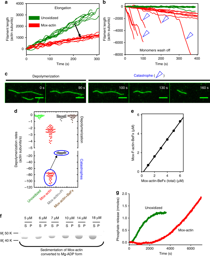
Fig. 1
Mical-oxidation of actin induces its nucleotide state-dependent catastrophic disassembly. a Mical oxidation of actin inhibits filament elongation. Average elongations rates of unoxidized and Mical-oxi...
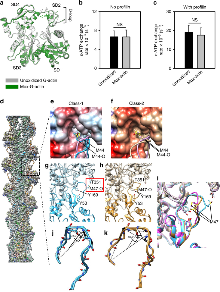
Fig. 2
Atomic structures of monomeric and filamentous Mical-oxidized actin. a Overlay of the crystal structures of Mical-oxidized (Mox) Ca-ATP-G-actin (2.7u2009u00c5 resolution, green) and unoxidized (PDB# 1...
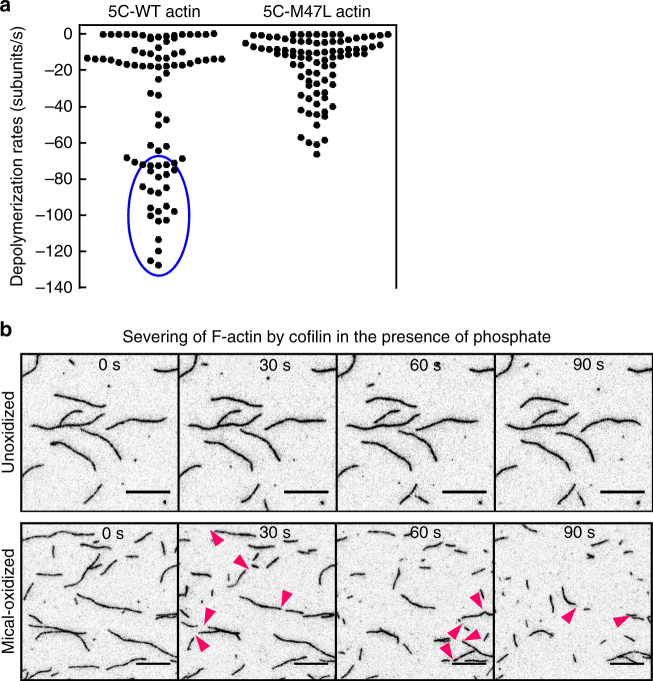
Fig. 3
Mical oxidation of both M44 and M47 residues on actin drives its catastrophic disassembly and cofilin severing. a Mical oxidation of bothu2014M44 and M47u2014contributes to F-actin destabilization. Si...
Figure images are served from the NIH/NLM PubMed Central Open Access Subset or Europe PMC; copyright remains with the publishers and authors.
💬 Discussion
0 commentsNo comments yet. Be the first to start a discussion!
Leave a Comment