Abstract
Abstract Microbes are critical components of ecosystems and provide vital services (e.g., photosynthesis, decomposition, nutrient recycling). From the diverse roles microbes play in natural ecosystems, high levels of functional diversity result. Quantifying this diversity is challenging, because it is weakly associated with morphological differentiation. In addition, the small size of microbes hinders morphological and behavioral measurements at the individual level, as well as interactions between individuals. Advances in microbial community genetics and genomics, flow cytometry and digital analysis of still images are promising approaches. They miss out, however, on a very important aspect of populations and communities: the behavior of individuals. Video analysis complements these methods by providing in addition to abundance and trait measurements, detailed behavioral information, capturing dynamic processes such as movement, and hence has the potential to describe the interactions between individuals. We introduce BEMOVI, a package using the R and ImageJ software, to extract abundance, morphology, and movement data for tens to thousands of individuals in a video. Through a set of functions BEMOVI identifies individuals present in a video, reconstructs their movement trajectories through space and time, and merges this information into a single database. BEMOVI is a modular set of functions, which can be customized to allow for peculiarities of the videos to be analyzed, in terms of organisms features (e.g., morphology or movement) and how they can be distinguished from the background. We illustrate the validity and accuracy of the method with an example on experimental multispecies communities of aquatic protists. We show high correspondence between manual and automatic counts and illustrate how simultaneous time series of abundance, morphology, and behavior are obtained from BEMOVI. We further demonstrate how the trait data can be used with machine learning to automatically classify individuals into species and that information on movement behavior improves the predictive ability.
🔬 Techniques
🧬 Organisms
🏭 Microscope Brands
💻 Software Details
💻 Code & Software
💾 Data Repositories
🏛️ Research Organizations (ROR)
Affiliated research institutions:
📋 Methods
Species and experimental conditions
Microcosms of protists are widely used model systems in ecology and evolution (Altermatt et al. 2015 ). For the following experiments illustrating the use of the BEMOVI package, a set of nine small, single-celled ciliates was used: Paramecium caudatum , Paramecium aurelia , Blepharisma japonicum , Colpidium striatum , Colpidium campylum , Cyclidium glaucoma , Tetrahymena thermophila , Didinium nasutum , and Loxocephalus sp., which show variation in morphology (Giometto et al. 2013 ) as well as in movement behavior (Carrara et al. 2012 ). The aquatic microcosms used followed a similar setup as Petchey ( 2000 ), and for detailed and comprehensive methods, please see Altermatt et al. ( 2015 ). For each experiment, ciliates were cultured in jars of 240 mL volume covered by an aluminum cap to allow air exchange but prevent contamination. Each jar contained 100 mL of bacterized medium before ciliates were added. The medium consisted of protist pellet medium (Carolina Biological Supplies, Burlington, NC) at a concentration of 0.55 g per liter of Chalkley’s medium, as well as two wheat seeds for slow nutrient release. The medium was then inoculated with three species of bacteria ( Serratia fonticola , Brevibacillus brevis and Bacillus subtilis ). Bacteria were cultured for a day at 37 ° C. Ciliates were then added to the jars and kept in temperature-controlled incubators at 20 ° C for the remainder of the experiments. Three experiments were run to test and illustrate the performance and scope of the BEMOVI package: Monocultures of eight ciliate species (previous list except for Didinium nasutum ) were assembled, and videos taken to characterize the variation among species in terms of morphology and movement behavior. Mono- and mixed cultures of Colpidium striatum , Didinium nasutum , Paramecium caudatum , and Tetrahymena thermophila were assembled and ten independent samples assessed by automatic and manual counts, in order to compare the abundances per experimental unit (the individual microcosm). Colpidium striatum , Paramecium caudatum , and Tetrahymena thermophila were followed over 28 days in monocultures and mixed cultures by automatic and manual counts (nine sampling days), again to compare these two methods of observation.
Show full methods section
Species and experimental conditions
Microcosms of protists are widely used model systems in ecology and evolution (Altermatt et al. 2015 ). For the following experiments illustrating the use of the BEMOVI package, a set of nine small, single-celled ciliates was used: Paramecium caudatum , Paramecium aurelia , Blepharisma japonicum , Colpidium striatum , Colpidium campylum , Cyclidium glaucoma , Tetrahymena thermophila , Didinium nasutum , and Loxocephalus sp., which show variation in morphology (Giometto et al. 2013 ) as well as in movement behavior (Carrara et al. 2012 ). The aquatic microcosms used followed a similar setup as Petchey ( 2000 ), and for detailed and comprehensive methods, please see Altermatt et al. ( 2015 ). For each experiment, ciliates were cultured in jars of 240 mL volume covered by an aluminum cap to allow air exchange but prevent contamination. Each jar contained 100 mL of bacterized medium before ciliates were added. The medium consisted of protist pellet medium (Carolina Biological Supplies, Burlington, NC) at a concentration of 0.55 g per liter of Chalkley’s medium, as well as two wheat seeds for slow nutrient release. The medium was then inoculated with three species of bacteria ( Serratia fonticola , Brevibacillus brevis and Bacillus subtilis ). Bacteria were cultured for a day at 37 ° C. Ciliates were then added to the jars and kept in temperature-controlled incubators at 20 ° C for the remainder of the experiments. Three experiments were run to test and illustrate the performance and scope of the BEMOVI package: Monocultures of eight ciliate species (previous list except for Didinium nasutum ) were assembled, and videos taken to characterize the variation among species in terms of morphology and movement behavior. Mono- and mixed cultures of Colpidium striatum , Didinium nasutum , Paramecium caudatum , and Tetrahymena thermophila were assembled and ten independent samples assessed by automatic and manual counts, in order to compare the abundances per experimental unit (the individual microcosm). Colpidium striatum , Paramecium caudatum , and Tetrahymena thermophila were followed over 28 days in monocultures and mixed cultures by automatic and manual counts (nine sampling days), again to compare these two methods of observation.
Microscope and video setup
All videos were taken using a stereomicroscope (Leica M205 C) with a 25× magnification mounted with a digital CMOS camera (Hamamatsu Orca C11440 , Hamamatsu Photonics, Japan). Dark field illumination (LED ring light stage controlled by a Schott VisiLED MC 1500) was used such that the ciliates, usually transparent in bright field microscopy, appear white on black background; this greatly facilitates segmentation. For details how to setup the hardware for best segmentation results, please refer to Pennekamp and Schtickzelle ( 2013 ) or Dell et al. ( 2014 ). To sample a culture, we transferred 1 mL of culture into a Sedgewick Rafter cell (S52, SPI supplies, Westchester, PA), which was placed under the microscope objective. We took videos at a frame rate of 25 frames per second in the proprietary .cxd format. The BEMOVI package is limited in the video formats and it can currently read (.avi and .cxd, via the BIO-formats plug-in), but could easily be extended to accommodate any of the many other video formats readable by ImageJ and the BIO-formats plugin (Linkert et al. 2010 ). As tracking parameters, we specified a link range of five frames for all three experiments and a displacement of 20 pixels for experiments 1 and 3. In experiment 2, a higher displacement of 25 pixels was required to account for the fast moving Didinium nasutum . Trajectories were filtered by the filter_data function to get rid of artefacts such as spurious trajectories due to moving debris. Trajectories for analysis were required to show a minimum net displacement of at least 50 μ m, a duration of 0.2 sec, and a detection rate of 80% (for a trajectory with a duration of 10 frames, the individual has to be detected on at least eight frames) and a median step length of >2 μ m. In open systems where the viewing field is not restricted (i.e., individuals can swim in and out of the viewing field), automatic counts by BEMOVI are required on a by-frame basis and then averaged across the whole video. This avoids that occlusions resulting in multiple trajectories recorded for a single individual inflate the counts. For manual counting, a sample was taken from the culture and manually counted under a dissecting microscope by an experienced experimenter (as described in Altermatt et al. 2015 ). Such manual observations, albeit very time-consuming and limited to abundance measurements, are still the most widely used method in experimental micro- and mesocosm studies (Pennekamp and Schtickzelle 2013 ). Therefore, we chose them as the standard against which to compare the automatic measurements by BEMOVI. However, as an alternative methodology, manually counting individuals on the recorded video would have the advantage of disentangling different sources of error (e.g., intersample variability, compensation between false positives and false negatives in the automatic segmentation procedure of BEMOVI). It was not chosen here for the following reasons: first, we consider the microcosm, not the sample, as the relevant experimental unit; second, a specific example may not be particularly informative because different sources of error are likely to vary widely among case studies as they are highly dependent on the video settings, the environment (amount of debris present) or the movement behavior of the target species; third, manual observation of videos, required for quantifying frequencies of different types of error, would be very cumbersome, and also prone to error. As we considered the microcosm as the relevant experimental unit for all experiments, we took independent samples for manual and automatic counts; this has the drawback that samples were not paired, and thus, intersample variability may be confounded with tracking errors. However, the intersample variability also affects manual counting and should be overall comparable between methods as sampling is performed in a similar fashion.
Machine learning for automated species identification
We used supervised machine learning to train and classify individuals in mixed cultures. The random forest (RF) classifier is a widely used classification algorithm based on ensembles of decision trees (Breiman 2001 ). By constraining the number of observations and variables included when constructing individual decision trees, trees within an ensemble are decorrelated and the identity assigned to an individual is usually based on the majority vote of the ensemble (Cutler et al. 2007 ). We trained the RF on the properties of individuals from monocultures and consequently used the model to predict species identity in mixed cultures using the same traits but on unidentified individuals. We report the classification success (1 - out-of-bag error rate; reported as percentage), which states how well the model performs on observations not included in training the model (i.e., a cross-validation).
Species and experimental conditions
Microcosms of protists are widely used model systems in ecology and evolution (Altermatt et al. 2015 ). For the following experiments illustrating the use of the BEMOVI package, a set of nine small, single-celled ciliates was used: Paramecium caudatum , Paramecium aurelia , Blepharisma japonicum , Colpidium striatum , Colpidium campylum , Cyclidium glaucoma , Tetrahymena thermophila , Didinium nasutum , and Loxocephalus sp., which show variation in morphology (Giometto et al. 2013 ) as well as in movement behavior (Carrara et al. 2012 ). The aquatic microcosms used followed a similar setup as Petchey ( 2000 ), and for detailed and comprehensive methods, please see Altermatt et al. ( 2015 ). For each experiment, ciliates were cultured in jars of 240 mL volume covered by an aluminum cap to allow air exchange but prevent contamination. Each jar contained 100 mL of bacterized medium before ciliates were added. The medium consisted of protist pellet medium (Carolina Biological Supplies, Burlington, NC) at a concentration of 0.55 g per liter of Chalkley’s medium, as well as two wheat seeds for slow nutrient release. The medium was then inoculated with three species of bacteria ( Serratia fonticola , Brevibacillus brevis and Bacillus subtilis ). Bacteria were cultured for a day at 37 ° C. Ciliates were then added to the jars and kept in temperature-controlled incubators at 20 ° C for the remainder of the experiments. Three experiments were run to test and illustrate the performance and scope of the BEMOVI package: Monocultures of eight ciliate species (previous list except for Didinium nasutum ) were assembled, and videos taken to characterize the variation among species in terms of morphology and movement behavior. Mono- and mixed cultures of Colpidium striatum , Didinium nasutum , Paramecium caudatum , and Tetrahymena thermophila were assembled and ten independent samples assessed by automatic and manual counts, in order to compare the abundances per experimental unit (the individual microcosm). Colpidium striatum , Paramecium caudatum , and Tetrahymena thermophila were followed over 28 days in monocultures and mixed cultures by automatic and manual counts (nine sampling days), again to compare these two methods of observation.
Supporting Information Additional Supporting Information may be found in the online version of this article: Video S1. Video footage of microcosm sample in counting chamber. Video S2. Overlay video showing the individuals identified by BEMOVI and their respective trajectory labels. Video S3. Overlay video showing the individuals of two ciliate species with different colour codes and their respective trajectory labels. Table S1. Overview of traits/measures used in the automatic classification of species. Figure S1. Positive correlation between ciliate cell length measurements (collected from literature and online resources) and cell length measured by BEMOVI.
📊 Figures
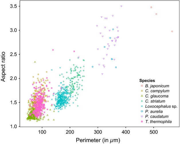
Figure 1
Morphological characteristics of eight ciliate species: cell perimeter and aspect ratio (major axis/minor axis of a fitted ellipse).
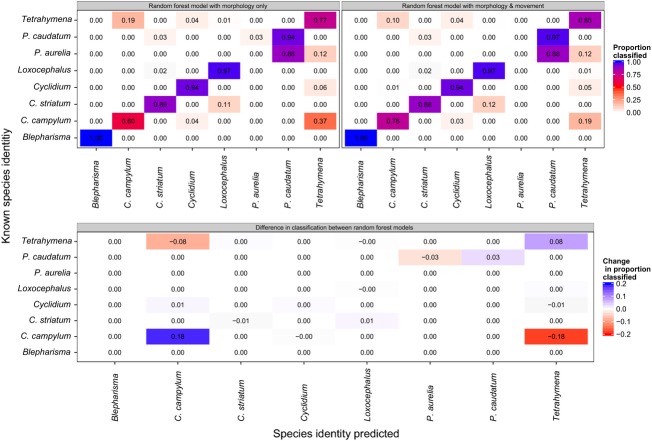
Figure 2
Confusion matrices between eight ciliate species to illustrate the improved classification success when both morphology and movement features are considered (>89%), compared to morphology only (84%). ...
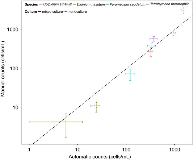
Figure 3
Comparison of manual and automatic estimates of population abundance in single (dashed bars) and mixed species cultures (solid bars). The u00b11 STD error bars are calculated for the 10 repeated sampl...
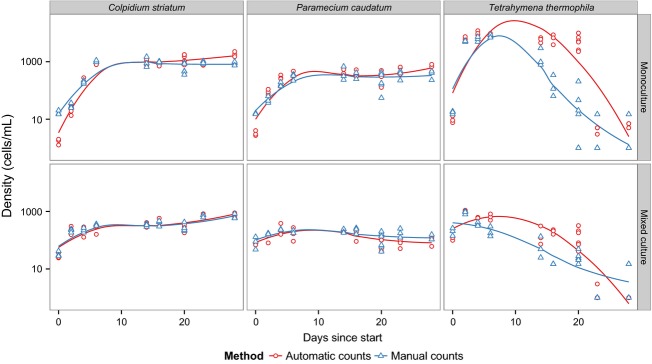
Figure 4
Population dynamics of three species of ciliates given by manual and automatic counting (fitted lines are local polynomial regressions) to illustrate the general growth dynamics. The mixed culture con...
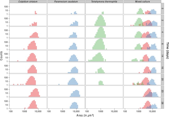
Figure 5
Body size changes through time (from first row to last row) in the mono- and mixed cultures during the experiment. At day 20, the medium was partly replaced by fresh medium perturbing the mono- and mi...
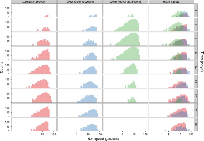
Figure 6
Changes in movement speed through time (from first row to last row) in the mono- and mixed cultures during the experiment. At day 20, the medium was partly replaced by fresh medium perturbing the mono...
Figure images are served from the NIH/NLM PubMed Central Open Access Subset or Europe PMC; copyright remains with the publishers and authors.
💬 Discussion
0 commentsNo comments yet. Be the first to start a discussion!
Leave a Comment