Abstract
Oral cancer is a serious and fatal disease. Cisplatin is the first line of chemotherapeutic agent for oral cancer therapy. However, the development of drug resistance and severe side effects cause tremendous problems clinically. In this study, we investigated the pharmacologic mechanisms of YC-1 on cisplatin-resistant human oral cancer cell line, CAR. Our results indicated that YC-1 induced a concentration-dependent and time-dependent decrease in viability of CAR cells analyzed by MTT assay. Real-time image analysis of CAR cells by IncuCyte™ Kinetic Live Cell Imaging System demonstrated that YC-1 inhibited cell proliferation and reduced cell confluence in a time-dependent manner. Results from flow cytometric analysis revealed that YC-1 promoted G0/G1 phase arrest and provoked apoptosis in CAR cells. The effects of cell cycle arrest by YC-1 were further supported by up-regulation of p21 and down-regulation of cyclin A, D, E and CDK2 protein levels. TUNEL staining showed that YC-1 caused DNA fragmentation, a late stage feature of apoptosis. In addition, YC-1 increased the activities of caspase-9 and caspase-3, disrupted the mitochondrial membrane potential (AYm) and stimulated ROS production in CAR cells. The protein levels of cytochrome c, Bax and Bak were elevated while Bcl-2 protein expression was attenuated in YC-1-treated CAR cells. In summary, YC-1 suppressed the viability of cisplatin-resistant CAR cells through inhibiting cell proliferation, arresting cell cycle at G0/G1 phase and triggering mitochondria-mediated apoptosis. Our results provide evidences to support the potentially therapeutic application of YC-1 on fighting against drug resistant oral cancer in the future.
🔬 Techniques
✨ Fluorophores
🧪 Sample Preparation
🏭 Microscope Brands
🧪 Reagent Suppliers
🏛️ Research Organizations (ROR)
Affiliated research institutions:
📋 Methods
2.1.
Chemicals and reagents
Cisplatin, propidium iodide (PI) and thiazolyl blue tetrazolium bromide (MTT) were purchased from Sigma-Aldrich (St. Louis, MO, USA). Trypsin-EDTA was purchased from BioConcept (Allschwil/BL, Switzerland). Fetal bovine serum (FBS), L-glutamine, penicillin G, 2’, 7’-dichlorodihydrofluorescein diacetate (H 2 DCFDA) and 3, 3-dihexyloxa-carbocyanine iodide [DiOC 6 (3)] were obtained from Thermo Fisher Scientific (Carlsbad, CA, USA). Caspase-3 and caspase-9 activity assay kits were purchased from R&D Systems Inc. (Minneapolis, MN, USA). The primary antibodies against Bcl-2, Bax, cytochrome c , Apaf-1, AIF, p21, cyclin A, cyclin D, cyclin E, CDK 2, β-actin and the goat anti-rabbit or anti-mouse IgG-horseradish peroxidase (HRP) secondary antibodies were purchased from GeneTex, (Hsinchu, Taiwan). Pan-caspase inhibitor (z-VAD-fmk) and enhanced chemiluminescence (ECL) detection kit (Immobilon Western Chemiluminescent HRP Substrate) were purchased from Merck Millipore (Billerica, MA, USA). YC-1 was designed and synthesized as detailed in the previous study [ 21 ]. 2.2.
Cell culture
The cisplatin-resistant cell line (CAR) was developed by treating CAL 27 cell line, a parental human tongue squamous cell carcinoma (American Type Culture Collection, Manassas, VA, USA) with 10-80 μM of cisplatin. CAR cells are characterized by its stable resistance to cisplatin as previously described [ 1 , 18 , 85 , 86 ]. The cells were cultured in Dulbecco’s modified Eagle’s medium (DMEM) fortified with 10% fetal bovine serum (FBS), 100 U/ml penicillin, 100 μg/ml streptomycin, and 2 mM L-glutamine (Thermo Fisher Scientific) and were incubated at 37 °C with a humidified 5% CO2 air. The cisplatin-resistant CAR cells were constantly cultured in medium containing 80 μM cisplatin unless otherwise indicated [ 1 , 18 , 85 , 86 ]. 2.3.
Show full methods section
2.1.
Chemicals and reagents
Cisplatin, propidium iodide (PI) and thiazolyl blue tetrazolium bromide (MTT) were purchased from Sigma-Aldrich (St. Louis, MO, USA). Trypsin-EDTA was purchased from BioConcept (Allschwil/BL, Switzerland). Fetal bovine serum (FBS), L-glutamine, penicillin G, 2’, 7’-dichlorodihydrofluorescein diacetate (H 2 DCFDA) and 3, 3-dihexyloxa-carbocyanine iodide [DiOC 6 (3)] were obtained from Thermo Fisher Scientific (Carlsbad, CA, USA). Caspase-3 and caspase-9 activity assay kits were purchased from R&D Systems Inc. (Minneapolis, MN, USA). The primary antibodies against Bcl-2, Bax, cytochrome c , Apaf-1, AIF, p21, cyclin A, cyclin D, cyclin E, CDK 2, β-actin and the goat anti-rabbit or anti-mouse IgG-horseradish peroxidase (HRP) secondary antibodies were purchased from GeneTex, (Hsinchu, Taiwan). Pan-caspase inhibitor (z-VAD-fmk) and enhanced chemiluminescence (ECL) detection kit (Immobilon Western Chemiluminescent HRP Substrate) were purchased from Merck Millipore (Billerica, MA, USA). YC-1 was designed and synthesized as detailed in the previous study [ 21 ]. 2.2.
Cell culture
The cisplatin-resistant cell line (CAR) was developed by treating CAL 27 cell line, a parental human tongue squamous cell carcinoma (American Type Culture Collection, Manassas, VA, USA) with 10-80 μM of cisplatin. CAR cells are characterized by its stable resistance to cisplatin as previously described [ 1 , 18 , 85 , 86 ]. The cells were cultured in Dulbecco’s modified Eagle’s medium (DMEM) fortified with 10% fetal bovine serum (FBS), 100 U/ml penicillin, 100 μg/ml streptomycin, and 2 mM L-glutamine (Thermo Fisher Scientific) and were incubated at 37 °C with a humidified 5% CO2 air. The cisplatin-resistant CAR cells were constantly cultured in medium containing 80 μM cisplatin unless otherwise indicated [ 1 , 18 , 85 , 86 ]. 2.3.
Cell viability assay
CAR cells (1 × 10 4 cells/per well) were seeded in 96-well plates in 100 μl medium with or without 25, 50, 75 and 100 μM of YC-1 for 24 h. After YC-1 treatment, DMEM containing 500 μg/ml of MTT was added and incubated at 37 °C for 4 h. The medium was then removed, and 100 μl DMSO was added to each well to dissolve the formed blue formazan crystals, followed by measuring the 570 nm absorbance of each well by the ELISA plate reader with a reference wavelength of 620 nm. For the caspase inhibition experiment, cells were pretreated with 15 μM z-VAD-fmk (a pan-caspase inhibitor) for 1 h before subjected to YC-1 administration. Cell morphological examination was observed and photographed by the IncuCyte ™ Kinetic Live Cell Imaging System (Essen BioScience, Ann Arbor, MI, USA) [ 87 – 89 ]. 2.4.
IncuCyte cell proliferation and confluence assay
To measure the cell confluence, a stable mixture of CAR cells (2 × 10 4 cells) were plated into a 96-well plate. The cells were then incubated with or without 25, 50, 75 and 100 μM of YC-1. Cell confluence relative to the control cells was determined by the IncuCyte ™ Kinetic Live Cell Imaging System (Essen BioScience) at a 2-h interval and up to 48 h [ 90 ]. 2.5.
Flow cytometry analysis of cell cycle distribution
CAR cells (2 × 10 5 cells/per well) were plated into the 12-well plates and then treated with 100 μM of YC-1 for 0, 12, 24, 36 and 48 h. The cells were then fixed, followed by staining with propidium iodide (PI) solution as previously described [ 91 , 92 ]. The cell cycle profiling and the data analysis were determined utilizing a Muse Cell Analyzer (Merck Millipore, Hayward, CA, USA) [ 93 – 98 ]. 2.6.
Immunoblotting analysis
CAR cells (1 × 10 7 /75-T flask) were treated with 0, 25, 50, 75 and 100 μM of YC-1 for 48 h. The cells were then harvested, and the total proteins in cell lysate were collected by SDS sample buffer. Briefly, protein sample from each treatment was subjected to electrophoresis on a 10% SDS-polyacrylamide gel (SDS-PAGE), followed by electro-transferring to a PVDF membrane. The transferred membranes were blocked in 20 mM Tris-buffered saline/0.05% Tween-20 solution containing 5% non-fat dry milk for 1 h at room temperature. The membrane was then probed with the primary antibodies against proteins associated with either cell cycle regulation or apoptosis at 4 °C overnight. Afterwards, the membranes were washed with Tris-buffered saline/Tween-20 and incubated with secondary antibodies conjugated with horseradish peroxidase (HRP). The blots were developed by an enhanced chemiluminescence kit (Immobilon Western HRP Substrate; Merck Millipore, Bedford, MA, USA), followed by X-ray film exposure [ 99 , 100 ]. 2.7.
TUNEL staining
CAR cells (2 × 10 5 cells/ per well) were seeded into 12-well plates and incubated with 0, 25, 50, 75 and 100 of YC-1 for 48 h. At the end of the treatment, apoptotic DNA fragmentation was detected using the In Situ Cell Death Detection kit, Fluorescein (Roche Diagnostics GmbH, Roche Applied Science, Mannheim, Germany) according to the protocol by the manufacturer [ 101 – 104 ]. 2.8. Assays for caspase-3 and caspase-9 activities CAR cells (2 × 10 5 cells/ per well) were seeded into 6-well plates and incubated with 0, 25, 50, 75 and 100 of YC-1 for 48 h. At the end of the treatment, cells were harvested and cell lysates were assessed in accordance with the manufacturer’s instruction provided in the caspase-3 and caspase-9 Colorimetric Assay kits (R&D Systems Inc.). Cell lysate protein was then incubated for 1 h at 37 °C with specific caspase-3 substrate (DEVD-pNA) or caspase-9 substrate (LEHD-pNA) in the reaction buffer (provided in the kits). The OD 405 of the released pNA in each sample was measured as previously described [ 86 , 105 ]. 2.9. Detection of ROS generation and mitochondrial membrane potential (ΔΨm) CAR cells (2 × 10 5 cells/ per well) were seeded into 6-well plates and incubated with 0, 25, 50, 75 and 100 of YC-1 for 48 h. At the end of the treatment, cells were harvested and incubated with 10 μM H 2 DCFDA and 4 nM DiOC 6 at 37 °C for 30 min for H 2 O 2 detection and A¥m, respectively. The mean fluorescence intensity (MFI) was quantified by BD CellQuest Pro software (BD Biosciences, San Jose, CA, USA) after analysis by flow cytometry [ 86 , 105 , 106 ]. 2.10.
Statistical analysis
All the statistical results are presented as the mean ± sd for at least three separate experiments. Statistical analysis of data was done using one-way ANOVA followed by Student’s t-test. *** P < 0.001 was considered statistically significant.
Supplementary video Effects of YC-1 on cell confluence in CAR cells. cclls were incubatcd with or without 100 μM of YC-1. The dyuamic ccll imaging was 3en by the IneuC’yte™ Kinetic Live Cell Imaging System at a 2 h interval and up to 48 h
📊 Figures
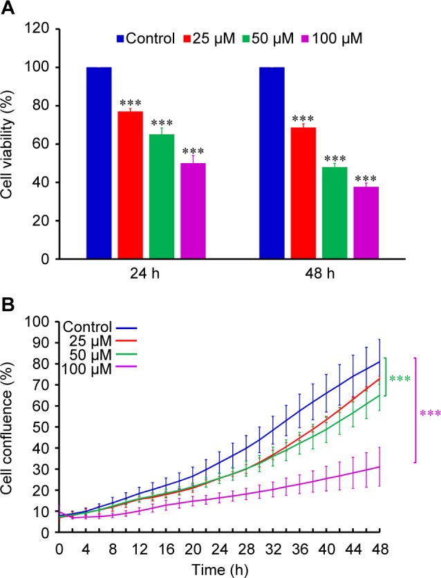
Fig. 1
Effects of YC-1 on cell viability and cell confluence in CAR cells. Cells were incubated with 0, 25, 50 and 100u00a0u03bcM of YC-1 for various duration. (A) The cell viability was determined by MTT as...
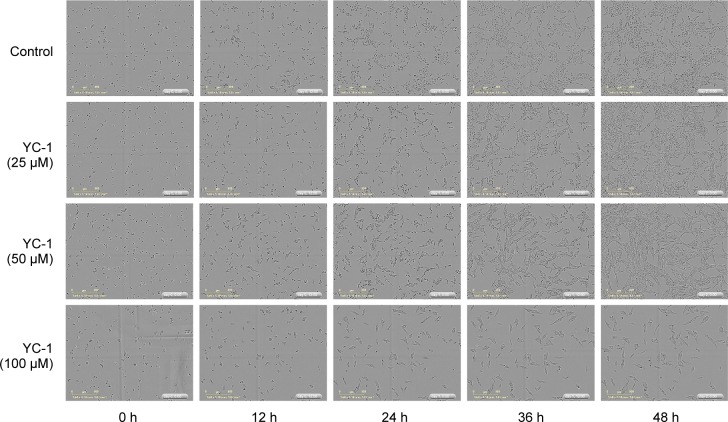
Fig. 2
Effects of YC-1 on cell morphology and confluence of CAR cells. Cells were incubated with 0, 25, 50 and 100 of YC-1 for 0, 12, 24, 36 and 48u00a0h. The cell morphology and density was determined by th...
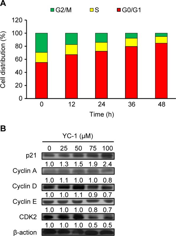
Fig. 3
Effects of YC-1 on cell cycle distribution and the levels of G 0 /G 1 proteins of CAR cells. (A) Cells were incubated with 100u00a0u03bcM of YC-1 for 0, 12, 24, 36 and 48u00a0h. The cell cycle distrib...
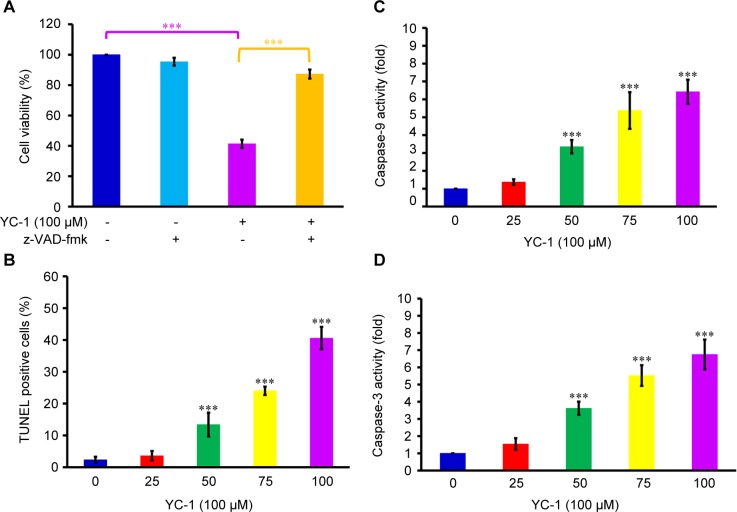
Fig. 4
Effects of YC-1 on DNA fragmentation, caspase-9 and caspase-3 activities in CAR cells. (A) Cells were incubated with 100u00a0u03bcM of YC-1 with or without z-VAD-fmk for 48u00a0h. The cell viability w...
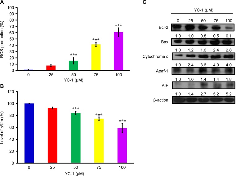
Fig. 5
Effects of YC-1 on ROS, mitochondrial membrane potential (u2206u03a8m) and the levels of apoptosis-related proteins in CAR cells. Cells were incubated with 0, 25, 50, 75 and 100 of YC-1 for 48u00a0h. ...
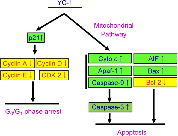
Fig. 6
Schematic diagram of proposed molecular mechanism of YC-1-induced G 0 /G 1 phase arrest and apoptosis in cisplatin- resistant human oral cancer CAR cells.
Figure images are served from the NIH/NLM PubMed Central Open Access Subset or Europe PMC; copyright remains with the publishers and authors.
💬 Discussion
0 commentsNo comments yet. Be the first to start a discussion!
Leave a Comment