Abstract
While single cell heterogeneity is present in all biological systems, most studies cannot address it due to technical limitations. Here we describe a nano-liter droplet microfluidic-based approach for stimulation and monitoring of surface and secreted markers of live single immune dendritic cells (DCs) as well as monitoring the live T cell/DC interaction. This nano-liter in vivo simulating microenvironment allows delivering various stimuli reagents to each cell and appropriate gas exchanges which are necessary to ensure functionality and viability of encapsulated cells. Labeling bioassay and microsphere sensors were integrated into nano-liter reaction volume of the droplet to monitor live single cell surface markers and secretion analysis in the time-dependent fashion. Thus live cell stimulation, secretion and surface monitoring can be obtained simultaneously in distinct microenvironment, which previously was possible using complicated and multi-step in vitro and in vivo live-cell microscopy, together with immunological studies of the outcome secretion of cellular function.
🧬 Organisms
✨ Fluorophores
🔬 Cell Lines
🏭 Microscope Brands
🧪 Reagent Suppliers
💻 Software Details
🏛️ Research Organizations (ROR)
Affiliated research institutions:
📋 Methods
Materials Rat FITC-anti-CD86 antibody [GL1] was obtained from Abcam and used at concentration of 1 μg/10 6 cells. Biotin-anti-IL-6 Ab (504601) for bead conjugation was obtained from BioLegend and FITC-anti-IL-6 Ab (11-7061-81) for IL-6 detection was obtained from eBioscience and used at concentration of 1 μg/mL. Microfluidic device fabrication Microfluidic flow chambers were fabricated using soft lithography at the MGH/CEM Facility. Negative photo resist SU-8 2100 (MicroChem, Newton, MA) was deposited onto clean silicon wafers to a thickness of 150 μm, and patterned by exposure to UV light through a transparency photomask (CAD/Art Services, Bandon, OR). The Sylgard 184 poly(dimethylsiloxane) (PDMS) (Dow Corning, Midland, MI) was mixed with crosslinker (ratio 10:1), poured onto the photoresist patterns, degassed thoroughly and cured for 12 hours at 75°C. Next, we peeled the PDMS devices off the wafer and bonded to glass slides after oxygen-plasma activation of both surfaces. The device is composed of a droplet forming nozzle and a storage array for 10 3 droplets. To improve the wetting of the channels with mineral oil in the presence (1%, w/w) of the surfactant (span80), prior to the experiments the microfluidic channels were treated with Pico-Sur™ 2 (Dolomite Microfluidics, UK) by filling the channels with the solution as received and then flushing them with air. Tygon Micro Bore PVC Tubing 100f, 0.010" ID, 0.030" OD, 0.010" Wall (Small Parts Inc, FL, USA) were connected to the channels and to the syringes. We used 1 ml syringes to load the fluids into the devices, while syringe pumps (Harvard Apparatus, USA) controlled the flow rates. Microsphere sensors preparation ProActive® Streptavidin Coated Microspheres (10 μm) (Bang Laboratories Inc., USA) were conjugated with the biotinylatedanti-mouse IL6 (Abcam®, MA, USA) according to the manufacturer's protocol. Unbounded active sites were blocked with StarlingBlock™ (thermo Scientific, USA) for one hour. Next, we washed the beads with the Phosphate Buffered Saline (PBS) (Sigma, USA), diluted to the final concentration 0.5 mg ml −1 , and stored at 4°C.
Show full methods section
Materials Rat FITC-anti-CD86 antibody [GL1] was obtained from Abcam and used at concentration of 1 μg/10 6 cells. Biotin-anti-IL-6 Ab (504601) for bead conjugation was obtained from BioLegend and FITC-anti-IL-6 Ab (11-7061-81) for IL-6 detection was obtained from eBioscience and used at concentration of 1 μg/mL. Microfluidic device fabrication Microfluidic flow chambers were fabricated using soft lithography at the MGH/CEM Facility. Negative photo resist SU-8 2100 (MicroChem, Newton, MA) was deposited onto clean silicon wafers to a thickness of 150 μm, and patterned by exposure to UV light through a transparency photomask (CAD/Art Services, Bandon, OR). The Sylgard 184 poly(dimethylsiloxane) (PDMS) (Dow Corning, Midland, MI) was mixed with crosslinker (ratio 10:1), poured onto the photoresist patterns, degassed thoroughly and cured for 12 hours at 75°C. Next, we peeled the PDMS devices off the wafer and bonded to glass slides after oxygen-plasma activation of both surfaces. The device is composed of a droplet forming nozzle and a storage array for 10 3 droplets. To improve the wetting of the channels with mineral oil in the presence (1%, w/w) of the surfactant (span80), prior to the experiments the microfluidic channels were treated with Pico-Sur™ 2 (Dolomite Microfluidics, UK) by filling the channels with the solution as received and then flushing them with air. Tygon Micro Bore PVC Tubing 100f, 0.010" ID, 0.030" OD, 0.010" Wall (Small Parts Inc, FL, USA) were connected to the channels and to the syringes. We used 1 ml syringes to load the fluids into the devices, while syringe pumps (Harvard Apparatus, USA) controlled the flow rates. Microsphere sensors preparation ProActive® Streptavidin Coated Microspheres (10 μm) (Bang Laboratories Inc., USA) were conjugated with the biotinylatedanti-mouse IL6 (Abcam®, MA, USA) according to the manufacturer's protocol. Unbounded active sites were blocked with StarlingBlock™ (thermo Scientific, USA) for one hour. Next, we washed the beads with the Phosphate Buffered Saline (PBS) (Sigma, USA), diluted to the final concentration 0.5 mg ml −1 , and stored at 4°C.
Cell isolation and stimulation 6–8 week old female
C57BL/6J mice were obtained from the Jackson Laboratories.
Bone marrow dendritic cells
(BMDCs) were collected from femora and tibiae and plated on non-tissue culture treated plastic dishes in RPMI medium (Gibco, Carlsbad, CA, Invitrogen, Carlsbad, CA), supplemented with 10% FBS, L-glutamine, penicillin/streptomycin, MEM non-essential amino acids, HEPES, sodium pyruvate, β-mercaptoethanol, and GM-CSF (15 ng/mL; Peprotech, Rocky Hill, NJ). At day 7 floating cells were collected and used as GM-CSF derived dendritic cells. Cells were stimulated with LPS (rough, ultra-pure E. coli K12 strain LPS, 100 ng/ml; Invitrogen, San Diego, CA).
Image analysis
Fluorescence images of droplets were captured on a Zeiss 200 Axiovert microscope using an AxioCAM MRm digital camera. For the cell studies, fluorescence signals were captured separately using appropriate filter sets for FITC. Image processing and analysis was conducted using ImageJ software.
Materials Rat FITC-anti-CD86 antibody [GL1] was obtained from Abcam and used at concentration of 1 μg/10 6 cells. Biotin-anti-IL-6 Ab (504601) for bead conjugation was obtained from BioLegend and FITC-anti-IL-6 Ab (11-7061-81) for IL-6 detection was obtained from eBioscience and used at concentration of 1 μg/mL.
📊 Figures
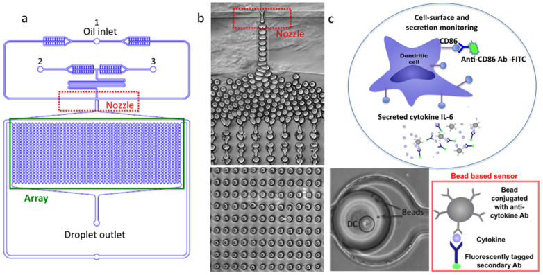
Figure 1
(a) Illustration of the nano-liter droplet-based technology. (b) Droplet generation in the PDMS device. (c) Schematic illustration of microfluidic reaction droplet for monitoring cell surface and secr...
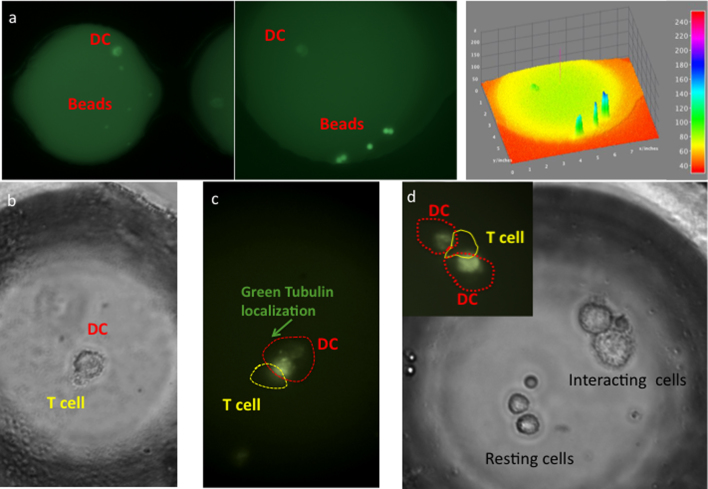
Figure 2
(a) DCs and bead-based sensors and reagents co-encapsulated in nano-liter reaction droplet containing anti-CD86 fluorescently tagged Abs for cell surface analysis. 1,2. Fluorescence images of droplets...
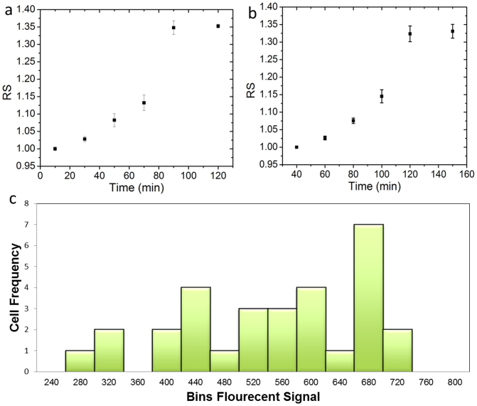
Figure 3
(a) The increase in accumulative CD86 expression over the time in response to LPS stimulation. (b) The increase in IL-6 secretion over the time in response to LPS stimulation. RS stands for relative s...
Figure images are served from the NIH/NLM PubMed Central Open Access Subset or Europe PMC; copyright remains with the publishers and authors.
💬 Discussion
0 commentsNo comments yet. Be the first to start a discussion!
Leave a Comment