Abstract
AbstractQuantification of cellular antigens and their interactions via antibody-based detection methods are widely used in scientific research. Accurate high-throughput quantitation of these assays using general image analysis software can be time consuming and challenging, particularly when attempted by users with limited image processing and analysis knowledge. To overcome this, we have designed Andy’s Algorithms, a series of automated image analysis pipelines for FIJI, that permits rapid, accurate and reproducible batch-processing of 3,3′-diaminobenzidine (DAB) immunohistochemistry, proximity ligation assays (PLAs) and other common assays. Andy’s Algorithms incorporates a step-by-step tutorial and optimization pipeline to make batch image analysis simple for the untrained user and adaptable across laboratories. Andy’s algorithms provide a simpler, faster, standardized work flow compared to existing programs, while offering equivalent performance and additional features, in a free to use open-source application of FIJI. Andy’s Algorithms are available at GitHub, publicly accessed athttps://github.com/andlaw1841/Andy-s-Algorithm.
🔬 Techniques
✨ Fluorophores
🧪 Sample Preparation
🔬 Cell Lines
🏭 Microscope Brands
🧪 Reagent Suppliers
🎨 Filters
💻 Software Details
💻 Code & Software
💾 Data Repositories
🏛️ Research Organizations (ROR)
Affiliated research institutions:
📋 Methods
Mice
All mice were housed in specific pathogen-free conditions at the Garvan Institute, with all animal procedures approved by the Garvan/St Vincent’s Animal Ethics and Experimentation Committee (Approval #14/27). All animal experiments were performed in accordance with the NSW Animal Research Act 1985 (PDF), NSW Animal Research Legislation 2010 and the Australian code of practice for the care and use of animals for scientific purposes, 8th Edition 2013. Immune-compromised NOD.Cg-Prkdc scid Il2rg tm1Wjl /SzJ were housed in SPF conditions in a 12-hour:12-hour light:dark cycle and given food and water ad libitum. Intraductal injections were modified from a previously described protocol without a Y incision in the abdomen 39 . For cross-sectional studies of tumour metastasis, mice were again randomized into DOX-treated or control-treated groups and sacrificed at 9 weeks (MDA-MB-231 xenografts) or 12 weeks (MDA-MB-468 xenografts) post tumor cell inoculation. Mice were then euthanized with CO 2 asphyxiation and the lungs were harvested and fixed in 10% neutral buffered formalin for 4 hours in 10% buffered formalin at room temperature. Immunohistochemistry DAB IHC validation data in Fig. 1 was performed on 4 µm lung sections from 3–4 mice bearing human breast cancer xenografts subjected to immunohistochemistry using an antibody to anti-high molecular weight cytokeratin (model 1) or anti-human vimentin (model 2) with detailed methodology described in 22 . After fixation the lungs were cut into 4 µm lung sections, baked for 4 hours at 60 °C, deparaffinised and antigens retrieved using pH9 retrieval solution (S2367) in a pressure cooker for 30 seconds. Endogenous peroxidases were blocked using a 3% H 2 O 2 solution and then incubated with primaries antibodies against high molecular weight cytokeratin (1:100, Leica 34BE12) for lungs bearing MDA-MB-468 metastases and against Vimentin (1:400, Leica NCL-L-VIM-V9) for lungs bearing MDA-MB-231 metastases for 30 minutes at room temperature. Sections were washed and then incubated with secondary peroxidase conjugated antibody (Envision mouse K4007) before application of the DAB chromagen substrate (K3468). All reagents were from DAKO unless otherwise specified and immunohistochemistry was performed on a DAKO autostainer. 20 images from each lung bearing breast cancer metastases (n = 3–4 mice was acquired from each on a Leica DM4000 light microscope an objective of 20X and was used further image analysis. A table of the optimised image parameters used for the IHC image analysis of lung metastatic deposits is provided in Supp. Table 4 . Breast cancer tissue microarrays Immunohistochemistry was used to assay MCL-1 protein expression using a mouse monoclonal antibody to MCL-1 (ThermoFischer (Pierce) MA5–13932) on TMAs constructed from tumors from a cohort of 292 patients diagnosed with invasive ductal breast carcinoma described in 40 . Immunohistochemistry was performed as above except antigen retrieval (pH9 S2367) was performed in a pressure cooker for 4 minutes and primary antibody concentration was performed at a 1:20 dilution. The cohort consists of cases of invasive ductal carcinoma of no special type, median age 54 (range 24–87), with a median follow-up of 64 months (range 0–152.1). Of these, 68.6% were ER + , 57.1% were PR + , 18.7% were HER-2 amplified (by FISH) and 43.3% were lymph node-positive. Endocrine therapy (TAM) was given to 49.3% of patients and chemotherapy (AC or CMF) to 38%. MCL-1 protein could only be detected in a subset of 246 of these cases due to missing or folded cores. To examine the performance of Andy’s DAB+ IHC pipeline on this cohort, 11 cores with nuclear and 16 cores with cytoplasmic expression were randomly selected for analysis and compared to the pathologists manual estimate. A table of the optimised image parameters used for the IHC image analysis of TMAs is provided in Supp. Table 5 .
Show full methods section
Mice
All mice were housed in specific pathogen-free conditions at the Garvan Institute, with all animal procedures approved by the Garvan/St Vincent’s Animal Ethics and Experimentation Committee (Approval #14/27). All animal experiments were performed in accordance with the NSW Animal Research Act 1985 (PDF), NSW Animal Research Legislation 2010 and the Australian code of practice for the care and use of animals for scientific purposes, 8th Edition 2013. Immune-compromised NOD.Cg-Prkdc scid Il2rg tm1Wjl /SzJ were housed in SPF conditions in a 12-hour:12-hour light:dark cycle and given food and water ad libitum. Intraductal injections were modified from a previously described protocol without a Y incision in the abdomen 39 . For cross-sectional studies of tumour metastasis, mice were again randomized into DOX-treated or control-treated groups and sacrificed at 9 weeks (MDA-MB-231 xenografts) or 12 weeks (MDA-MB-468 xenografts) post tumor cell inoculation. Mice were then euthanized with CO 2 asphyxiation and the lungs were harvested and fixed in 10% neutral buffered formalin for 4 hours in 10% buffered formalin at room temperature. Immunohistochemistry DAB IHC validation data in Fig. 1 was performed on 4 µm lung sections from 3–4 mice bearing human breast cancer xenografts subjected to immunohistochemistry using an antibody to anti-high molecular weight cytokeratin (model 1) or anti-human vimentin (model 2) with detailed methodology described in 22 . After fixation the lungs were cut into 4 µm lung sections, baked for 4 hours at 60 °C, deparaffinised and antigens retrieved using pH9 retrieval solution (S2367) in a pressure cooker for 30 seconds. Endogenous peroxidases were blocked using a 3% H 2 O 2 solution and then incubated with primaries antibodies against high molecular weight cytokeratin (1:100, Leica 34BE12) for lungs bearing MDA-MB-468 metastases and against Vimentin (1:400, Leica NCL-L-VIM-V9) for lungs bearing MDA-MB-231 metastases for 30 minutes at room temperature. Sections were washed and then incubated with secondary peroxidase conjugated antibody (Envision mouse K4007) before application of the DAB chromagen substrate (K3468). All reagents were from DAKO unless otherwise specified and immunohistochemistry was performed on a DAKO autostainer. 20 images from each lung bearing breast cancer metastases (n = 3–4 mice was acquired from each on a Leica DM4000 light microscope an objective of 20X and was used further image analysis. A table of the optimised image parameters used for the IHC image analysis of lung metastatic deposits is provided in Supp. Table 4 . Breast cancer tissue microarrays Immunohistochemistry was used to assay MCL-1 protein expression using a mouse monoclonal antibody to MCL-1 (ThermoFischer (Pierce) MA5–13932) on TMAs constructed from tumors from a cohort of 292 patients diagnosed with invasive ductal breast carcinoma described in 40 . Immunohistochemistry was performed as above except antigen retrieval (pH9 S2367) was performed in a pressure cooker for 4 minutes and primary antibody concentration was performed at a 1:20 dilution. The cohort consists of cases of invasive ductal carcinoma of no special type, median age 54 (range 24–87), with a median follow-up of 64 months (range 0–152.1). Of these, 68.6% were ER + , 57.1% were PR + , 18.7% were HER-2 amplified (by FISH) and 43.3% were lymph node-positive. Endocrine therapy (TAM) was given to 49.3% of patients and chemotherapy (AC or CMF) to 38%. MCL-1 protein could only be detected in a subset of 246 of these cases due to missing or folded cores. To examine the performance of Andy’s DAB+ IHC pipeline on this cohort, 11 cores with nuclear and 16 cores with cytoplasmic expression were randomly selected for analysis and compared to the pathologists manual estimate. A table of the optimised image parameters used for the IHC image analysis of TMAs is provided in Supp. Table 5 .
Proximity ligation assays
PLA validation data in Fig. 3 was performed in fixed cells using antibodies to CDK2 and Cyclin E1 with detailed methodology described in 37 . Cells were fixed with 4% PFA/PBS for 20 minutes at room temperature, with or without methanol post-fixation (−20 °C for 20 min). Samples were blocked with 1% BSA/PBS, stained with the indicated antibodies and counterstained with ToPro3/DAPI (Jackson ImmunoResearch Laboratories). PFA fixed cells were subjected to the Duolink Proximity Ligation Assay (Sigma) as described by the manufacturer using antibodies CDK2 (M2, D12) and cyclin E1 (HE12) (Santa Cruz Biotechnology). Confocal microscopy was performed on Leica DMRBE/DMIRE2. Images were analysed with Imaris where individual spots were defined with a variable and initial size estimate of 0.5 μm. 7 composite (foci, nuclear and cytoplasmic) images were used for image analysis. Images were processed with Adobe Photoshop, and adjusted for optimal brightness/contrast. Minimal gamma changes were made to enable visualisation of overlaid signals. A table of the optimised image parameters used for the PLA image analysis is provided in Supp. Table 6 .
Statistics
All statistics were performed in Prism7 for MacOSX. An ANOVA and a Pearson’s R 2 correlation coefficient was used to compare between Andy’s DAB+ IHC and PLA algorithms and the general image analysis programs CellPofiler 15 , ilastik 11 and Imaris. A Chi-squared distribution was used to determine whether there was any significant variation between the output of Andy’s DAB+ IHC and pathological scoring. Instructions to load Andy’s algorithms in FIJI The image processing steps for the DAB+ IHC and PLA algorithms are illustrated in Figs 1A and 3A and Supp. Figure 1 and 4 and for the H&E and 3D Colony forming image analysis are illustrated in Supp. Figure 2A and 3A . To download, install and run Andy’s Algorithms, follow the following steps: Download and install FIJI or update FIJI (version 1.51 k or later) [ https://fiji.sc ] Download Andy’s Algorithms (eg DAB_IHC_v2.40.ijm, PLA_v2.40.ijm, HandE_v2.40.ijm and 3D_colony_v2.40.ijm) from https://github.com/andlaw1841/Andy-s-Algorithm Go to Plugins > Macros > Install the Algorithm of choice in the menu bar of FIJI Select the Andy’s Algorithm preference (eg DAB_IHC_v2.40.ijm) to install the algorithm Go to Plugins > Macros. An option to select the algorithm will now be in the dropdown menu. Click run. The algorithm will be temporally installed into the toolbar of FIJI and closes when FIJI is exited. Simply reinstall the algorithm when you open FIJI again. The algorithm is designed for single images and not Z-stacks. Please convert image stacks to single files before running each algorithm. Sample images are provided at https://github.com/andlaw1841/Andy-s-Algorithm to assist users with the optimization of the image processing and analysis settings.
Data availability
The datasets generated during and/or analysed during the current study are included in this published article (and its Supplementary Information files). Andy’s DAB_IHC_2.40.ijm, PLA_2.40.ijm, HandE_2.40.ijm and 3D_colony_2.40.ijm) from https://github.com/andlaw1841/Andy-s-Algorithm . All images used in the current study will be made available at Figshare at https://figshare.com/ .
Electronic supplementary material Supplementary Figure Legends, Tables and Figures
📊 Figures
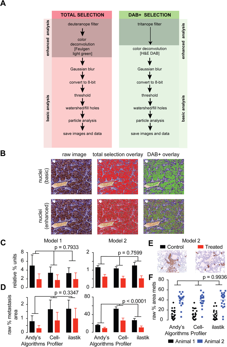
Figure 1
A new pipeline for image quantification of DAB+ IHC. ( A ) Flow chart depicting the image processing steps within the DAB+ algorithm for the selection of all (hematoxylin and DAB+) and positive cells ...
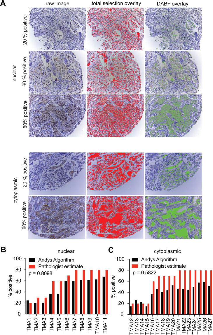
Figure 2
Andyu2019s DAB+ IHC algorithm can be used to score breast cancer TMAs. ( A ) Representative raw IHC images (left panels) depicting 20u201380% nuclear or cytoplasmic expression of MCL-1 using cores sel...
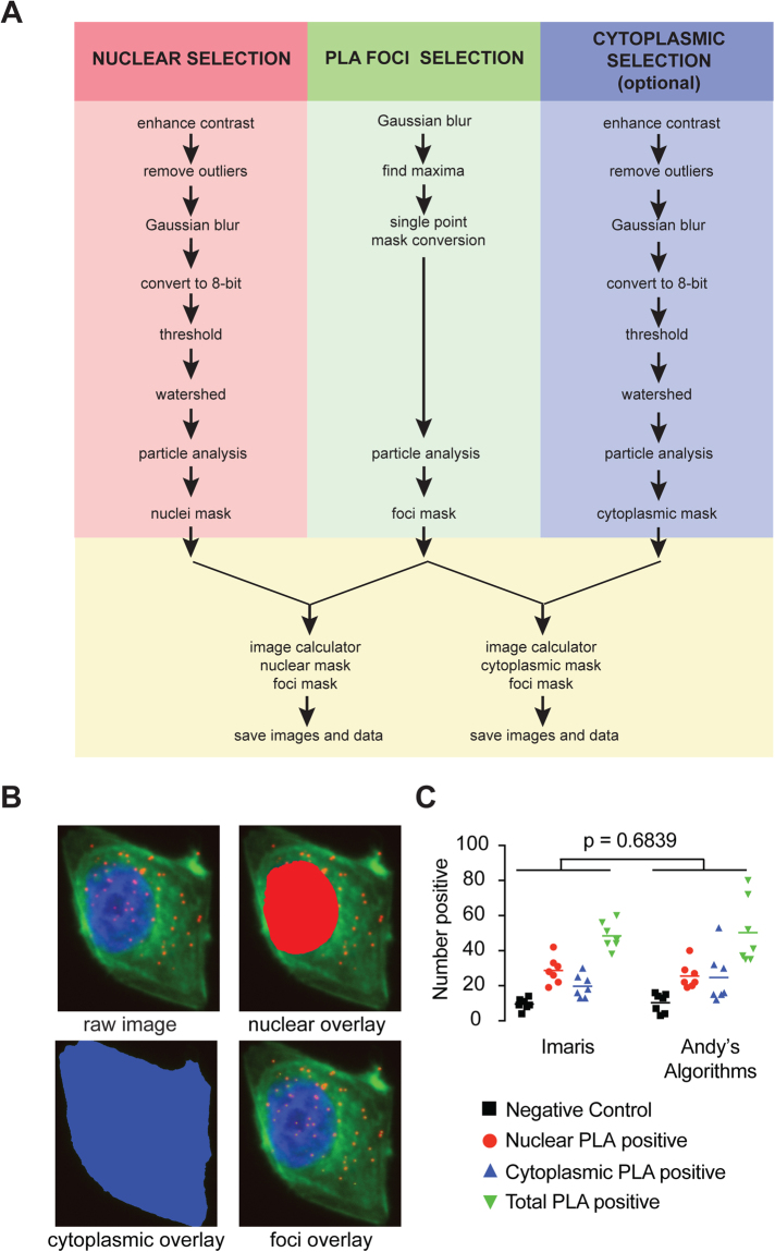
Figure 3
A new pipeline for image quantification for proximity ligation assays. ( A ) Flow chart depicting the image processing steps within the PLA particle analysis algorithm for the selection of all positiv...
Figure images are served from the NIH/NLM PubMed Central Open Access Subset or Europe PMC; copyright remains with the publishers and authors.
💬 Discussion
0 commentsNo comments yet. Be the first to start a discussion!
Leave a Comment