Abstract
Multimodal nonlinear optical microscopy is a valuable tool to study complex biological samples. We present an easy-to-operate approach to perform coherent anti-Stokes Raman scattering (CARS), two-photon fluorescence (TPF), second harmonic generation (SHG), and third-harmonic generation (THG) imaging using a single laser source composed of an 80 MHz femtosecond (fs) laser, an optical parametric oscillator (OPO), and a PPLN crystal for frequency doubling. The platform allows vibrationally resonant CARS imaging of CH-rich myelin sheath in fresh spinal tissues and lipid bodies in live cells. Multimodal nonlinear optical imaging and microspectroscopy analysis of fresh liver tissues are demonstrated.
🔬 Techniques
🔭 Microscopes
💻 Software
🧪 Sample Preparation
🏭 Microscope Brands
🧪 Reagent Suppliers
🔴 Lasers
📷 Detectors
🎨 Filters
💻 Software Details
🏛️ Research Organizations (ROR)
Affiliated research institutions:
📋 Methods
Sample preparation TiO 2 nanopowder (
📊 Figures
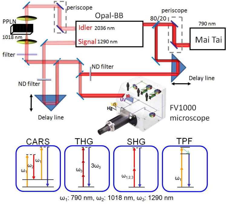
Fig. 1
Schematic diagram of a multimodal NLO microscope using a fs laser source. The Mai Tai output at 790 nm (u03c9 1 ) is split into two beams, with 80% of power being used to pump the Opal-BB. The idler o...
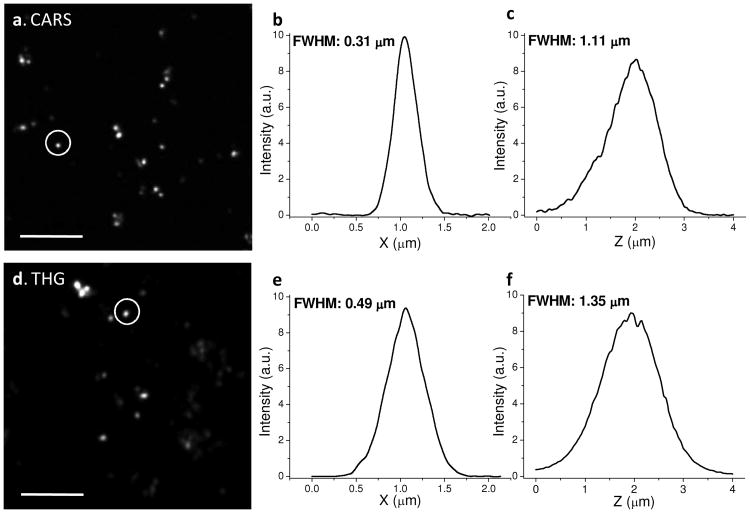
Fig. 2
Spatial resolution. (a) Epi-detected CARS image of TiO 2 nanoparicles, excited at 790 and 1018 nm. (b-c) Lateral and axial intensity profiles along the circled TiO 2 nanoparticle in (a). (d) Epi-detec...
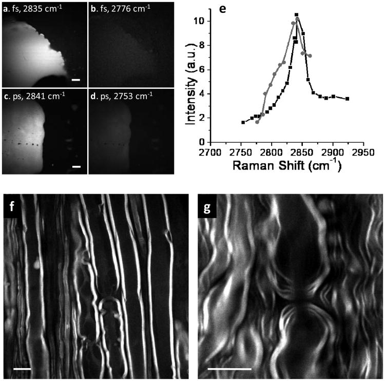
Fig. 3
Vibrational contrast of C-H rich objects. (a-d) The forward-detected CARS images of a subcutaneous fat tissue by the fs (a-b) and ps (c-d) lasers, respectively. The Raman shift is marked in each image...
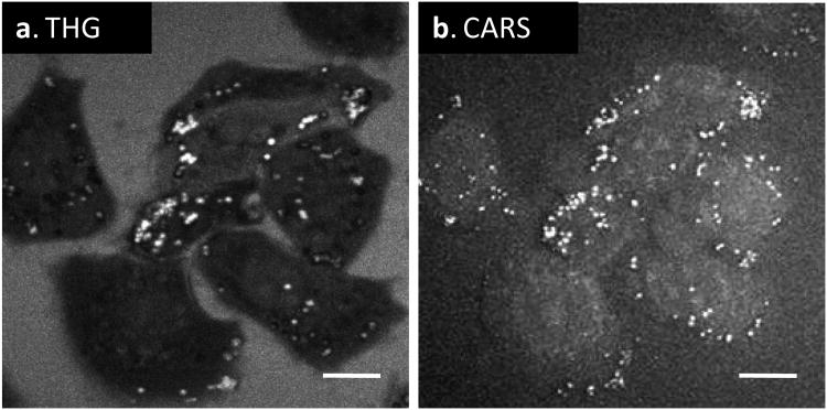
Fig. 4
Comparison of forward-detected THG (a) and CARS (b) images of lipid droplets inside live KB cells. Scale bars = 10 u03bcm.
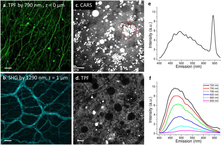
Fig. 5
Multimodal nonlinear optical imaging of fresh liver tissues from high fat fed mice. (a) TPF image excited at 790 nm and detected in the 450-550 nm region. Fibrous structure on the liver surface produc...
Figure images are served from the NIH/NLM PubMed Central Open Access Subset or Europe PMC; copyright remains with the publishers and authors.
💬 Discussion
0 commentsNo comments yet. Be the first to start a discussion!
Leave a Comment