Abstract
Abstract The kinin B1 receptor is an inducible receptor not normally expressed but induced by inflammatory stimuli and plays a major role in neutrophil recruitment, particularly in response to the cytokine IL-1β. However, the exact mechanism involved in this response is unclear. The aim of this study was to dissect the molecular mechanism involved, in particular to determine whether specific ELR-CXCL chemokines (specific neutrophil chemoattractants) played a role. Using intravital microscopy, we demonstrated that IL-1β-induced leukocyte rolling, adherence, and emigration in mesenteric venules of wild-type (WT) mice, associated with an increase in B1 receptor mRNA expression, were substantially attenuated (>80%) in B1 receptor knockout mice (B1KO). This effect in B1KO mice was correlated with a selective down-regulation of IL-1β-induced CXCL5 mRNA and protein expression compared with WT mice. Furthermore a selective neutralizing CXCL5 Ab caused profound suppression of leukocyte emigration in IL-1β-treated WT mice. Finally, treatment of human endothelial cells with IL-1β enhanced mRNA expression of the B1 receptor and the human (h) CXCL5 homologues (hCXCL5 and hCXCL6). This response was suppressed by ∼50% when cells were pretreated with the B1 receptor antagonist des-Arg9-[Leu8]-bradykinin while treatment with des-Arg9-bradykinin, the B1 receptor agonist, caused a concentration-dependent increase in hCXCL5 and hCXCL6 mRNA expression. This study unveils a proinflammatory pathway centered on kinin B1 receptor activation of CXCL5 leading to leukocyte trafficking and highlights the B1 receptor as a potential target in the therapeutics of inflammatory disease.
🔬 Techniques
🧪 Sample Preparation
🔬 Cell Lines
🧪 Reagent Suppliers
💾 Data Repositories
🏛️ Research Organizations (ROR)
Affiliated research institutions:
📋 Methods
MATERIELS and METHODS Animals
All experiments were conducted according to the Animals (Scientific Procedures) Act of 1986 (United Kingdom). Male C57BL6J wild type (WT) or B1KO ( 10 ) mice (C57BL6 background) at 5 weeks of age (10-15g) were used in all experiments. Intravital Microscopy Male WT and B1KO mice received either murine IL-1β (5ng/mouse, i.p., PeproTech, UK) or saline vehicle. After 4h mice were anesthetized with diazepam (60mg/kg subcutaneously) and Hypnorm® (0.7mg/kg fentanyl citrate and 20mg/kg fluanisone intramuscularly) and the mesenteric vascular bed prepared for viewing by intra-vital microscopy. Mesenteries were superfused with bicarbonate-buffered solution at 37°C (132mM NaCl, 4.7mM KCl, 1.2mM MgSO , 17.9mM NaHCO 3, 2.0mM CaCl 2 , pH 7.4, gassed with 5% CO 2 , 95% N 2 ) at a rate of 2ml/min. The temperature of the stage was maintained at 37°C. The extent of the inflammatory response elicited by IL-1β was analyzed by counting the number of white blood cells rolling per min. Cell adhesion was quantified by counting, for each vessel, the number of adherent neutrophils in a 100μm length, and leukocyte emigration from the microcirculation into the tissue was quantified by counting the number of cells that had emigrated up to 50μm away from the wall of 100μm vessel segments. Venular blood flow was calculated from the product of mean RBC velocity (V mean = centerline velocity/1.6) and microvascular cross-sectional area, assuming a cylindrical geometry. Wall shear rate was calculated by the Newtonian definition: shear rate = 8,000× (V mean /diameter). A minimum of 3 postcapillary venules (diameter between 20-40μm; length of at least 100μm) were observed for each mouse. To evaluate the role of CXCL5 in regulating leukocyte recruitment, a selective neutralizing monoclonal antibody or control IgG (20μg per animal, i.p., PeproTech, UK) was injected into the tail vein 30min prior to IL-1β treatment. After 4h, leukocyte rolling, adhesion and emigration were measured as described above. Myeloperoxidase (MPO) assay MPO activity was determined in mesenteric tissue as an index of neutrophil accumulation ( 14 ). Mesenteric tissue, collected 4h after IL-1β treatment, was homogenized in 1mL of a 0.5% hexa-decyl-trimethyl ammonium bromide (HTAB) in MOPS buffer (10mM, pH 7). After homogenization, samples were centrifuged at 4000g for 20min at 4°C and the supernatant collected for determination of MPO levels as previously described ( 15 ). Data are expressed as U/g of total protein content in the tissue determined by Bradford assay. Real-Time Quantitative RT-PCR of murine mesenteric tissue Chemokine mRNA expression was determined by real-time quantitative RT-PCR. Briefly mesenteric tissue was removed from saline or IL-1β (2h)-treated mice as above, snap frozen in liquid nitrogen and stored at −80°C until use. Samples were homogenised and total RNA isolated using a NucleoSpin RNA II purification kit (Macherey-Nagel) and then stored at −80°C until use. cDNA was synthesized from 1μg of total RNA with M-MLV reverse transcriptase (Promega, UK) using oligo-dt nucleotides. The following primers were used for mouse: CXCL1 (TGA GCT GCG CTG TCA GTG CCT and AGA AGC CAG CGT TCA CCA GA), CXCL2 (GAG CTT GAG TGT GAC GCC CCC AGG and GTT AGC CTT GCC TTT GTT CAG TAT C), CXCL5 (GCA TTT CTG TTG CTG TTC ACG CTG and CCT CCT TCT GGT TTT TCA GTT TAG C), CXCL7 (TGG GCC TGA TCC TTG TTG CGC AND GCA CCG TTT TTT GTC CAT TCT TCA G), B1 recepto r (TGG AGT TGA ACG TTT TGG GTT T and GTG AGG ATC AGC CCC ATT GT) and βactin (GAA ATC GTG CGT GAC ATC AAA G and TGT AGT TTC ATG GAT GCC ACA G). Standard curves for these molecules were generated to determine amplification efficiencies of target and reference genes. Quantitative PCR was performed on ABI Prism 7900 with 100nM of primers and 20ng of cDNA. Chemokine/receptor expression was normalized to ß-actin and expressed as relative value using the comparative Ct method (2-ΔΔCt) according to the manufacturer’s instructions. The levels of mRNA expression of genes of interest were normalised to saline control.
Show full methods section
MATERIELS and METHODS Animals
All experiments were conducted according to the Animals (Scientific Procedures) Act of 1986 (United Kingdom). Male C57BL6J wild type (WT) or B1KO ( 10 ) mice (C57BL6 background) at 5 weeks of age (10-15g) were used in all experiments. Intravital Microscopy Male WT and B1KO mice received either murine IL-1β (5ng/mouse, i.p., PeproTech, UK) or saline vehicle. After 4h mice were anesthetized with diazepam (60mg/kg subcutaneously) and Hypnorm® (0.7mg/kg fentanyl citrate and 20mg/kg fluanisone intramuscularly) and the mesenteric vascular bed prepared for viewing by intra-vital microscopy. Mesenteries were superfused with bicarbonate-buffered solution at 37°C (132mM NaCl, 4.7mM KCl, 1.2mM MgSO , 17.9mM NaHCO 3, 2.0mM CaCl 2 , pH 7.4, gassed with 5% CO 2 , 95% N 2 ) at a rate of 2ml/min. The temperature of the stage was maintained at 37°C. The extent of the inflammatory response elicited by IL-1β was analyzed by counting the number of white blood cells rolling per min. Cell adhesion was quantified by counting, for each vessel, the number of adherent neutrophils in a 100μm length, and leukocyte emigration from the microcirculation into the tissue was quantified by counting the number of cells that had emigrated up to 50μm away from the wall of 100μm vessel segments. Venular blood flow was calculated from the product of mean RBC velocity (V mean = centerline velocity/1.6) and microvascular cross-sectional area, assuming a cylindrical geometry. Wall shear rate was calculated by the Newtonian definition: shear rate = 8,000× (V mean /diameter). A minimum of 3 postcapillary venules (diameter between 20-40μm; length of at least 100μm) were observed for each mouse. To evaluate the role of CXCL5 in regulating leukocyte recruitment, a selective neutralizing monoclonal antibody or control IgG (20μg per animal, i.p., PeproTech, UK) was injected into the tail vein 30min prior to IL-1β treatment. After 4h, leukocyte rolling, adhesion and emigration were measured as described above. Myeloperoxidase (MPO) assay MPO activity was determined in mesenteric tissue as an index of neutrophil accumulation ( 14 ). Mesenteric tissue, collected 4h after IL-1β treatment, was homogenized in 1mL of a 0.5% hexa-decyl-trimethyl ammonium bromide (HTAB) in MOPS buffer (10mM, pH 7). After homogenization, samples were centrifuged at 4000g for 20min at 4°C and the supernatant collected for determination of MPO levels as previously described ( 15 ). Data are expressed as U/g of total protein content in the tissue determined by Bradford assay. Real-Time Quantitative RT-PCR of murine mesenteric tissue Chemokine mRNA expression was determined by real-time quantitative RT-PCR. Briefly mesenteric tissue was removed from saline or IL-1β (2h)-treated mice as above, snap frozen in liquid nitrogen and stored at −80°C until use. Samples were homogenised and total RNA isolated using a NucleoSpin RNA II purification kit (Macherey-Nagel) and then stored at −80°C until use. cDNA was synthesized from 1μg of total RNA with M-MLV reverse transcriptase (Promega, UK) using oligo-dt nucleotides. The following primers were used for mouse: CXCL1 (TGA GCT GCG CTG TCA GTG CCT and AGA AGC CAG CGT TCA CCA GA), CXCL2 (GAG CTT GAG TGT GAC GCC CCC AGG and GTT AGC CTT GCC TTT GTT CAG TAT C), CXCL5 (GCA TTT CTG TTG CTG TTC ACG CTG and CCT CCT TCT GGT TTT TCA GTT TAG C), CXCL7 (TGG GCC TGA TCC TTG TTG CGC AND GCA CCG TTT TTT GTC CAT TCT TCA G), B1 recepto r (TGG AGT TGA ACG TTT TGG GTT T and GTG AGG ATC AGC CCC ATT GT) and βactin (GAA ATC GTG CGT GAC ATC AAA G and TGT AGT TTC ATG GAT GCC ACA G). Standard curves for these molecules were generated to determine amplification efficiencies of target and reference genes. Quantitative PCR was performed on ABI Prism 7900 with 100nM of primers and 20ng of cDNA. Chemokine/receptor expression was normalized to ß-actin and expressed as relative value using the comparative Ct method (2-ΔΔCt) according to the manufacturer’s instructions. The levels of mRNA expression of genes of interest were normalised to saline control.
Measurement of CXCL5 protein expression
Mesenteric tissue of saline and IL-1β-treated (4h) WT and B1KO mice were collected, homogenised and supernatants collected. Mouse CXCL5 protein levels were determined by ELISA (Duoset R&D Systems) according to the manufacturer’s protocol. CXCL5 levels were expressed relative to total protein concentration of the supernatant samples. Human Umbilical Vein Endothelial Cell (HUVEC) Culture HUVEC were cultured to passage 3 in EGMII medium (Cambrex, UK). Confluent cells were treated with vehicle (saline) or IL-1β (1ng/ml, 0-24h). In some experiments, the B1 antagonist Lys-[Leu 8 ]des-Arg 9 -BK (10μM) was added to the medium 15 min prior to IL-1β treatment and the reaction stopped after 8h. In a further series of experiments cells were incubated with the B1 agonist Lys-des-Arg 9 -BK (1–10,000nM) for 4h either directly or following a 24h pre-treatment with IL-1β.
Real-Time Quantitative RT-PCR of endothelial cells
HUVEC from the above experiments were washed with sterile PBS, collected by scraping and samples kept at −80°C until mRNA extraction. The human CXCL5 homologues hCXCL5 and hCXCL6, and kinin B1 receptor mRNA expression were determined as described above. The following primers were used: hCXCL5 (GAG AGC TGC GTT GCG TTT G and TTT CCT TGT TTC CAC CGT CCA) hCXCL6 (GGT CCT GTC TCT GCT GTG C and GGG AGG CTA CCA CTT CCA) hB1 receptor (ACG CCT TCA TTT TCT GCC TG and GCT GGC TCT GGT TGG AGG AT) kininogen (AGA CAC GGC ATT CAG TAC TTT AAC A and TGG GCC CGT TTT ACT TCA TT) kallikrein ( GGG TCG CCA CAA CTT GTT TG and GCT GTA GTC CTC GTC TGC TT) and GAPDH (CAT GTT CGT CAT GGG TGT GAA and ATG GAC TGT GGT CAT GAG TCC TT).
Western blotting of endothelial cells
Following treatment cells outlined above cells were washed with ice-cold PBS, scraped and lysed in ice-cold lysis buffer (20mM Tris-HCl [pH 7.4], 50mM NaCl, 50mM NaF, 5mM EDTA, and 20mM Na 4 P 2 O 7 ·10H 2 O). Supernatants were collected and protein concentration determined by Bradford assay. Samples (20μg) were subjected to eletrophoresis using an 8% polyacrylamide gel followed by electrotransfer to nitrocellulose membrane. To detect B1 receptor, nitrocellulose were incubated with the polyclonal B1 receptor antibody (A15C ( 16 ); kind gift of Dr Jean-Loup Bascands, dilution 1:5000) overnight at 4°C and then a secondary peroxidase-coupled goat anti–rabbit antibody (1/2000, Dako). Visualization of bands was achieved by chemiluminescence (ECL kit; Amersham Pharmacia Biotech). Selectivity of the antibody was determined by preadsorption of the antibody to its corresponding peptide at a concentration of 10μg/ml at 4°C overnight. The autoradiogaphic bands were semiquantified and normalised to α-tubulin levels.
Statistical analysis
Values are given as means ± SE where n represents the number of animals or the number of experiments conducted for cells. Statistical comparisons were conducted using paired or unpaired Student’s t test for 2 groups or one way ANOVA for more than 2 groups. Differences were considered significant when p < 0.05.
📊 Figures

Fig 1
IL-1u03b2-induced PMN recruitment is abolished in B1KO mice
(A) Leukocyte-endothelial cell interactions in mouse mesenteric postcapillary venules in vivo in response to IL-1u03b2 (5ng, i.p.) in WT and B1KO mice were measured by intravital microscopy. The diffe...

Fig 2
IL-1u03b2-induced ELR-CXCL chemokine mRNA expression is attenuated in B1KO mice
mRNA expression of CXCL1, CXCL7, CXCL2 and CXCL5 assessed using quantitative real-time RT-PCR of mesenteric tissue from WT and B1KO mice treated with saline or IL-1u03b2 (5ng, i.p., 2h).The data are e...
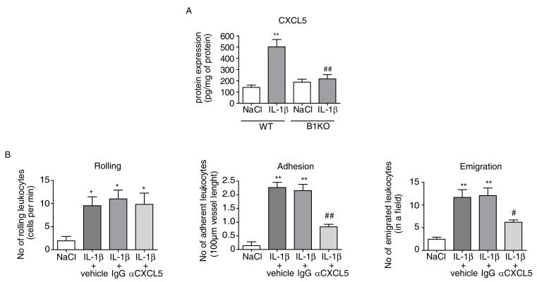
Fig 3
CXCL5 plays a major role in IL-1u03b2-induced leukocyte recruitment
(A) CXCL5 protein was measured by ELISA in mesenteric tissue removed from WT and B1KO mice 4h after treatment with saline or with IL-1u03b2 (5ng, i.p.). The data are expressed as CXCL5 per mg of total...

Fig 4
Temporal relationship between endothelial kinin B1 receptor, hCXCL5, hCXCL6 and kallikrein/kininogen expression in IL-u03b2-treated endothelial cells
(A) Time course (0, 4h, 8h and 24h) of B1 receptor, hCXCL5 and hCXCL6 mRNA expression in response to IL-1u03b2 (1ng/mL) in HUVEC. Expression of B1 receptor, hCXCL5 and hCXCL6 were measured by quantita...
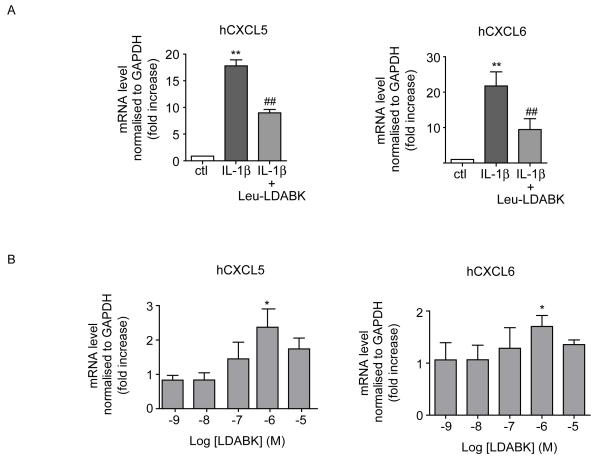
Fig 5
Kinin B1 receptor-induced hCXCL5 and hCXCL6 expression in HUVEC
(A) Effect of B1 antagonist (Lys-[Leu 8 ]des-Arg 9 -BK at 10u03bcM, 15min pre-treatment at t=0) on IL-1u03b2 (1ng/mL, 8h)-induced hCXCL5 and hCXCL6 mRNA expression in HUVEC. (B) Effect of B1 agonist (...
Figure images are served from the NIH/NLM PubMed Central Open Access Subset or Europe PMC; copyright remains with the publishers and authors.
💬 Discussion
0 commentsNo comments yet. Be the first to start a discussion!
Leave a Comment