Abstract
The mammalian cell nucleus contains a variety of organelles or nuclear bodies which contribute to key nuclear functions. Promyelocytic leukemia nuclear bodies (PML NBs) are involved in the regulation of apoptosis, antiviral responses, the DNA damage response and chromatin structure, but their precise biochemical function in these nuclear pathways is unknown. One strategy to tackle this problem is to assess the biophysical properties of the component parts of these macromolecular assemblies in living cells. In this study we determined PML NB assembly dynamics by live cell imaging, combined with mathematical modeling. For the first time, dynamics of PML body formation were measured in cells lacking endogenous PML. We show that all six human nuclear PML isoforms are able to form nuclear bodies in PML negative cells. All isoforms exhibit individual exchange rates at NBs in PML positive cells but PML I, II, III and IV are static at nuclear bodies in PML negative cells, suggesting that these isoforms require additional protein partners for efficient exchange. PML V turns over at PML Nbs very slowly supporting the idea of a structural function for this isoform. We also demonstrate that SUMOylation of PML at Lysine positions K160 and/or K490 are required for nuclear body formation in vivo.We propose a model in which the isoform specific residence times of PML provide both, structural stability to function as a scaffold and flexibility to attract specific nuclear proteins for efficient biochemical reactions at the surface of nuclear bodies.MCS code: 92C37.
🔬 Techniques
🔭 Microscopes
💻 Software
✨ Fluorophores
🧪 Sample Preparation
🔬 Cell Lines
🏭 Microscope Brands
🧪 Reagent Suppliers
💻 Software Details
🏛️ Research Organizations (ROR)
Affiliated research institutions:
📋 Methods
Cell Culture and Transfection Mouse 3T3-PML +/+ and 3T3-PML -/- cells [ 26 ], kindly provided by T. Hofmann (DKFZ Heidelberg) were cultured in Dulbecco' modified Eagle's medium (DMEM) supplemented with 10% fetal calf serum in a 10% CO 2 atmosphere at 37°C. For live cell imaging experiments, cells were seeded on 42 mm glass dishes (Saur Laborbedarf, Reutlingen, Germany) and transfected with plasmid DNA one to two days before observation using FuGENE-HD Transfection reagent (Roche, Basel, Switzerland) according to the manufacturer' protocol. Plasmids The GFP-PML expression constructs have been described in detail previously [ 25 ].
Western blots
Whole cell extracts were produced from transiently or stably transfected cell lines, electrophoresed on SDS-PAGE and transferred to Protran nitrocellulose membrane (Schleicher & Schuell, Dassel, Germany). The membrane was incubated with primary antibodies (in PBS-T) and developed with a peroxidase conjugated secondary species-specific antibody (Jackson Immunoresearch, West Grove, PA, USA). Signal was detected using the ECL reagent (Amersham, Uppsala, Sweden) on imaging film (Biomax, Kodak, Stuttgart, Germany). Anti-GFP monoclonal antibody was from Santa-Cruz Biotechnology (Heidelberg, Germany). Anti-mouse PML monoclonal antibody (# 05-718), non-cross reactive with human PML protein, was purchased from Upstate.
Immunocytochemistry and Microscopy
Cells grown on 15 mm diameter coverslips were fixed with 4% formaldehyde for 10 minutes and permeabilized with 0.25% Triton-X100 for 3 minutes. Diluted anti-mouse PML mAB was incubated on cells for 45 minutes. After 3 washing steps with PBS, an anti-mouse secondary antibody coupled to Cy3 (Jackson Immunosearch, West Grove, USA) was incubated on cells for 45 minutes, followed by a DNA-staining step using ToPro3 or DAPI (Invitrogen, Carlsbad, USA) for 10 minutes and mounting with Prolong Gold antifade mounting medium (Invitrogen, Carlsbad, USA). For microscopy, a LSM 510Meta or LSM710 laser scanning confocal microscope (Carl Zeiss, Jena, Germany) was used.
Show full methods section
Cell Culture and Transfection Mouse 3T3-PML +/+ and 3T3-PML -/- cells [ 26 ], kindly provided by T. Hofmann (DKFZ Heidelberg) were cultured in Dulbecco' modified Eagle's medium (DMEM) supplemented with 10% fetal calf serum in a 10% CO 2 atmosphere at 37°C. For live cell imaging experiments, cells were seeded on 42 mm glass dishes (Saur Laborbedarf, Reutlingen, Germany) and transfected with plasmid DNA one to two days before observation using FuGENE-HD Transfection reagent (Roche, Basel, Switzerland) according to the manufacturer' protocol. Plasmids The GFP-PML expression constructs have been described in detail previously [ 25 ].
Western blots
Whole cell extracts were produced from transiently or stably transfected cell lines, electrophoresed on SDS-PAGE and transferred to Protran nitrocellulose membrane (Schleicher & Schuell, Dassel, Germany). The membrane was incubated with primary antibodies (in PBS-T) and developed with a peroxidase conjugated secondary species-specific antibody (Jackson Immunoresearch, West Grove, PA, USA). Signal was detected using the ECL reagent (Amersham, Uppsala, Sweden) on imaging film (Biomax, Kodak, Stuttgart, Germany). Anti-GFP monoclonal antibody was from Santa-Cruz Biotechnology (Heidelberg, Germany). Anti-mouse PML monoclonal antibody (# 05-718), non-cross reactive with human PML protein, was purchased from Upstate.
Immunocytochemistry and Microscopy
Cells grown on 15 mm diameter coverslips were fixed with 4% formaldehyde for 10 minutes and permeabilized with 0.25% Triton-X100 for 3 minutes. Diluted anti-mouse PML mAB was incubated on cells for 45 minutes. After 3 washing steps with PBS, an anti-mouse secondary antibody coupled to Cy3 (Jackson Immunosearch, West Grove, USA) was incubated on cells for 45 minutes, followed by a DNA-staining step using ToPro3 or DAPI (Invitrogen, Carlsbad, USA) for 10 minutes and mounting with Prolong Gold antifade mounting medium (Invitrogen, Carlsbad, USA). For microscopy, a LSM 510Meta or LSM710 laser scanning confocal microscope (Carl Zeiss, Jena, Germany) was used.
Fluorescence Correlation Spectroscopy Measurements
Fluorescence correlation spectroscopy
(FCS) measurements were performed at 37°C on a LSM 510Meta/ConfoCor2 combi system using a C-Apochromat infinity-corrected 1.2 NA 40× water objective (Carl Zeiss, Jena, Germany) as described in detail elsewhere [ 25 ]. Briefly, GFP-tagged proteins were spot-illuminated with the 488 nm line of a 20 mW Argon laser at 5.5 Ampere tube current attenuated by an acousto-optical tunable filter (AOTF) to 0.1%. The detection pinhole had a diameter of 70 μm and emission was recorded through a 505-530 nm band-path filter. For the measurments, 10 × 30 time series of 10 s each were recorded with a time resolution of 1 μs and then superimposed for fitting to an anomalous diffusion model in three dimensions with triplett function [ 27 ] using Origin Software (OriginLab, Northhampton, MA, USA). The diffusion coefficients and anomaly parameters were extracted from fit curves as previously described [ 25 ]. Fluorescence Recovery after Photobleaching Fluorescence Recovery after Photobleaching (FRAP) experiments were carried out on a Zeiss LSM 510Meta confocal microscope (Carl Zeiss, Jena, Germany). One or two image stacks were taken before the bleach pulse and 50-70 image stacks after bleaching of "regions of interest" (ROIs) containing one nuclear body each at 0.05% laser transmission to minimize scan bleaching. Image aquisition frequency was adapted to the recovery rate of the respective GFP fusion protein, usually a 20 second interval was applied. The pinhole was adjusted to 1 airy unit. The image stacks were maximum-projected into a single plane from which relative fluorescence intensities within the ROIs were quantitated according to [ 25 ] using Excel (Microsoft, Redmond, WA, USA) and Origin software (OriginLab, Northhampton, MA, USA).
Reaction-diffusion model
For the mathematical model, the structural complexity of a PML body has been approximated assuming that molecules undergoing binding and unbinding to and from the body do so at the surface, and molecules situated more towards the inside of the body cannot unbind before moving to the surface. There is thus a reservoir of tightly bound "inner" molecules and one of loosely bound "outer" ones. Exchange between these reservoirs is modeled by linear kinetics, i. e. the more molecules there are, the more will move inside or out with rate constants k in and k out , respectively. Binding and unbinding to the PML body is treated similarly, with rate constants k on and k off , respectively. The experimental set-up provided that bleach ROI and FRAP ROI is similar, and so are assumed to be the same for modelling purposes. Diffusion inside and out of the ROI was modelled as a linear two-way process with a rate constant proportional 2D/r 2 , where D is the diffusion coefficient measured by FCS, and r is the ROI's radius. This constant yields the effective exchange rate through the boundary of a circular area. The model considers only fluorescent molecules of one type at a time, so no interactions between different molecular species are considered. It distinguishes between molecules outside the ROI, inside the ROI but diffusing freely, loosely bound at the surface of the PML body, and tightly bound inside the body. The fluorescence outside the ROI is all but unchanged by the bleaching pulse and subsequent diffusion, so this value was used to normalize all concentrations in the equations. Describing normalized concentrations of the fluorescent protein in free diffusion (x), loosely bound (y) and tightly bound (z), the reaction system results in the model equations The differential equations were numerically solved using an explicit Runge-Kutta formula (method ode45 in MATLAB). To fit the parameters of the model, an Evolution Strategy with Covariance Matrix Adaptation (CMA-ES) was employed [ 28 ]. We determined the ratio p = 20 of steady state fluorescence in the body vs. the background by confocal microscopy of GFP-PML isoforms and pixel intensity evaluation using MetaMorph software (Molecular Devices, Sunnyvale, USA). This enabled us to express the observable fluorescence in the ROI as Since the amount of bleached molecules is small compared to the overall amount of molecules in the nucleus, it is plausible to assume that after a sufficiently long time, fluorescence returns to the value measured before the photobleach. This constraint removes one degree of freedom from the model, yielding These values were used to normalize the concentrations used in the model. The mathematical model treats normalized concentrations of fluorescent molecules in free diffusion (x), loosely bound (y) and tightly bound (z) to the PML body. It is important to note that the concentration values x, y and z have been normalized by the background fluorescence outside the region of interest. Since we have determined the ratio p between equilibrium fluorescence inside the body and outside the ROI, and because the data used to fit the model was given in relative fluorescence intensity with an equilibrium value of 1, the background flourescence is given by 1/ p . Therefore, the observed RFI value w is related to the model concentrations by Over a very long time, all molecular species in the PML-body will eventually turn over, so that it is reasonable to assume w (t) tends towards 1 for large t. This means that for equilibrium conditions, in which , w(t) = 1. In this case, we have and thus, from the first three lines, we get Finally, taking this together with the conditition x + y + z = p , we find This enabled us to remove one degree of freedom, k on , from the model.
📊 Figures
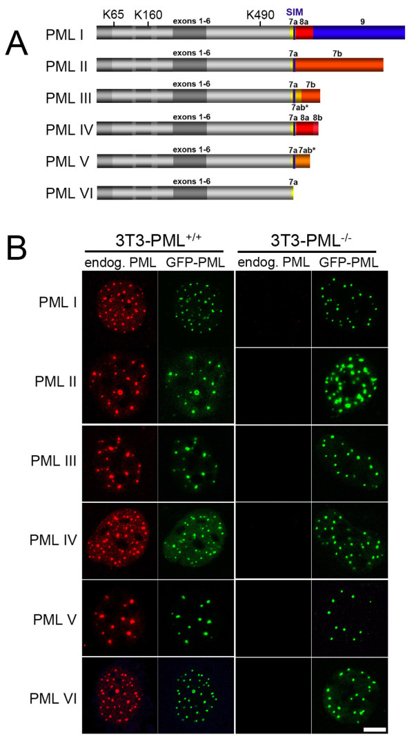
Figure 1
All PML protein isoforms form nuclear bodies in cells lacking endogenous PML . (A) Schematic depiction of the domain structure of PML isoforms (data taken from [ 16 ]). All PML isoforms share a common...
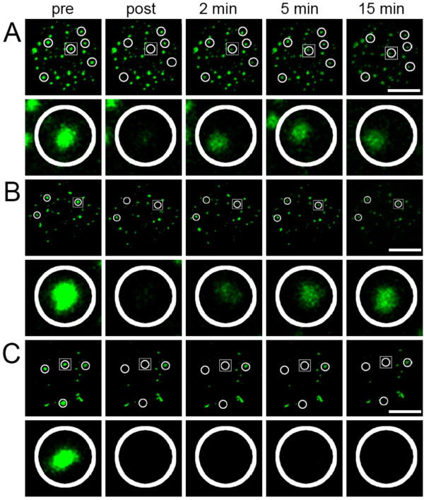
Figure 2
FRAP to determine exchange of PML isoforms at nuclear bodies . (A) FRAP experiment of GFP-tagged PML II in 3T3-PML +/+ cells. Fluorescence was bleached in circled areas containing a nuclear body as in...
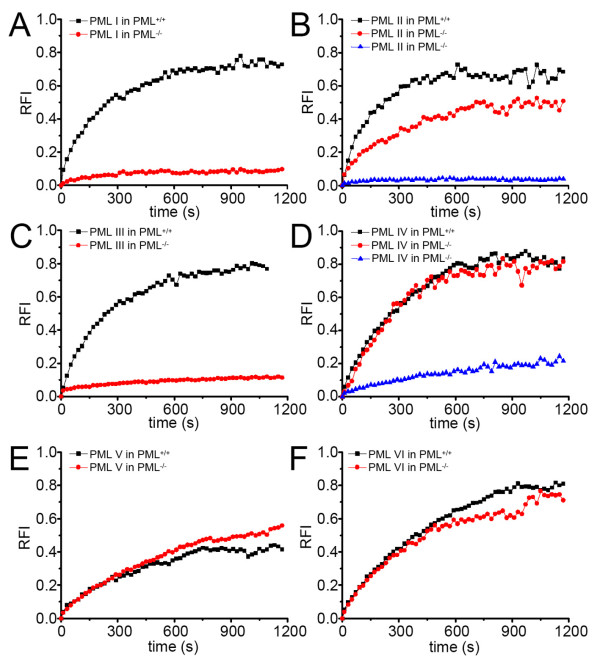
Figure 3
Quantitation of FRAP experiments . FRAP experiments as described in Figure 2 were performed for all GFP-PML isoforms in PML positive (black curves) and PML negative cells (red curves) as indicated. A ...
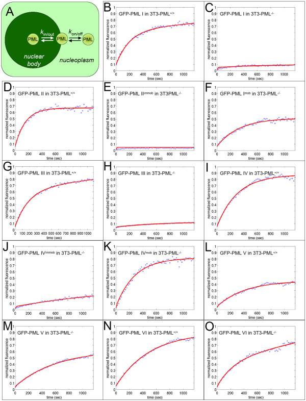
Figure 4
Kinetic model for molecule exchange at PML nuclear bodies . (A) Molecules with the potential to interact with PML nuclear bodies move by diffusion in the nucleoplasm outside nuclear bodies. Upon stoch...
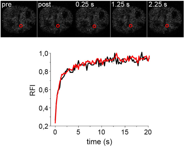
Figure 5
PML SUMOylation sites K160 and K490 are required for PML body formation . The upper part shows a FRAP experiment of GFP-tagged mutant PML IV (K160/490R) in 3T3-PML -/- cells. Fluorescence was bleached...
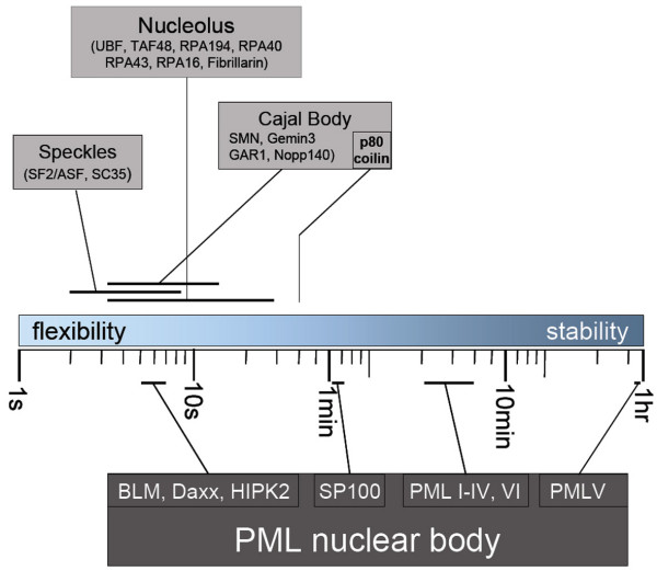
Figure 6
Assembly dynamics of subnuclear domains . The residence times of components of the indicated subnuclear domains are depicted on a logarithmic scale between 1 second and 1 hour. Fast or slow exchanging...
Figure images are served from the NIH/NLM PubMed Central Open Access Subset or Europe PMC; copyright remains with the publishers and authors.
💬 Discussion
0 commentsNo comments yet. Be the first to start a discussion!
Leave a Comment