Abstract
Accelerating imaging speed in optical microscopy is often realized at the expense of image contrast, image resolution, and detection sensitivity--a common predicament for advancing high-speed and high-throughput cellular imaging. We here demonstrate a new imaging approach, called asymmetric-detection time-stretch optical microscopy (ATOM), which can deliver ultrafast label-free high-contrast flow imaging with well delineated cellular morphological resolution and in-line optical image amplification to overcome the compromised imaging sensitivity at high speed. We show that ATOM can separately reveal the enhanced phase-gradient and absorption contrast in microfluidic live-cell imaging at a flow speed as high as ~10 m/s, corresponding to an imaging throughput of ~100,000 cells/sec. ATOM could thus be the enabling platform to meet the pressing need for intercalating optical microscopy in cellular assay, e.g. imaging flow cytometry--permitting high-throughput access to the morphological information of the individual cells simultaneously with a multitude of parameters obtained in the standard assay.
🔬 Techniques
💻 Software
🧪 Sample Preparation
🔬 Cell Lines
🧪 Reagent Suppliers
💻 Software Details
🏛️ Research Organizations (ROR)
Affiliated research institutions:
📋 Methods
Illumination and imaging optics of ATOM The pulsed beam of a home-built ytterbium-doped mode-locked laser (repetition rate = 26 MHz; center wavelength = 1064 nm) with a 3-dB bandwidth of ~10 nm and a pulse width of 4 ps is spatially dispersed in 1D by a transmission holographic grating (1200 lines/mm) to generate a spectral shower which is then focused by an objective lens (numerical aperture (NA) = 0.66) for illumination. Another identical objective lens (NA = 0.66) and a mirror are added behind the sample in order to operate ATOM in a double-pass transmission mode ( Fig. 2(a) ). The double-passed spectral shower is then collected by a dispersive fiber using a fiber collimator lens (NA = 0.25). Time multiplexing the time-stretch temporal signals The double-passed spectrally-encoded light is split into two different paths by a beam splitter (power splitting ratio = 45:55). One of the beams (beam A in Fig. 2(a) ) is incident to the fiber collimator lens with an angle range of +4°, whereas the other beam is time-delayed by ~3.6 ns with respect to beam A and is incident to the same fiber collimator lens with an angle range of −4° (beam B in Fig. 2(a) ) The tilting angles of the two beams are independently controlled by the two steering mirrors, as shown in Fig. 2(a) . The imaging region of interest is mainly near the center of the microfluidic channel, within a size of ~30 μm (i.e. the cell size in most of our experiments), which corresponds to a wavelength bandwidth of ~5 nm and thus a temporal width of ~1.75 ns (with GVD = 0.35 ns/nm). Therefore, a time delay of 3.6 ns guarantees no temporal overlap between the two pulses. The pulses are then time-stretched within a dispersive fiber module, consisting of a 5-km single-mode fiber (SMF) in 1 μm (Nufern) and a standard telecommunication SMF (Corning, SMF28), which acts as a few-mode fiber 22 . The total GVD achieved is ~0.35 ns/nm. The time-stretch pulses are also amplified by a fiber-based SOA (Superlum) which achieves an on-off gain as high as ~500. Finally, the signal is detected by a photo-detector (Picometrix, electrical bandwidth: 8 GHz) and a real-time oscilloscope (Agilent Technologies, sampling rate: 40 GS/s). Having a bandwidth of 10 nm, the stretched pulse has a temporal width of ~4 ns (with GVD = 0.35 ns/nm). Therefore, given the laser repetition rate of 26 MHz (i.e. a period of ~40 ns), the line-scan duty cycle in the current ATOM system is ~2 × (4/40)% = 20%. The factor of 2 refers to the two time-multiplexed replicas.
Show full methods section
Illumination and imaging optics of ATOM The pulsed beam of a home-built ytterbium-doped mode-locked laser (repetition rate = 26 MHz; center wavelength = 1064 nm) with a 3-dB bandwidth of ~10 nm and a pulse width of 4 ps is spatially dispersed in 1D by a transmission holographic grating (1200 lines/mm) to generate a spectral shower which is then focused by an objective lens (numerical aperture (NA) = 0.66) for illumination. Another identical objective lens (NA = 0.66) and a mirror are added behind the sample in order to operate ATOM in a double-pass transmission mode ( Fig. 2(a) ). The double-passed spectral shower is then collected by a dispersive fiber using a fiber collimator lens (NA = 0.25). Time multiplexing the time-stretch temporal signals The double-passed spectrally-encoded light is split into two different paths by a beam splitter (power splitting ratio = 45:55). One of the beams (beam A in Fig. 2(a) ) is incident to the fiber collimator lens with an angle range of +4°, whereas the other beam is time-delayed by ~3.6 ns with respect to beam A and is incident to the same fiber collimator lens with an angle range of −4° (beam B in Fig. 2(a) ) The tilting angles of the two beams are independently controlled by the two steering mirrors, as shown in Fig. 2(a) . The imaging region of interest is mainly near the center of the microfluidic channel, within a size of ~30 μm (i.e. the cell size in most of our experiments), which corresponds to a wavelength bandwidth of ~5 nm and thus a temporal width of ~1.75 ns (with GVD = 0.35 ns/nm). Therefore, a time delay of 3.6 ns guarantees no temporal overlap between the two pulses. The pulses are then time-stretched within a dispersive fiber module, consisting of a 5-km single-mode fiber (SMF) in 1 μm (Nufern) and a standard telecommunication SMF (Corning, SMF28), which acts as a few-mode fiber 22 . The total GVD achieved is ~0.35 ns/nm. The time-stretch pulses are also amplified by a fiber-based SOA (Superlum) which achieves an on-off gain as high as ~500. Finally, the signal is detected by a photo-detector (Picometrix, electrical bandwidth: 8 GHz) and a real-time oscilloscope (Agilent Technologies, sampling rate: 40 GS/s). Having a bandwidth of 10 nm, the stretched pulse has a temporal width of ~4 ns (with GVD = 0.35 ns/nm). Therefore, given the laser repetition rate of 26 MHz (i.e. a period of ~40 ns), the line-scan duty cycle in the current ATOM system is ~2 × (4/40)% = 20%. The factor of 2 refers to the two time-multiplexed replicas.
Image processing of ATOM
The individual time-stretch waveforms are first subtracted and normalized by the background pulse (which has the spectral shape of the laser source). The final 2D ATOM image is obtained by digitally stacking all the pulses, i.e. the line scans, along the flow direction. The differential phase-gradient and absorption contrasts are further obtained by calculating the difference and sum between the two neighboring time-stretch waveforms, (having opposite phase-gradient contrasts), respectively. The digital signal processing and image reconstruction are done off-line by a custom program in MATLAB.
Imaging the fixed MIHA cells with ATOM
For the ATOM images shown in Fig. 3 , the MIHA cells are fixed on a glass slide, which is scanned perpendicular to the spectral shower direction during imaging by ATOM. The MIHA sample is scanned for 70 lines with 1 μm step size by a mechanical scanning stage. 2D images are obtained by digitally stacking the 1D line-scans. Averaging of 25 sequential line-scans is taken for realizing an effective single-shot line-scan rate of 1 MHz. Microfluidic channel design and fabrication We designed and fabricated a PDMS microfluidic channel platform in which the balance between the inertial lift force and the viscous drag force is achieved for manipulating the positions of the individual cells and focusing them in ultrafast flow inside the channel. This microfluidic technique is essential for ensuring robust imaging by ATOM at the record high microfluidic flow speed (as high as ~10 m/s). Detailed fabrication steps are depicted in Supplementary Fig. S3 . The microfluidic platform consists of two parts: a focusing section followed by an imaging section (see Supplementary Fig. S4(a) ). The focusing section consists of multiple pairs of connected curved channels with radii of curvature 400 μm and 1000 μm, respectively. There are 16 turns in total. (see Supplementary Fig. S4(a) ). The width (150 μm) and height (50 μm) of the channel were chosen such that the channel is suitable for focusing cells with a size ranging from ~5–30 μm. In the imaging section in which the spectral shower is illuminated onto the channel, the channel width is narrowed to 45 μm to further boost the flow speed. Note that laminar flow condition is still satisfied at such an ultrafast flow. The Reynolds number of our current microfluidic channel design is at 600, which is far below the limit of 2000, beyond which the turbulence flow occurs 32 . The thicknesses of the top and bottom channel walls have been minimized to accommodate the high NA objective lens, which typically has a working distance of less than 1 mm (see Supplementary Fig. S4(b) ). Preparation of mammalian cell lines – MIHA, BEL-7402 and HeLa cells The BEL-7402 cells, HeLa cells, and MIHA cells were cultured on 100 mm cell culture dish (Corning) in DMEM-HG supplemented with 10% fetal bovine serum (FBS), 100 U/ml penicillin and 100 μg/ml streptomycin. Sodium pyruvate was added to the culture media when culturing MIHA cells. Cells were grown in a humidified incubator at 37°C and 5% CO 2 . After reaching confluence, cells in cell culture dish were trypsinized to suspend in aqueous environment. Trypsin was removed by centrifuging the cell sample at 250 g for 5 minutes. Glutaraldehyde was added to re-suspend the cells and fix the cells. Glutaraldehyde was removed by centrifuging the cell sample by 250 g for 5 minutes. Phosphate buffered saline (PBS) was added to re-suspend the cells which are then loaded to the microfluidic channel for ATOM experiments.
Preparation of cell lines
(THP-1), and human whole blood samples THP-1, a human monocytic cell line obtained from patients with acute monocytic leukemia, was cultured in Roswell Park Memorial Institute (RPMI) medium 1640 (Gibco) supplemented with 10% fetal bovine serum (Hyclone, Thermo scientific), penicillin streptomycin (Gibco), 2 mM Glutamax™ (Gibco) at 37°C, 95% humidity and 5% CO 2 until confluence was achieved. Optimal conditions were maintained until the cells were utilized for the experiment. Fresh blood was collected from the right median cubital artery of a healthy donor amounting to 3 mililitres and kept at 2–8°C in an ethylene diamine tetraacetic acid (EDTA) anticoagulated evacuated tube (Greiner Bio-one). Blood was drawn at least eight hours prior to the experiment.
Supplementary Material Supplementary Information Asymmetric-detection time-stretch optical microscopy (ATOM) for ultrafast high-contrast cellular imaging in flow
📊 Figures
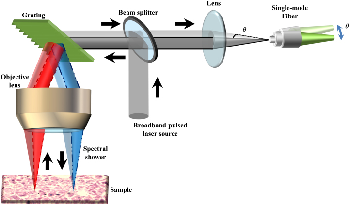
Figure 1
Key approach of enabling phase-gradient contrast in ATOM.
In a typical configuration of time-stretch optical microscopy, the spatial information of the specimen is first mapped to the spectrum of a broadband laser pulsed beam by using a diffraction grating t...
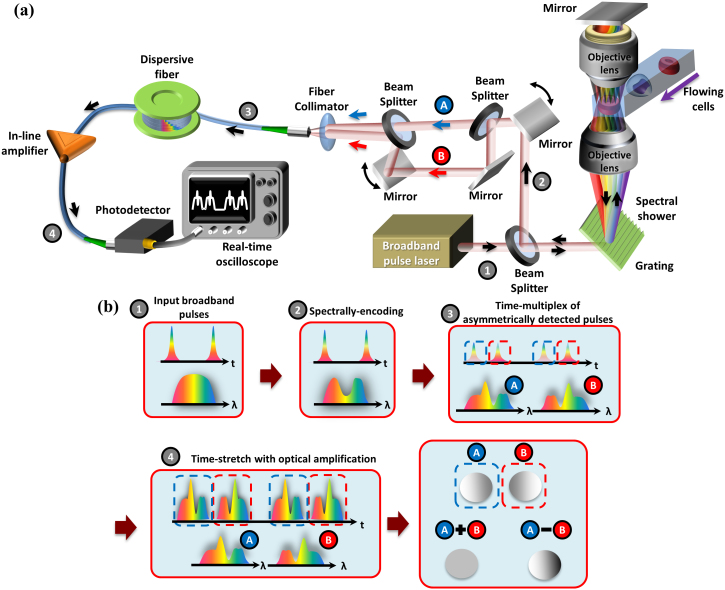
Figure 2
General schematic of an ATOM system.
(a) A broadband light pulsed beam (The temporal pulse train and the corresponding spectrum are shown in box 1 of (b)) is first spatially dispersed by a diffraction grating to generate a 1D spectral sh...
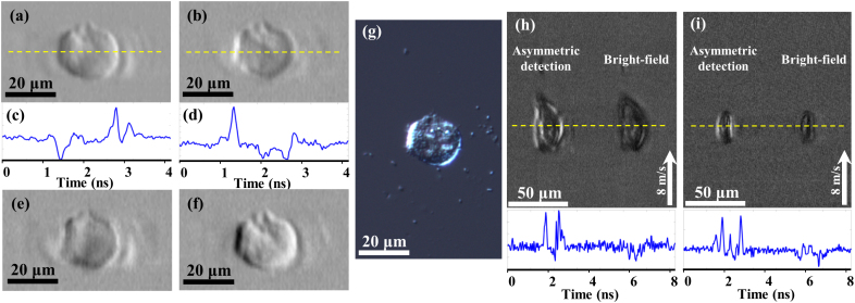
Figure 3
Basic performance of ATOM.
(a, b) Two single-angle ATOM images of a MIHA cells fixed on a glass slide, which show the opposite phase-gradient contrasts, respectively. (c, d) The corresponding line profiles (yellow dotted lines)...
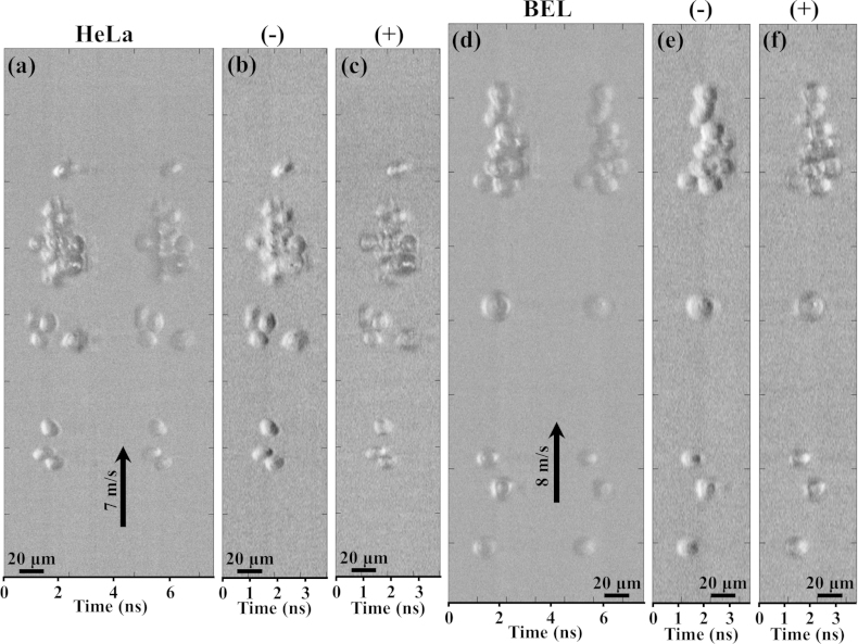
Figure 4
Imaging of mammalian cells by ATOM in ultrafast flow.
(a) Time-multiplexed single-angle ATOM images of stain-free HeLa cells flowing at a speed of 7u2005m/s. In this time-multiplexed image, it appears that two groups of identical cells are flowing in par...
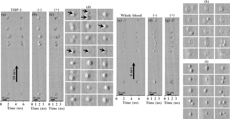
Figure 5
Imaging of stain-free THP-1 and normal human blood cells (from whole blood) by ATOM in ultrafast flow.
(a) Time-multiplexed single-angle ATOM images of the stain-free THP-1 cells flowing at a speed of 10u2005m/s. (b) Dual-angle (differential) ATOM image obtained by subtraction of the two opposite-contr...
Figure images are served from the NIH/NLM PubMed Central Open Access Subset or Europe PMC; copyright remains with the publishers and authors.
💬 Discussion
0 commentsNo comments yet. Be the first to start a discussion!
Leave a Comment