Abstract
The synthesis quality of artificial inorganic nanocrystals is most often assessed by transmission electron microscopy (TEM) for which high-throughput advances have dramatically increased both the quantity and information richness of metal nanoparticle (mNP) characterization. Existing automated data analysis algorithms of TEM mNP images generally adopt a supervised approach, requiring a significant effort in human preparation of labeled data that reduces objectivity, efficiency, and generalizability. We have developed an unsupervised algorithm AutoDetect-mNP for automated analysis of TEM images that objectively extracts morphological information on convex mNPs from TEM images based on their shape attributes, requiring little to no human input in the process. The performance of AutoDetect-mNP is tested on two data sets of bright field TEM images of Au nanoparticles with different shapes and further extended to palladium nanocubes and cadmium selenide quantum dots, demonstrating that the algorithm is quantitatively reliable and can thus serve as a generalizable measure of the morphology distributions of any mNP synthesis. The AutoDetect-mNP algorithm will aid in future developments of high-throughput characterization of mNPs and the future advent of time-resolved TEM studies that can investigate reaction mechanisms of mNP synthesis and reactivity.
🔬 Techniques
🔭 Microscopes
💻 Software
✨ Fluorophores
🧪 Sample Preparation
🏭 Microscope Brands
🧪 Reagent Suppliers
💻 Software Details
💾 Data Repositories
🏛️ Research Organizations (ROR)
Affiliated research institutions:
📋 Methods
Chemicals and Materials
Hexadecyltrimethylammonium bromide (CTAB, >98.0%) and sodium oleate (NaOL, >97.0%) were purchased from TCI America. Hydrogen tetrachloroaurate trihydrate (HAuCl 4 ·3H 2 O, ≥99.9%), l -ascorbic acid (BioXtra, ≥99.0%), silver nitrate (AgNO 3 , ≥99.0%), sodium borohydride (NaBH 4 , 99.99%), sodium iodide (NaI, ≥99.5%), sodium hydroxide (NaOH, ≥ 97.0%), sodium citrate tribasic dihydrate (≥99.0%), and hydrochloric acid (36.5–38.0 wt %%) were obtained from Sigma-Aldrich (USA). NaBH 4 powder was stored in an argon glovebox. HAuCl 4 ·3H 2 O, l -ascorbic acid, and AgNO 3 were stored in a vacuum desiccator at room temperature. Hexadecyltrimethylammonium chloride (CTAC, >95.0%) was purchased from TCI America. Palladium(II) chloride (PdCl 2 , 99%) and potassium iodide (KI, >99.0%) were obtained from Sigma-Aldrich (USA). Cadmium oxide (CdO) 99.99% Aldrich; oleic acid 90% (OA) technical grade Aldrich; oleylamine (OAm) technical grade 70% Aldrich; octadecene 90% (ODE) technical grade Aldrich; trioctylphosphine oxide (TOPO) 99% Aldrich; sulfur Aldrich; octadecylphosphonic acid (ODPA) 99%, PCI Synthesis; n -trioctylphosphine 97% (TOP) Strem; 0.2 M Cd(oleate) 2 in ODE was prepared by degassing appropriate amounts of CdO, OA, and ODE under vacuum at 110 °C at until all gases and water had evolved. The flask was switched to argon and heated to 240 °C until a clear (slightly yellow) solution solution was obtained. The flask was then cooled to 110 °C and degassed a second time to remove additional water. Deionized water (DI-water, Milipore, Milford, MA, USA) was used for all aqueous solution. All the glassware used for the synthesis of Au NPs was thoroughly cleaned using freshly prepared aqua regia (3:1 volume ratio of HCl and HNO 3 , respectively) followed by fully rinsing with copious amounts of DI-water. All chemicals were of reagent grade and used without further purification unless specified otherwise.
Show full methods section
Chemicals and Materials
Hexadecyltrimethylammonium bromide (CTAB, >98.0%) and sodium oleate (NaOL, >97.0%) were purchased from TCI America. Hydrogen tetrachloroaurate trihydrate (HAuCl 4 ·3H 2 O, ≥99.9%), l -ascorbic acid (BioXtra, ≥99.0%), silver nitrate (AgNO 3 , ≥99.0%), sodium borohydride (NaBH 4 , 99.99%), sodium iodide (NaI, ≥99.5%), sodium hydroxide (NaOH, ≥ 97.0%), sodium citrate tribasic dihydrate (≥99.0%), and hydrochloric acid (36.5–38.0 wt %%) were obtained from Sigma-Aldrich (USA). NaBH 4 powder was stored in an argon glovebox. HAuCl 4 ·3H 2 O, l -ascorbic acid, and AgNO 3 were stored in a vacuum desiccator at room temperature. Hexadecyltrimethylammonium chloride (CTAC, >95.0%) was purchased from TCI America. Palladium(II) chloride (PdCl 2 , 99%) and potassium iodide (KI, >99.0%) were obtained from Sigma-Aldrich (USA). Cadmium oxide (CdO) 99.99% Aldrich; oleic acid 90% (OA) technical grade Aldrich; oleylamine (OAm) technical grade 70% Aldrich; octadecene 90% (ODE) technical grade Aldrich; trioctylphosphine oxide (TOPO) 99% Aldrich; sulfur Aldrich; octadecylphosphonic acid (ODPA) 99%, PCI Synthesis; n -trioctylphosphine 97% (TOP) Strem; 0.2 M Cd(oleate) 2 in ODE was prepared by degassing appropriate amounts of CdO, OA, and ODE under vacuum at 110 °C at until all gases and water had evolved. The flask was switched to argon and heated to 240 °C until a clear (slightly yellow) solution solution was obtained. The flask was then cooled to 110 °C and degassed a second time to remove additional water. Deionized water (DI-water, Milipore, Milford, MA, USA) was used for all aqueous solution. All the glassware used for the synthesis of Au NPs was thoroughly cleaned using freshly prepared aqua regia (3:1 volume ratio of HCl and HNO 3 , respectively) followed by fully rinsing with copious amounts of DI-water. All chemicals were of reagent grade and used without further purification unless specified otherwise.
Synthesis of Gold Nanorods
Two types of AuNRs were synthesized by a facile seed-mediated growth involving a binary surfactant mixture. The seed solution was prepared as follows: 10 mL of 0.1 M CTAB solution was mixed with 100 μL of 25 mM HAuCl 4 in a 20 mL scintillation vial under vigorous stirring. A 600 μL portion of ice cooled 10 mM NaBH 4 was rapidly injected into the Au-CTAB solution and stirred for 2 min. Upon the addition of NaBH 4 , the color of the seed solution turned yellow-brownish. Afterward, the seed solution was left undisturbed at 28 °C for 30 min prior to use in the following step. The growth solution was obtained by first mixing 3.6 g of CTAB and 0.4936 g of NaOL in 196 mL of DI-water in a 500 mL Erlenmeyer flask. The solution was heated with occasional agitation until all the CTAB was dissolved. The mixture was allowed to cool down to 30 °C and 4 mM AgNO 3 referred to Table 2 was then added under stir at 700 rpm for 15 min. Afterward, 4 mL of 25 mM HAuCl 4 was added to the mixture and kept undisturbed at 28 °C for 90 min. The yellowish color of the growth solution turned to colorless. A certain amount of HCl ( Table 2 ) was added to the solution, and the mixture was stirred at 400 rpm for 15 min. Finally, 500 μL of 0.064 M ascorbic acid was injected into the growth solution, and the mixture was vigorously stirred at 1200 rpm for 30 s. An 80 μL portion of the seed solution was then injected, and the solution was stirred for 30 s before it was left undisturbed at 28 °C for 12 h to complete the growth process. A 40 mL portion of the final products were isolated by centrifugation at 8000 rpm for 15 min followed by careful removal of the supernatant. A 10 mL portion of DI-water was added to the pellet, and the mixture was sonicated briefly to disperse the pellet. Table 2 Amount of Reagents Used for the Synthesis of Au Nanorods sample 4 mM AgNO 3 (mL) HCl (mL) long rods 1.45 0.84 short rods 9.6 1.2 Synthesis of Gold Triangular Prisms Homogeneous gold triangular prisms were also synthesized by seed-mediated method. The citrate ligand based seed solution was prepared as follows: 500 μL of 10 mM sodium citrate solution was mixed with 250 μL of 10 mM HAuCl 4 and 18.95 mL of DI-water in a 20 mL scintillation vial under vigorous stirring. A 300 μL portion of ice cooled 10 mM NaBH 4 was rapidly injected into the Au-citrate solution and stirred for 1 min. Upon the addition of NaBH 4 , the color of the seed solution turned yellow-brownish. Afterward, the seed solution was stirred at 40–45 °C for 15 min and left undisturbed at room temperature prior to use in the following step. For the growing process, the UV–vis spectra of the seed solution were taken using 1 cm quartz cuvette to determine the concentration of the seed. The extinction coefficient of the seed solution is 9.696 × 10 6 M –1 cm –1 at its wavelength of maximum optical density, 504 nm. In order to synthesize the triangular prism with edge length in 80 nm, the final concentration of the seed in the growth solution should be 97.7 pM. Prior to preparing the growth solution, the stock mixture of 0.05 M CTAB and 50 μM NaI was prepared at room temperature. The solution was heated with occasional agitation until all the CTAB was dissolved. The mixture was allowed to cool down to 30 °C. The growth solution was prepared by mixing 9 mL of previously made CTAB/NaI stock mixture, 250 μL of 10 mM HAuCl 4 solution, 50 μL of 100 mM NaOH solution, and 50 μL of 100 mM ascorbic acid solution. Finally, a certain amount of seed solution was added under vigorous stirring. The nanoparticle solution was then heated around 40 °C for 30 min and was cooled down to room temperature. For the purification process of Au nanoprisms, 0.6 mL of the growing solution was mixed with 0.4 mL of 1 M NaCl solution into 1.5 mL Eppendorf tubes. The mixture was left undisturbed for 4 h and centrifuged twice at 1000 rpm for 15 s. The supernatant was carefully removed, and 0.6 mL of DI-water was added to the pellet and was sonicated briefly to disperse the pellet.
Synthesis of Pd Nanocubes
Palladium nanocubes were synthesized with a method similar to a previous report. 52 A stock solution of 10 mM H 2 PdCl 4 was prepared by dissolving 0.1773 g of PdCl 2 in 10 mL of 200 mM HCl. A 50 mL aqueous growth solution was prepared in a 100 mL round-bottom flask containing 12.5 mM CTAC and 2 mM KBr, and 2.5 mL of 10 mM H 2 PdCl 4 was added to this solution. The solution was heated to 95 °C and held at this temperature for 5 min. The solution was stirred with a magnetic stir-bar, and subsequently 400 μL of 100 mM l -ascorbic acid was injected into the solution with a micropipette to initiate growth. The reaction was allowed to proceed for 30 min at 95 °C to complete the growth process. The solution turned into a dark brown color during the growth process. The final products were cooled to room temperature and isolated by centrifugation at 8000 rpm for 15 min followed by removal of the supernatant. The resulting pellet was dispersed in 5 mL of water by sonication. Synthesis of CdSe/CdS QDs CdSe cores were synthesized using a modified version of a previously published procedure. 53 Typically, 120 mg of CdO, 560 mg of ODPA, and 6 g of TOPO were combined and degassed at 150 °C under vacuum for 1 h. The reaction was then heated to 320 °C under argon and held at that temperature until the solution turned clear, indicative of Cd-ODPA complexation. At 320 °C, 3 g of TOP was injected, and then the solution was heated to 360 °C, at which point a solution of Se (120 mg) dissolved in TOP (0.72 g) was quickly injected. The reaction was stopped after approximately 4 min and quickly cooled. The QDs were purified from free ligand and excess precursors via precipitation with acetone and redispersion in hexanes several times. Sizing and concentrations were determined using previously established calibration curves. 54 Samples were prepared according to the work of Ondry et al. 55 Samples with a nominal shell thickness of 6 monolayers were synthesized as follows. A 100 nmol portion of wurtzite CdSe seeds (570 nm first exciton), 3 mL ODE, and 3 mL OAm were loaded into a 50 mL three-neck, round-bottom flask and degassed at 110 °C for ∼30 min. Under Ar, the reaction was heated to 310 °C. At 240 °C, slow injection of a 3.14 mL of 0.2 M Cd(oleate) 2 in ODE solution and 3.14 mL of 0.2 M TOP-S solution in TOP in separate syringes commenced at a rate of 1 mL/h. TOP-S in TOP was prepared by stirring 20 mg of S in 2.6 g TOP in a glovebox for ∼30 min. After the injection completed, the reaction was kept at 310 °C for 10 min and then rapidly cooled to room temperature. Nanocrystals were isolated from the reaction by precipitating the nanocrystals with acetone and redissolving in hexanes 2×. Finally the nanocrystals were centrifuged at 8000 rpm in hexanes to remove any remaining insoluble impurities.
Preparation of TEM Samples
To prepare the stock sample of Au NPs for TEM analysis, a 50 μL aliquot of the nanoparticle solution was added to 1 mL of DI-water. The sample was centrifuged at 5500 rpm for 8 min, the supernatant was carefully removed, and the isolated product was resuspended in 50 μL of DI-water. We added 200 μL of DI-water to the sample to dilute by a factor of 5 to prevent overlapping particles on the grid. A 5 μL portion of this solution was pipetted onto a standard carbon TEM grid (Electron Microscopy Sciences, CF-400-Cu). The TEM grid with the sample was fully dried in a vacuum desiccator at room temperature before TEM imaging. In order to calculate the concentration of Au nanorods, we referred to the theoretical extinction coefficient of the AuNR reported by Park et al. 56 Each extinction coefficient of long and short AuNR was extracted to 1.4 × 10 10 L mol –1 cm –1 at 1059 nm for long rods, and 1.1 × 10 10 L mol –1 cm –1 at 698 nm for short rods. The optical density of the solution was collected using a Shimadzu UV-3600 UV–vis spectrophotometer with 1 nm resolution. The path length of the cuvette was 1 cm. To make a 50:50 ratio of short and long AuNRs, the concentration of each sample was fixed to 90 pM. Pd nanocubes samples for TEM analysis were prepared by pipetting 1 μL of the nanocube solution onto a standard carbon TEM grid (Electron Microscopy Sciences, CF-400-CU). The sample was fully dried in a vacuum desiccator before being imaged. CdSe/CdS QD samples were prepared at the liquid–air interface. 57 Briefly, 1 mL of anhydrous DMF was placed in a 1 cm 2 square Teflon well. Next CdSe/CdS QDs were diluted in octane to an appropriate concentration to achieve monolayer coverage and 100 μL of the diluted nanocrystal solution in octane was floated on top of the DMF layer. The well was covered with a glass slide to slow solvent evaporation and was allowed to sit for at least 8 h. The samples were transferred via scooping from below to an amorphous carbon coated TEM grid for microscopy. Samples were placed in a vacuum oven under house vacuum at ∼50 °C to remove any remaining subphase solvent.
TEM Imaging
Images of Au NPs were acquired using a FEI Tecnai T20 transmission electron microscope equipped with a Gatan RIO16IS camera and a LaB 6 filament. All images were recorded under 200 kV accelerating voltage. Drift correction feature of the imaging software was enabled during the acquisition of all images. For each sample, images were taken from a series of evenly spaced locations on the sample grid, typically resulting in 50–100 images of the sample containing >1000 recognizable NPs. Experimental parameters during the imaging process can impact the performance of the algorithm. The efficiency of particle detection can depend on the resolutions and contrasts of the TEM images. Therefore, optimizing experimental parameters during imaging to improve image resolution and contrast is recommended. We observed that magnification and camera exposure time are two of the most important imaging parameters to fine-tune. Matlab codes for real-time image analysis during imaging are included in the Github repository for AutoDetect-mNP to help the users optimize experimental parameters during imaging. For the data showed in this work, long rods were imaged at 43k× magnification with 1 s exposure time; short rods were imaged at 19k× magnification with 1 s exposure time; mixtures were imaged at 43k× magnification with 1 s exposure time; triangular prisms were imaged at 26k× magnification with 1.5 s exposure time. AutoDetect-mNP Algorithm Unprocessed TEM images in dm4 format were loaded into Matlab by the ReadDMFile function available from MathWorks file exchange. Particle detection was performed by Matlab’s built-in function imsegkmeans . Imsegkmeans segments the image by performing a K -means clustering on the pixel intensities of each image. Shape descriptors for each particles were calculated by the regionprops function in Matlab. The definition for each shape descriptors is as follows: Area is defined as the total area, in squared nanometers, of the region encompassed by the edge of the particle; eccentricity is defined as the eccentricity of the smallest ellipse that encapsulates the particle; aspect ratio is defined as the ratio between the major and minor axis lengths of the particle; circularity is defined as the reciprocal of the first Hu’s moment invariant of the particle; 43 , 44 solidity and convexity are defined as the ratio of the areas and perimeters, respectively, between the particle and the smallest convex polygon that encapsulates the particle. Hu’s moment invariants of the particles were calculated by codes available from the MathWorks file exchange. UECS was implemented using Matlab codes adapted from those published by Park et al. (available from https://aml.engr.tamu.edu/2001/09/01/publications/ ). 30 The UECS algorithm attempts to resolve convex components from nonconvex shapes by iteratively eroding the shapes until convex shapes are obtained. If convex shapes are not obtained after 90% of the area of the original particle has been eroded, the particle will be discarded. Each convex-shaped marker obtained at the end of UECS iterations was then dilated for the same number of times as it has been eroded to recover its original shape. K -means clustering and naive Bayes classifier used for unsupervised classification are well established clustering algorithms and are implemented in Matlab. An example demonstrating using P max ( K ) to decide the optimal number of classes is shown in Figure 6 . In Figure 6 , circularity of the Au nanorods was used as an example to demonstrate the selection of optimal K . In this case, K = 3 is selected as the optimal K for this data set, which agrees with the observation that the data set roughly consists of three normal distributions. Of note, particles resolved by UECS were excluded during the automated classification process to avoid skewing of classification results by potential artifacts generated by UECS. Instead, these particles were assigned classes after the normal particles are clustered into different classes. Figure 6 Unsupervised clustering of extracted features and selection of optimal number of clusters ( K ), using circularity of Au nanorods as an example.
Chemicals and Materials
Hexadecyltrimethylammonium bromide (CTAB, >98.0%) and sodium oleate (NaOL, >97.0%) were purchased from TCI America. Hydrogen tetrachloroaurate trihydrate (HAuCl 4 ·3H 2 O, ≥99.9%), l -ascorbic acid (BioXtra, ≥99.0%), silver nitrate (AgNO 3 , ≥99.0%), sodium borohydride (NaBH 4 , 99.99%), sodium iodide (NaI, ≥99.5%), sodium hydroxide (NaOH, ≥ 97.0%), sodium citrate tribasic dihydrate (≥99.0%), and hydrochloric acid (36.5–38.0 wt %%) were obtained from Sigma-Aldrich (USA). NaBH 4 powder was stored in an argon glovebox. HAuCl 4 ·3H 2 O, l -ascorbic acid, and AgNO 3 were stored in a vacuum desiccator at room temperature. Hexadecyltrimethylammonium chloride (CTAC, >95.0%) was purchased from TCI America. Palladium(II) chloride (PdCl 2 , 99%) and potassium iodide (KI, >99.0%) were obtained from Sigma-Aldrich (USA). Cadmium oxide (CdO) 99.99% Aldrich; oleic acid 90% (OA) technical grade Aldrich; oleylamine (OAm) technical grade 70% Aldrich; octadecene 90% (ODE) technical grade Aldrich; trioctylphosphine oxide (TOPO) 99% Aldrich; sulfur Aldrich; octadecylphosphonic acid (ODPA) 99%, PCI Synthesis; n -trioctylphosphine 97% (TOP) Strem; 0.2 M Cd(oleate) 2 in ODE was prepared by degassing appropriate amounts of CdO, OA, and ODE under vacuum at 110 °C at until all gases and water had evolved. The flask was switched to argon and heated to 240 °C until a clear (slightly yellow) solution solution was obtained. The flask was then cooled to 110 °C and degassed a second time to remove additional water. Deionized water (DI-water, Milipore, Milford, MA, USA) was used for all aqueous solution. All the glassware used for the synthesis of Au NPs was thoroughly cleaned using freshly prepared aqua regia (3:1 volume ratio of HCl and HNO 3 , respectively) followed by fully rinsing with copious amounts of DI-water. All chemicals were of reagent grade and used without further purification unless specified otherwise.
Supplementary Material au0c00030_si_001.pdf
📊 Figures
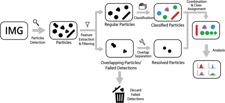
Figure 1
Schemenof the AutoDetect-mNP algorithm. The algorithm can be dividedninto four parts: particle detection, feature extraction, filteringnand resolution of irregularly shaped particles, and classificati...
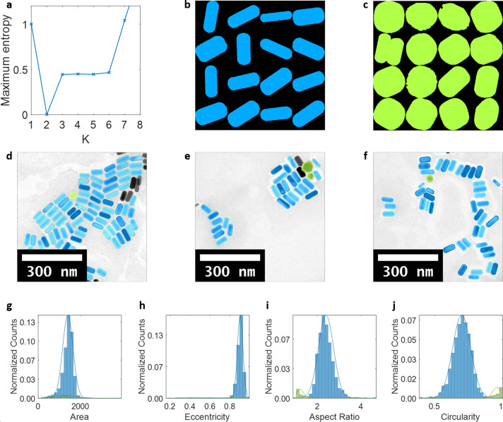
Figure 2
Detection and classification of Au NPsnof different morphologiesnin short rods. (a) Maximum entropy as a function of the number ofnclasses in which K = 2 was found to be the optimalnnumber of classes....
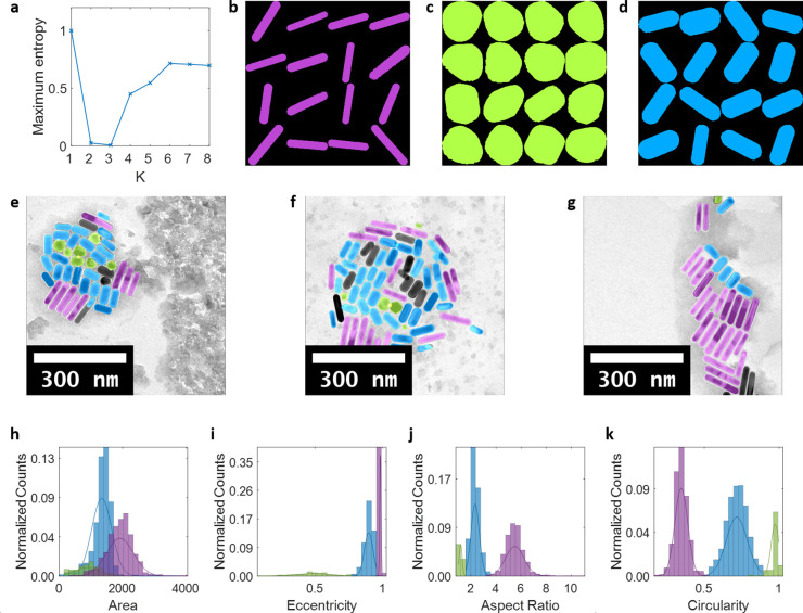
Figure 3
Classificationnresults of a 1:1 mixture of Au nanorods with differentnaspect ratios. (a) Maximum entropy as a function of the number ofnclasses in which K = 3 was found to be the optimalnnumber of cla...
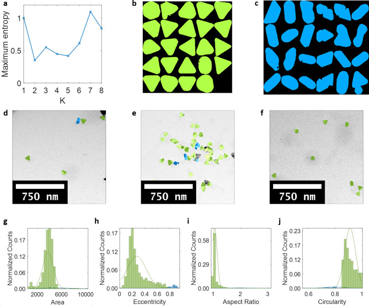
Figure 4
Detection and classificationnof Au NPs of different morphologiesnin a sample of triangular prisms. (a) Maximum entropy as a functionnof the number of classes in which K = 2 was foundnto be the optimal...
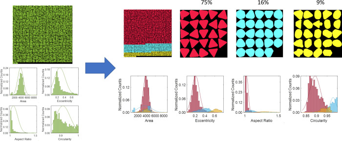
Figure 5
Further classification of the Au triangular prisms class. Distributionsnand relative population of particles in each class (red pure triangles,ncyan symmetrically truncated triangles, yellow asymmetri...
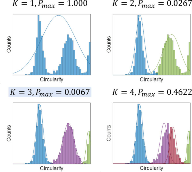
Figure 6
Unsupervisednclustering of extracted features and selection ofnoptimal number of clusters ( K ), using circularitynof Au nanorods as an example.
Figure images are served from the NIH/NLM PubMed Central Open Access Subset or Europe PMC; copyright remains with the publishers and authors.
💬 Discussion
0 commentsNo comments yet. Be the first to start a discussion!
Leave a Comment