Abstract
Abstract Introduction Analysis of autoantibodies (AAB) by indirect immunofluorescence (IIF) is a basic tool for the serological diagnosis of systemic rheumatic disorders. Automation of autoantibody IIF reading including pattern recognition may improve intra- and inter-laboratory variability and meet the demand for cost-effective assessment of large numbers of samples. Comparing automated and visual interpretation, the usefulness for routine laboratory diagnostics was investigated. Methods Autoantibody detection by IIF on human epithelial-2 (HEp-2) cells was conducted in a total of 1222 consecutive sera of patients with suspected systemic rheumatic diseases from a university routine laboratory (n = 924) and a private referral laboratory (n = 298). IIF results from routine diagnostics were compared with a novel automated interpretation system. Results Both diagnostic procedures showed a very good agreement in detecting AAB (kappa = 0.828) and differentiating respective immunofluorescence patterns. Only 98 (8.0%) of 1222 sera demonstrated discrepant results in the differentiation of positive from negative samples. The contingency coefficients of chi-square statistics were 0.646 for the university laboratory cohort with an agreement of 93.0% and 0.695 for the private laboratory cohort with an agreement of 90.6%, P < 0.0001, respectively. Comparing immunofluorescence patterns, 111 (15.3%) sera yielded differing results. Conclusions Automated assessment of AAB by IIF on HEp-2 cells using an automated interpretation system is a reliable and robust method for positive/negative differentiation. Employing novel mathematical algorithms, automated interpretation provides reproducible detection of specific immunofluorescence patterns on HEp-2 cells. Automated interpretation can reduce drawbacks of IIF for AAB detection in routine diagnostics providing more reliable data for clinicians.
🔬 Techniques
✨ Fluorophores
🧪 Sample Preparation
🏭 Microscope Brands
🏛️ Research Organizations (ROR)
Affiliated research institutions:
📋 Methods
Autoantibody detection by IIF on human epithelial-2 (HEp-2) cells was conducted in a total of 1222 consecutive sera of patients with suspected systemic rheumatic diseases from a university routine laboratory (n = 924) and a private referral laboratory (n = 298). IIF results from routine diagnostics were compared with a novel automated interpretation system.
Materials and methods
Consecutive serum samples of 924 patients with a suspected diagnosis of systemic rheumatic diseases were referred to the routine laboratory at the Department of Rheumatology and Clinical Immunology of the Charité Universitätsmedizin Berlin. ANAs were determined using a HEp-2 cell-based assay. Samples with a titer of 1 in 320 or higher were scored as positive and subsequently tested for AABs against ENA. Samples with a titer of 1 in 80 or 1 in 160 were scored as weakly positive. Moreover, to assess the performance of the automated interpretation in a different setting, 288 consecutive serum samples were tested from a private referral laboratory. This laboratory receives mainly samples from general practitioners and small- and medium-sized hospitals to provide serological findings for the clarification of suspected rheumatic symptoms. Final diagnoses are usually not reported to the laboratory. The study was approved by the local ethics committee (EA1/001/06). Written informed consent was obtained from each patient.
Detection of anti-nuclear antibodies by HEp-2 cell assay
ANAs in patient samples were assessed by commercial ANA assays in accordance with the instructions of the manufacturer (GA Generic Assays GmbH, Dahlewitz, Germany). Briefly, samples diluted in phosphate-buffered saline were incubated on HEp-2 cells fixed on glass slides in a moisture chamber for 30 minutes at room temperature (RT). The screening dilution was 1 in 160, except for individuals younger than 14 years old, in whom a screening dilution of 1 in 80 was applied. After washing, bound AABs were detected by incubation with fluorescein isothiocyanate-conjugated sheep anti-human immunoglobulin for 30 minutes at RT. Subsequently, slides were washed, embedded with 4',6-diamidino-2-phenylindol (DAPI)-containing medium, and assessed either visually with a fluorescence microscope (Axiovert 40; Carl Zeiss, Jena, Germany, and Eurostar; Euroimmun AG, Lübeck, Germany) or automatically with the interpretation system (AKLIDES ® ; Medipan GmbH, Dahlewitz, Germany). Observers conducting the visual assessment were DR, M-GW, TB, RK, and BL.
Show full methods section
Autoantibody detection by IIF on human epithelial-2 (HEp-2) cells was conducted in a total of 1222 consecutive sera of patients with suspected systemic rheumatic diseases from a university routine laboratory (n = 924) and a private referral laboratory (n = 298). IIF results from routine diagnostics were compared with a novel automated interpretation system.
Materials and methods
Consecutive serum samples of 924 patients with a suspected diagnosis of systemic rheumatic diseases were referred to the routine laboratory at the Department of Rheumatology and Clinical Immunology of the Charité Universitätsmedizin Berlin. ANAs were determined using a HEp-2 cell-based assay. Samples with a titer of 1 in 320 or higher were scored as positive and subsequently tested for AABs against ENA. Samples with a titer of 1 in 80 or 1 in 160 were scored as weakly positive. Moreover, to assess the performance of the automated interpretation in a different setting, 288 consecutive serum samples were tested from a private referral laboratory. This laboratory receives mainly samples from general practitioners and small- and medium-sized hospitals to provide serological findings for the clarification of suspected rheumatic symptoms. Final diagnoses are usually not reported to the laboratory. The study was approved by the local ethics committee (EA1/001/06). Written informed consent was obtained from each patient.
Detection of anti-nuclear antibodies by HEp-2 cell assay
ANAs in patient samples were assessed by commercial ANA assays in accordance with the instructions of the manufacturer (GA Generic Assays GmbH, Dahlewitz, Germany). Briefly, samples diluted in phosphate-buffered saline were incubated on HEp-2 cells fixed on glass slides in a moisture chamber for 30 minutes at room temperature (RT). The screening dilution was 1 in 160, except for individuals younger than 14 years old, in whom a screening dilution of 1 in 80 was applied. After washing, bound AABs were detected by incubation with fluorescein isothiocyanate-conjugated sheep anti-human immunoglobulin for 30 minutes at RT. Subsequently, slides were washed, embedded with 4',6-diamidino-2-phenylindol (DAPI)-containing medium, and assessed either visually with a fluorescence microscope (Axiovert 40; Carl Zeiss, Jena, Germany, and Eurostar; Euroimmun AG, Lübeck, Germany) or automatically with the interpretation system (AKLIDES ® ; Medipan GmbH, Dahlewitz, Germany). Observers conducting the visual assessment were DR, M-GW, TB, RK, and BL.
Automated interpretation of HEp-2 cell assay data
The concept of the automated interpretation system AKLIDES ® for evaluation of ANAs including pattern recognition is based on IIF using HEp-2 cells (Figure 1 ) [ 18 , 19 ]. Briefly, IIF patterns of serum samples were assessed automatically on HEp-2 cells (GA Generic Assays GmbH) by using a motorized inverse microscope (Olympus IX81; Olympus Corporation, Tokyo, Japan) with a motorized scanning stage (Märzhäuser Wetzlar GmbH & Co. KG, Wetzlar, Germany), 400-nm and 490-nm light-emitting diodes (CoolLED Ltd., Andover, UK), and a grey-scale camera (Kappa, Gleichen, Germany). The interpretation system is controlled by the specially designed software (AKLIDES ® ), which consists of modules for device and autofocus control, image analysis, and pattern recognition algorithms. The novel autofocus based on Haralick's image characterization of objects through grey-scale transition used DAPI as fluorescent dye for object recognition and focusing. To eliminate artifacts, an additional qualitative image analysis was performed by dividing the image into subobjects of equal size. Figure 1 Flowchart of automated human epithelial (HEp-2) cell assay interpretation by the automated reading system [ 18 ]. The fundamental analysis chain of the image processing by the automated system is divided into acquisition, quality control, segmenting, object description, and object classification. Segmented objects were described by boundary, regional, topological, and texture/surface descriptors. Digital features were combined to rules, analogous to rules defined by experts. Object segmentation was conducted by histogram-based threshold algorithm followed by watershed transformation [ 20 ]. Segmented objects were characterized by regional, topological, and texture/surface descriptors. More than 1,400 object-describing criteria were implemented. Mitotic cells were identified by DAPI staining. Classification was achieved through the combination of structure and texture characteristics by definition of rules for each pattern. Immunofluorescence image data were evaluated according to the following hierarchy: (a) positivity, (b) localization of staining (nuclear, cytoplasmic, chromatin of mitotic cells), and (c) determination of nuclear patterns: homogeneous (homogeneous or speckled pattern with specific staining of the metaphase chromatin), speckled (fine, medium, or coarse speckled staining of interphase nuclei), nucleolar (homogeneous or speckled staining of nucleoli with weak nuclear staining or without nuclear staining), centromere (more than 30 nuclear dots in the interphase nucleus and metaphase chromatin), and multiple nuclear dots (multiple dots, fewer than 30 nuclear dots in the interphase nucleus). A reactivity index (RI) was calculated by combining absolute image intensity, contrast, and number of grey-scale levels of the total image for the assessment of image data. Since RI is influenced by exposure time, which depends on the highest image signal after exclusion of artifacts, even patterns with weak absolute signals like centrioles or nuclear dots can be detected. The determination of threshold values for the differentiation of positive signals was conducted on the basis of RI values of 200 normal blood donors. With this software, the following six main patterns can be detected readily on HEp-2 cells: cytoplasmic, homogeneous, speckled, nucleolar, centromere, and multiple nuclear dots. Further stratification of nuclear or cytoplasmic patterns was performed by retrospective visual examination if required for discussion of differing results. Given an average workload of about 50 determinations a day, the system provides sufficient data storage capacity for 1 year.
Statistical analysis
Chi-square test was used to check the relationship between the two classification systems. To test for the strength of agreement, inter-rater agreement statistics was conducted [ 21 ]. McNemar test was performed to check the difference for paired proportions. P values of less than 0.05 were considered significant. Calculations were performed by using MedCalc statistical software (MedCalc, Mariakerke, Belgium).
📊 Figures
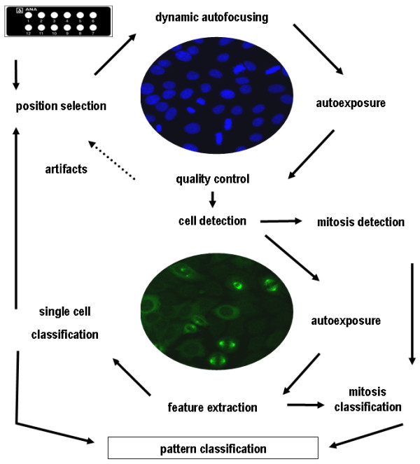
Figure 1
Flowchart of automated human epithelial (HEp-2) cell assay interpretation by the automated reading system [ 18 ]. The fundamental analysis chain of the image processing by the automated system is divi...
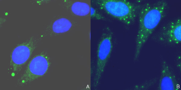
Figure 2
Immunofluorescence patterns of two sera (a, b) which were both scored as negative by visual examination but demonstrated positive cytoplasmic staining by AKLIDES u00ae system . Green color: fluorescei...
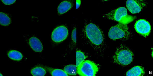
Figure 3
Immunofluorescence patterns of two sera (a, b) which were both assessed as positive with speckled pattern by AKLIDES u00ae system but revealed staining of the nuclear membrane by visual examination . ...
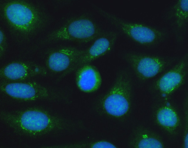
Figure 4
Immunofluorescence pattern with staining of the Golgi complex, which was identified by AKLIDES u00ae system as cytoplasmic speckled pattern . Green color: fluorescein isothiocyanate staining of autoan...
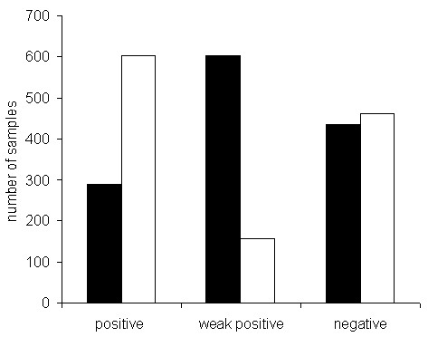
Figure 5
Comparison of positive and negative findings of 1,222 patient sera referred to a routine university laboratory (white bars) and a private laboratory (black bars) . Negative samples demonstrated titers...
Figure images are served from the NIH/NLM PubMed Central Open Access Subset or Europe PMC; copyright remains with the publishers and authors.
💬 Discussion
0 commentsNo comments yet. Be the first to start a discussion!
Leave a Comment