Abstract
Abstract Background Alzheimer’s disease (AD) and Parkinson’s disease (PD) are characterized by brain accumulation of aggregated amyloid-beta (Aβ) and alpha-synuclein (αSYN), respectively. In order to develop effective therapies, it is crucial to understand how the Aβ/αSYN aggregates can be cleared. Compelling data indicate that neuroinflammatory cells, including astrocytes and microglia, play a central role in the pathogenesis of AD and PD. However, how the interplay between the two cell types affects their clearing capacity and consequently the disease progression remains unclear. Methods The aim of the present study was to investigate in which way glial crosstalk influences αSYN and Aβ pathology, focusing on accumulation and degradation. For this purpose, human-induced pluripotent cell (hiPSC)-derived astrocytes and microglia were exposed to sonicated fibrils of αSYN or Aβ and analyzed over time. The capacity of the two cell types to clear extracellular and intracellular protein aggregates when either cultured separately or in co-culture was studied using immunocytochemistry and ELISA. Moreover, the capacity of cells to interact with and process protein aggregates was tracked using time-lapse microscopy and a customized “close-culture” chamber, in which the apical surfaces of astrocyte and microglia monocultures were separated by a <1 mm space. Results Our data show that intracellular deposits of αSYN and Aβ are significantly reduced in co-cultures of astrocytes and microglia, compared to monocultures of either cell type. Analysis of conditioned medium and imaging data from the “close-culture” chamber experiments indicate that astrocytes secrete a high proportion of their internalized protein aggregates, while microglia do not. Moreover, co-cultured astrocytes and microglia are in constant contact with each other via tunneling nanotubes and other membrane structures. Notably, our live cell imaging data demonstrate that microglia, when attached to the cell membrane of an astrocyte, can attract and clear intracellular protein deposits from the astrocyte. Conclusions Taken together, our data demonstrate the importance of astrocyte and microglia interactions in Aβ/αSYN clearance, highlighting the relevance of glial cellular crosstalk in the progression of AD- and PD-related brain pathology.
🔬 Techniques
🔭 Microscopes
💻 Software
✨ Fluorophores
🧪 Sample Preparation
🔬 Cell Lines
🏭 Microscope Brands
🧪 Reagent Suppliers
💻 Software Details
🏛️ Research Organizations (ROR)
Affiliated research institutions:
📋 Methods
The aim of the present study was to investigate in which way glial crosstalk influences αSYN and Aβ pathology, focusing on accumulation and degradation. For this purpose, human-induced pluripotent cell (hiPSC)-derived astrocytes and microglia were exposed to sonicated fibrils of αSYN or Aβ and analyzed over time. The capacity of the two cell types to clear extracellular and intracellular protein aggregates when either cultured separately or in co-culture was studied using immunocytochemistry and ELISA. Moreover, the capacity of cells to interact with and process protein aggregates was tracked using time-lapse microscopy and a customized “close-culture” chamber, in which the apical surfaces of astrocyte and microglia monocultures were separated by a
📊 Figures
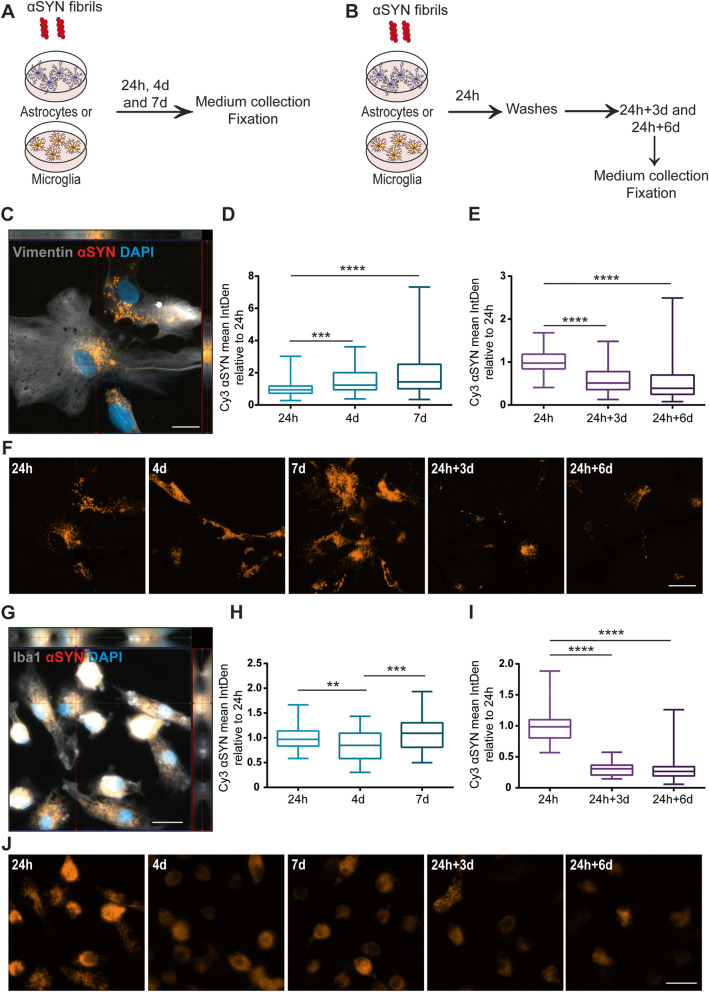
Fig. 1
In contrast to microglia, astrocytes accumulate u03b1SYN over time. Schematic figure of the study design illustrating that the cells were treated with u03b1SYN-F, either continuously ( a ) or with a 2...
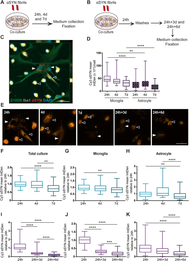
Fig. 2
u03b1SYN accumulation is reduced when microglia and astrocytes are co-cultured. Schematic figure of the study design illustrating that the cells were either treated with u03b1SYN-F constantly ( a ) or...
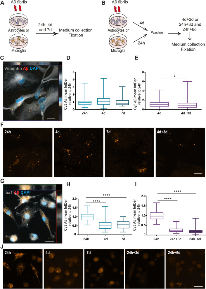
Fig. 3
Intracellular Au03b2 levels are reduced in microglia over time but remain stable in astrocytes. Schematic figure of the study design illustrating that the cells were either constantly treated with Au0...
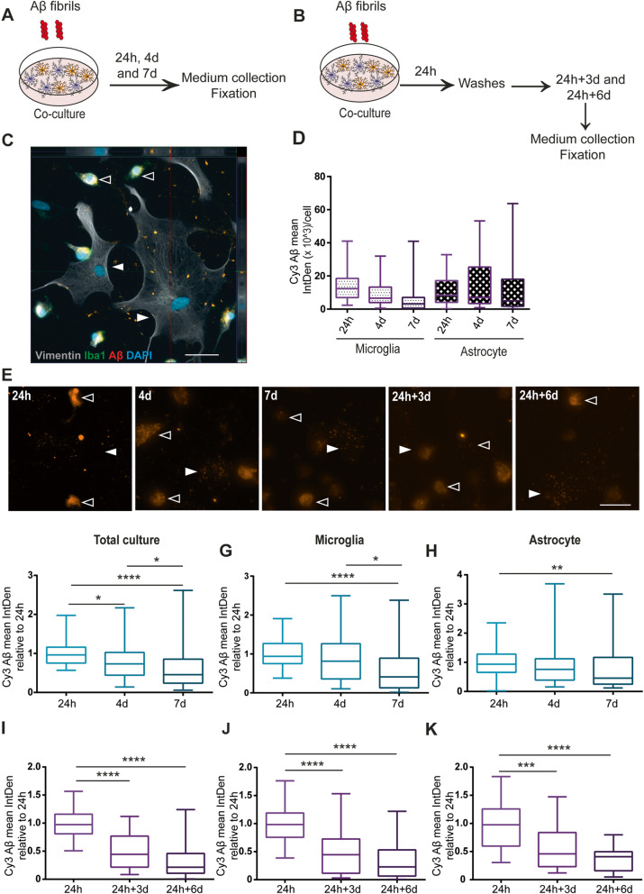
Fig. 4
Intracellular Au03b2 is reduced when astrocytes and microglia are cultured together. Schematic figure of the study design illustrating that the cells were either treated with Au03b2-F constantly ( a )...
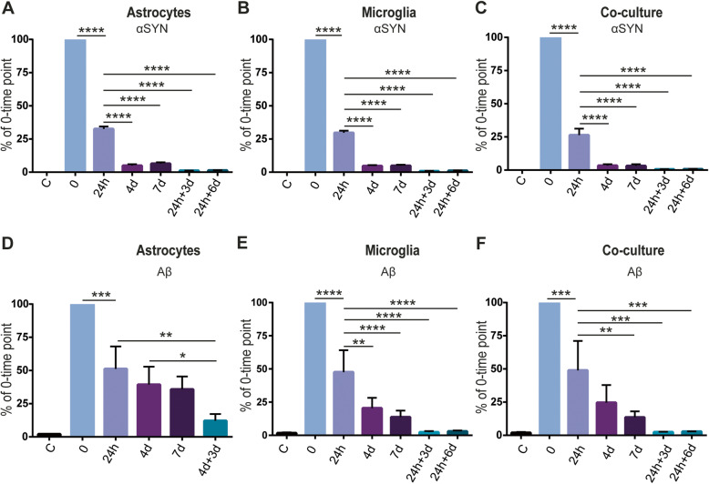
Fig. 5
Astrocytes and microglia clear equal amounts of u03b1SYN, but microglia clear extracellular Au03b2 more effectively. u03b1SYN levels in the medium revealed that both astrocytes and microglia engulfed ...
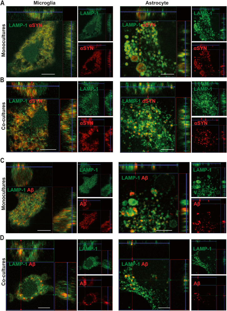
Fig. 6
Ingested u03b1SYN-F and Au03b2-F are located in LAMP1+ vesicles. Confocal microscopy demonstrated that intracellular deposits of u03b1SYN ( a , b ) and Au03b2 ( c , d ) were surrounded by LAMP1+ vesic...
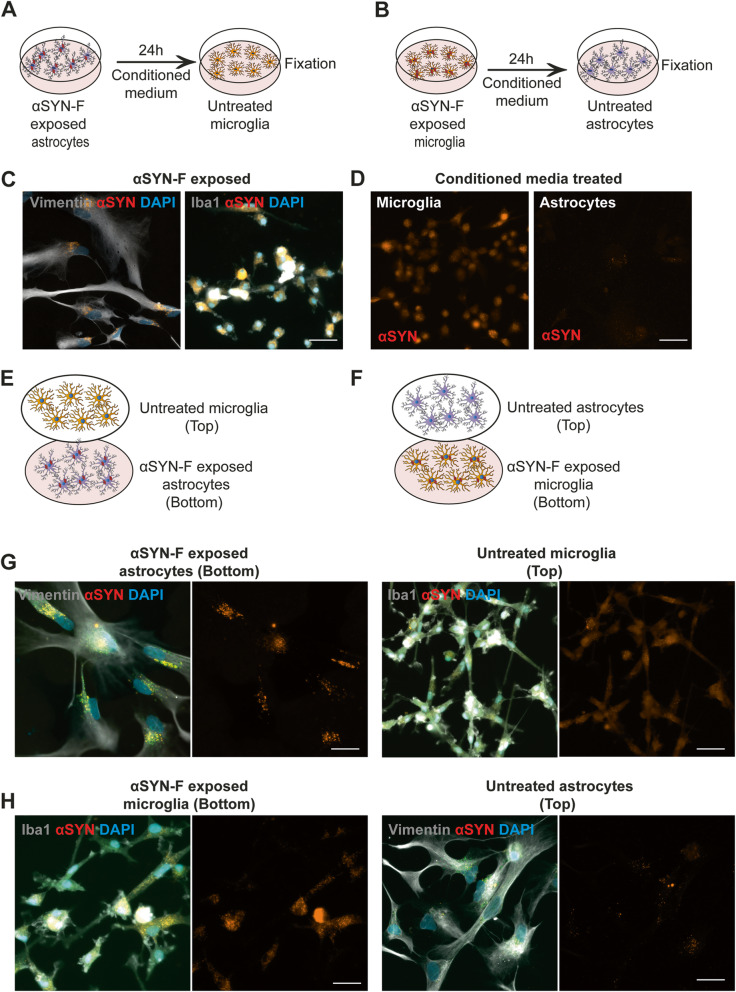
Fig. 7
Secreted u03b1SYN aggregates are transferred from astrocytes to microglia. Conditioned media experiments were performed to investigate if transfer of u03b1SYN occurred from one cell type to another vi...
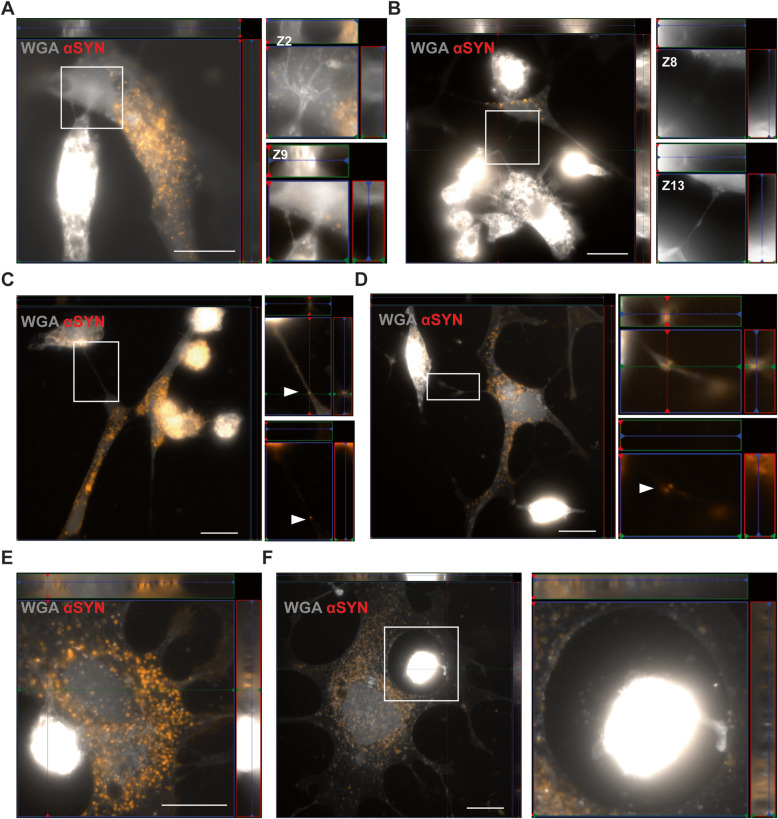
Fig. 8
Direct contact between microglia and astrocytes enables cell-to-cell transfer of u03b1SYN deposits. Astrocytes and microglia were found to have direct TNTs ( a , b ). Z-stacks of the zoomed regions of...
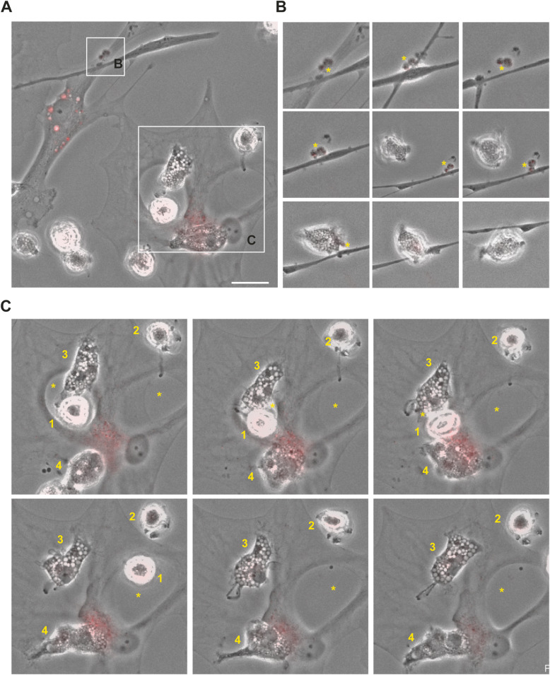
Fig. 9
Microglia can attract and clear intracellular u03b1SYN deposits from astrocytes via membrane contact. Time-lapse microscopy illustrated a complex interplay between astrocytes and microglia ( a ). Diff...
Figure images are served from the NIH/NLM PubMed Central Open Access Subset or Europe PMC; copyright remains with the publishers and authors.
💬 Discussion
0 commentsNo comments yet. Be the first to start a discussion!
Leave a Comment