Abstract
The ClpXP machinery is a two-component protease complex that performs targeted protein degradation in bacteria and mitochondria. The complex consists of the AAA+ chaperone ClpX and the peptidase ClpP. The hexameric ClpX utilizes the energy of ATP binding and hydrolysis to engage, unfold and translocate substrates into the catalytic chamber of tetradecameric ClpP, where they are degraded. Formation of the complex involves a symmetry mismatch, because hexameric AAA+ rings bind axially to the opposing stacked heptameric rings of the tetradecameric ClpP. Here we present the cryo-EM structure of ClpXP from Listeria monocytogenes. We unravel the heptamer-hexamer binding interface and provide novel insight into the ClpX-ClpP cross-talk and activation mechanism. Comparison with available crystal structures of ClpP and ClpX in different states allows us to understand important aspects of the complex mode of action of ClpXP and provides a structural framework for future pharmacological applications.
🔬 Techniques
🔭 Microscopes
🧬 Organisms
💻 Software
✨ Fluorophores
🧪 Sample Preparation
🏭 Microscope Brands
🧪 Reagent Suppliers
📷 Detectors
💻 Software Details
💾 Data Repositories
🏛️ Research Organizations (ROR)
Affiliated research institutions:
📋 Methods
Cloning
The cloning of pETDuet-1_ClpP1/2 and pET300_ClpX were described previously 32 . ClpX and ClpP1/2 point mutants, ClpP1/2 ΔC-3 , ClpP1/2 ΔC-4 and ClpP1/2 ΔC-5 were generated using the QuikChange™ technology. For ClpP1/2 ΔC-4 and ClpP1/2 ΔC-5 , the pETDuet-1_ClpP1/2 ΔC-3 plasmid was used as a template. ClpP1/2 ΔC-6 and ClpX ΔZBD (E183Q) were obtained with primers containing non-overlapping sequences 58 . All primers are listed in Supplementary Table 1 Protein overexpression and purification ClpP1/2 and its mutants’ variants were overexpressed and purified as follows. The proteins were overexpressed in E. coli BL21(DE3) bearing a pETDuet-1 vector with C-terminally Strep-II-tagged ClpP1 and C-terminally His 6 -tagged ClpP2 32 . The bacteria were grown in LB medium until OD 600 0.6 at 37 °C. Following induction with 1 mM isopropyl-β- D -thiogalactoside (IPTG), the bacteria were incubated at 37 °C for 6 h. After harvest, the cells were sonicated on ice in lysis buffer (20 mM MOPS, 300 mM KCl, 1% CHAPS, 10% glycerol, pH 7.5) and then kept at room temperature during the rest of the purification. The proteins from the cleared cell lysate were captured in a HisTrap HP 5 ml column (GE Healthcare) in His buffers (20 mM MOPS, 300 mM KCl, 10% glycerol, pH 7.5; +40 mM imidazole for washing) using an ÄKTA Purifier 10 system (GE Healthcare). The proteins were eluted by a 15 mL gradient from 40 mM to 300 mM imidazole, and the second elution peak was collected. A subsequent chromatography step was carried out on a StrepTrap HP 5 ml column (GE Healthcare) in Strep buffers (20 mM MOPS, 300 mM KCl, 10% glycerol, pH 7.5; +2.5 mM desthiobiotin for elution). A final gel filtration was performed on a Superdex200 pg 16/60 column (GE Healthcare) in ClpP SEC buffer (20 mM MOPS, 300 mM KCl, 15% glycerol, pH 7.0). In the case of the cystein-containing mutants, 1 mM TCEP was added to all buffers. ClpX(E183Q) and ClpX ΔZBD (E183Q) were overexpressed in E. coli BL21(DE3). An expression construct equipped with an N-terminal His 6 -tag and a TEV cleavage site in pET300 vector was used 32 . The bacteria were grown in LB medium to OD 600 0.6 at 37 °C. After induction with 0.5 mM IPTG, the cells were incubated overnight at 25 °C. After harvest, the cells were resuspended in ClpX lysis buffer (25 mM HEPES, 200 mM KCl, 1 mM DTT, 0.5 mM ATP, 5 mM MgCl 2 , 10 mM imidazole, 5% glycerol, pH 7.6) and lysed by ultrasonication. The cleared cell lysate was loaded on a 5 mL HisTrap HP column (GE Healthcare). The column was washed with ClpX wash buffer (25 mM HEPES, 200 mM KCl, 1 mM DTT, 5% glycerol, 40 mM imidazole, pH 7.6). The protein was eluted with ClpX elution buffer (25 mM HEPES, 200 mM KCl, 1 mM DTT, 5% glycerol, 300 mM imidazole, pH 7.6). The protein fractions were pooled, 1 mM EDTA and TEV protease [1.25 mg for ClpX(E183Q) and 3.75 mg for ClpX ΔZBD (E183Q)] were added and the reaction mixture was incubated at 10 °C overnight. Complete TEV cleavage was verified by intact-protein mass-spectrometry. The protein solution was loaded on a Superdex200 pg 16/60 column (GE Healthcare) and eluted in ClpX SEC buffer (25 mM HEPES, 200 mM KCl, 1 mM DTT, 0.5 mM ATP, 5 mM MgCl 2 , 5% glycerol, pH 7.6). ClpX(WT), ClpX(V264C), ClpX(I265C), ClpX(G266C) and ClpX(F267C) were overexpressed and purified similarly with the following modifications: the buffers contained 1 mM TCEP instead of DTT, and the ClpX wash buffer and ClpX elution buffer contained additionally 0.5 mM ATP and 5 mM MgCl 2 . The TEV digestion step was omitted. N-terminally Strep-II-tagged eGFP with a C-terminal SsrA tag (AGKEKQNLAFAA) was overexpressed in E. coli SG1146a (Δ clpP ) using pET55-Dest expression vector and purified by affinity chromatography and gel filtration as described previously 15 , 32 . Creatine kinase (product no. 10 127 566 001), lactate dehydrogenase (product no. 10 128 155 001) and pyruvate kinase (product no. 10 127 876 001) were purchased from Roche. Isolation of the ClpXP complex 4.4 nmol (ClpP1/2) 14 and 3.3 nmol ClpX 6 were incubated for 10 min at 37 °C in PZA buffer (25 mM HEPES, 200 mM KCl, 5 mM MgCl2, 1 mM DTT, 0.5 mM ATP, 15% glycerol, pH 7.6). The samples were loaded onto a Superose 6 increase 10/300 column (GE Healthcare) connected to an ÄKTA Purifier 10 system (GE Healthcare) and eluted at 0.2 mL/min flow rate. Samples were taken at 12 mL retention volume for EM and HDX-MS measurements. For cryoEM, the sample was diluted 1:3 with glycerol-free PZA buffer and 0.1% glutaraldehyde was added. The reaction was quenched after 30 s with 2 eq. Tris-HCl. For SDS-PAGE, 4.4 μg protein was loaded on a gel and stained with Coomassie blue after separation.
Show full methods section
Cloning
The cloning of pETDuet-1_ClpP1/2 and pET300_ClpX were described previously 32 . ClpX and ClpP1/2 point mutants, ClpP1/2 ΔC-3 , ClpP1/2 ΔC-4 and ClpP1/2 ΔC-5 were generated using the QuikChange™ technology. For ClpP1/2 ΔC-4 and ClpP1/2 ΔC-5 , the pETDuet-1_ClpP1/2 ΔC-3 plasmid was used as a template. ClpP1/2 ΔC-6 and ClpX ΔZBD (E183Q) were obtained with primers containing non-overlapping sequences 58 . All primers are listed in Supplementary Table 1 Protein overexpression and purification ClpP1/2 and its mutants’ variants were overexpressed and purified as follows. The proteins were overexpressed in E. coli BL21(DE3) bearing a pETDuet-1 vector with C-terminally Strep-II-tagged ClpP1 and C-terminally His 6 -tagged ClpP2 32 . The bacteria were grown in LB medium until OD 600 0.6 at 37 °C. Following induction with 1 mM isopropyl-β- D -thiogalactoside (IPTG), the bacteria were incubated at 37 °C for 6 h. After harvest, the cells were sonicated on ice in lysis buffer (20 mM MOPS, 300 mM KCl, 1% CHAPS, 10% glycerol, pH 7.5) and then kept at room temperature during the rest of the purification. The proteins from the cleared cell lysate were captured in a HisTrap HP 5 ml column (GE Healthcare) in His buffers (20 mM MOPS, 300 mM KCl, 10% glycerol, pH 7.5; +40 mM imidazole for washing) using an ÄKTA Purifier 10 system (GE Healthcare). The proteins were eluted by a 15 mL gradient from 40 mM to 300 mM imidazole, and the second elution peak was collected. A subsequent chromatography step was carried out on a StrepTrap HP 5 ml column (GE Healthcare) in Strep buffers (20 mM MOPS, 300 mM KCl, 10% glycerol, pH 7.5; +2.5 mM desthiobiotin for elution). A final gel filtration was performed on a Superdex200 pg 16/60 column (GE Healthcare) in ClpP SEC buffer (20 mM MOPS, 300 mM KCl, 15% glycerol, pH 7.0). In the case of the cystein-containing mutants, 1 mM TCEP was added to all buffers. ClpX(E183Q) and ClpX ΔZBD (E183Q) were overexpressed in E. coli BL21(DE3). An expression construct equipped with an N-terminal His 6 -tag and a TEV cleavage site in pET300 vector was used 32 . The bacteria were grown in LB medium to OD 600 0.6 at 37 °C. After induction with 0.5 mM IPTG, the cells were incubated overnight at 25 °C. After harvest, the cells were resuspended in ClpX lysis buffer (25 mM HEPES, 200 mM KCl, 1 mM DTT, 0.5 mM ATP, 5 mM MgCl 2 , 10 mM imidazole, 5% glycerol, pH 7.6) and lysed by ultrasonication. The cleared cell lysate was loaded on a 5 mL HisTrap HP column (GE Healthcare). The column was washed with ClpX wash buffer (25 mM HEPES, 200 mM KCl, 1 mM DTT, 5% glycerol, 40 mM imidazole, pH 7.6). The protein was eluted with ClpX elution buffer (25 mM HEPES, 200 mM KCl, 1 mM DTT, 5% glycerol, 300 mM imidazole, pH 7.6). The protein fractions were pooled, 1 mM EDTA and TEV protease [1.25 mg for ClpX(E183Q) and 3.75 mg for ClpX ΔZBD (E183Q)] were added and the reaction mixture was incubated at 10 °C overnight. Complete TEV cleavage was verified by intact-protein mass-spectrometry. The protein solution was loaded on a Superdex200 pg 16/60 column (GE Healthcare) and eluted in ClpX SEC buffer (25 mM HEPES, 200 mM KCl, 1 mM DTT, 0.5 mM ATP, 5 mM MgCl 2 , 5% glycerol, pH 7.6). ClpX(WT), ClpX(V264C), ClpX(I265C), ClpX(G266C) and ClpX(F267C) were overexpressed and purified similarly with the following modifications: the buffers contained 1 mM TCEP instead of DTT, and the ClpX wash buffer and ClpX elution buffer contained additionally 0.5 mM ATP and 5 mM MgCl 2 . The TEV digestion step was omitted. N-terminally Strep-II-tagged eGFP with a C-terminal SsrA tag (AGKEKQNLAFAA) was overexpressed in E. coli SG1146a (Δ clpP ) using pET55-Dest expression vector and purified by affinity chromatography and gel filtration as described previously 15 , 32 . Creatine kinase (product no. 10 127 566 001), lactate dehydrogenase (product no. 10 128 155 001) and pyruvate kinase (product no. 10 127 876 001) were purchased from Roche. Isolation of the ClpXP complex 4.4 nmol (ClpP1/2) 14 and 3.3 nmol ClpX 6 were incubated for 10 min at 37 °C in PZA buffer (25 mM HEPES, 200 mM KCl, 5 mM MgCl2, 1 mM DTT, 0.5 mM ATP, 15% glycerol, pH 7.6). The samples were loaded onto a Superose 6 increase 10/300 column (GE Healthcare) connected to an ÄKTA Purifier 10 system (GE Healthcare) and eluted at 0.2 mL/min flow rate. Samples were taken at 12 mL retention volume for EM and HDX-MS measurements. For cryoEM, the sample was diluted 1:3 with glycerol-free PZA buffer and 0.1% glutaraldehyde was added. The reaction was quenched after 30 s with 2 eq. Tris-HCl. For SDS-PAGE, 4.4 μg protein was loaded on a gel and stained with Coomassie blue after separation.
Electron microscopy
Sample quality was examined by negative stain EM. Sample from the respective fraction was further diluted to a concentration of 0.01-0.03 mg ml -1 and negative stain EM was performed as described previously 59 . Images were recorded with a JEOL JEM-1400 equipped with a 4K CMOS detector F416 (TVIPS) at a pixel size of 1.84 Å. For cryoEM, 4 μl of cross-linked ClpXP1/2 dimers at a concentration of 0.045 mg ml -1 were applied to a glow-discharged quantifoil 2/1 Cu grid with an additional 2nm thin carbon layer and after an incubation time of 45 sec, rapidly plunge-frozen using a CryoPlunge3 (Cp3, Gatan) at 90% humidity. To improve ice quality and thickness distribution, 0.01% Tween-20 was added shortly prior plunging. The quality of the grids was screened with a JEOL JEM 1400 and a FEI Tecnai Spirit, both equipped with a LaB 6 cathode and a 4K CMOS detector F416 (TVIPS). A cryoEM dataset was acquired on a FEI Titan KRIOS at 300 kV equipped with spherical aberration corrector and a Falcon III direct detector (linear mode) at a x112,807 magnification (x59,000 nominal magnification), corresponding to a pixel size of 1.1 Å. Each exposure was recorded with a total dose of ~114 electrons/Å 2 and a total exposure time of 2 sec (frame rate of 50 msec). A total of 3200 micrographs were collected using the EPU software (FEI).
Image processing and reconstruction
The frames were aligned, averaged and dose-weighted using unblur and sum_movie 60 . Unweighted full-dose images were further used to estimate the CTF parameters using CTER 61 (SPHIRE) 25 . Dose weighted full-dose images were used for all other steps of image processing. ClpXP1/2 dimers were picked automatically using EMAN2’s 62 neuralnet e2boxer. Further data processing was performed using the software package SPHIRE 25 . After inspection of micrographs using the CTF-assessment-GUI, 273,300 single particles were selected for further processing. The particle stack was subjected to 2D-clustering using ISAC2 (SPHIRE), resulting in a “clean” stack of 143,901 single particles producing stable and reproducible 2D-class averages. The 2D class-averages were used to calculate a 3D volume, using VIPER. After masking, this volume was used as the reference for a 3D refinement using Meridien (SPHIRE), which resulted in a 13 Å density map, as estimated by the “gold-standard” FSC. In agreement to the 2D clustering results (Supplementary Video 1), further 3D clustering using Sort3D (SPHIRE) confirmed that the ClpXP1/2 dimer is a continuously flexible structure ( Supplementary Figure 1g ). Independent refinement of the resulting subsets did not, however, further improve the resolution of the volume. We then manually picked the ClpXP1/2 monomers within each ClpXP1/2-dimer for 10 representative micrographs of the dataset and used these data to train crYOLO 63 , which then automatically selected 613,322 single particles. After 2D and 3D clustering, a final “clean” stack of 383.927 particles was used for further refinement. During the first rounds of the refinement, we applied local symmetrization of the reference after each refinement round, as previously described 64 , 65 i.e. after each refinement round the density of ClpP was symmetrized using D7 symmetry, whereas the density of ClpX was scaled in order to put an additional weight on this region during the asymmetric refinement. Finally, both densities (ClpX and ClpP) were combined and the resulting volume was used as a reference for the subsequent refinement iteration. This procedure was performed during the initial rounds in order to obtain global projection parameters. The user function was not applied during the local refinements. This resulted in a density map with an average resolution of 4 Å, where the resolution of the density decreases towards ClpX ( Supplementary Figure 2 ). The average resolution was calculated between two independently refined “half maps” at the 0.143 FSC criterion. The estimated accuracy of rotation and translation search during the last refinement round was estimated to 1.78° and 1.02 pixels, respectively. Local resolution was computed using the “Local Resolution” tool in SPHIRE. 3D clustering into four groups was performed using the RSORT3D tool of SPHIRE. However, according to the ANOVA analysis, the resulting volumes were not reproducible and were therefore not considered for further analysis. 3D Refinement and Clustering focusing on the density of ClpX, after removing the ClpP signal from the dataset, did also not result into further improvement of the ClpX density. The density of ClpP was auto-sharpened locally using phenix.auto_sharpen 66 and filtered to its average resolution of 3.9 Å. The ClpX desnity was filtered to an average resolution of 6.5 Å and sharpened with an ad-hoc b-factor of -240 Å 2 . Angular distribution plots were computed using SPHIRE. Sharpened 2D class averages were computed with 3500 members per group.
Atomic modelling
We built a homology model of ClpX with SWISS-MODEL 67 using ADP-bound E. coli ClpX (PDB-ID 3HWS, Chain A) and ATPγS-bound E. coli ClpX (PDB-ID 4I81, Chain B). We then used UCSF Chimera 68 to fit the structures of ClpX’s homology model and ClpP1/2 (PDBID 4RYF 32 into the cryo-EM density. We used the RosettaES protocol 69 to build the missing residues 9-16 for each ClpP2 subunit. Residues 1-2 were manually built in Coot 70 . With the complete model, we performed several iterative runs of molecular dynamics flexible fitting (MDFF) 71 and manual adjustment with Coot, paying particular attention to the fitting of the IGF loops. In the initial run, we applied 6-fold symmetry to ClpX, allowing regions poorly supported by the density to settle into reasonable conformations. This restraint was later removed. For the final iterations, we also included a step of real-space refinement in Phenix 72 , to decrease the number of Ramachandran outliers and to fit the atomic B-factors. The necessary files for the MDFF runs were set up with VMD 73 and all simulations were performed in NAMD 74 , using the CHARMM 36m force field 75 with the implicit solvation model implemented in NAMD. For the proper modeling of the structure with MDFF, we included all missing regions of the structures, even if their density does not allow full atomic modeling. After refinement, we removed all those from the final model. The quality of this model was assessed in Phenix, using the Molprobity 76 and EMRinger scores 77 as well as the overall geometry of the structure. Sequence conservation was analyzed using the ConSurfserver 78 . Analysis of the channel pathway was performed with ChExVis 79 . Electron density maps and models were visualized using Chimera 69 and Chimera X 80 .
Peptidase assay
In this assay, the degradation of a fluorogenic tripeptide was measured, for which ClpX was not required. 99 μL 1 μM ClpP1/2 was incubated in PZ buffer (25 mM HEPES, 200 mM KCl, 5 mM MgCl2, 1 mM DTT, 10% glycerol, pH 7.6) in flat bottom black 96-well plates for 15 min at 30 °C. 1 μL acetylalanyl-homoarginyl-2-aminooctanoyl-7-amino-4-carbamoylmethylcoumarin (Ac-Ala-hArg-2-Aoc-ACC) substrate (10 mM stock in DMSO) was added and the fluorescence was measured (380 nm, 430 nm) with an infinite M200Pro plate reader (Tecan) at 30 °C. Data were recorded in triplicate and two independent experiments were performed. Peptidase activity was determined by linear regression using Microsoft Excel and plots were made with GraphPad Prism 6.
Protease assay
Protease assays were carried out in flat bottom white 96-well plates in a final volume of 60 μL. (ClpP1/2) 14 (0.2 μM), ClpX 6 (0.4 μM) and ATP regeneration mix (4 mM ATP, 16 mM creatine phosphate, 20 U/mL creatine kinase) were pre-incubated for 15 min at 30 °C in PZ buffer. 0.8 μM eGFP-SsrA substrate was added and fluorescence was measured (485 nm, 535 nm) at 30 °C. Data were recorded in triplicate and at least two independent experiments were performed. Protease activity was determined by linear regression using Microsoft Excel and plots were made with GraphPad Prism 6. ATPase assay 90 μL 2 μM ClpX in ATPase buffer (100 mM HEPES, 200 mM KCl, 20mM MgCl 2 , 1 mM DTT, 1 mM NADH, 2 mM phosphoenolpyruvate, 50 U/mL lactate dehydrogenase, 50 U/mL pyruvate kinase, 5% glycerol, pH 7.5) was added to a flat bottom transparent 96-well plate and incubated for 15 min at 37 °C. The reaction was started by the addition of 10 μL 200 mM ATP in 100 mM HEPES, pH 7.5. Absorption at 340 nm was measured at 37 °C. Two independent experiments with three replicates each were carried out. ATPase activity was determined by linear regression using Microsoft Excel after subtraction of the background signal (measurement without ClpX), and the plot was made with GraphPad Prism 6. Hydrogen/deuterium exchange mass-spectrometry (HDX-MS) HDX-MS experiments were performed using an ACQUITY UPLC M-class system equipped with automated HDX technology (Waters). HDX kinetics were determined by taking data points at 0, 10, 60, 600, 1800 and 7200 s at 20 °C. At each data point of the kinetic, 3 µL of a solution of 30 µM „free” ClpP1/2 and „free” ClpX were analyzed and compared to the (ClpXP1/2) 2 complex (1.4 µM). The respective protein solutions were diluted automatically 1:20 into 99.9% D 2 O-containing buffer (25 mM HEPES, 200 mM KCl, 5 mM MgCl 2 , 0.5 mM ATP, 1 mM TCEP, 5% glycerol, pH 7.6). As reference, all samples were analyzed in H 2 O –containing buffers. The reaction mixture was quenched by the addition of 1:1 200 mM KH 2 PO 4 , 200 mM Na 2 HPO 4 , pH 2.3 (titrated with HCl) at 1 °C and 50 µL of the resulting sample were subjected to on-column peptic digest on a Waters Enzymate BEH pepsin column 2.1 × 30 mm at 20 °C. Peptides were separated by reverse phase chromatography at 0 °C in trapping mode using a Waters Acquity UPLC C18 1.7 µm Vangard 2.1 × 5 mm pre-column and a Waters Aquity UPLC BEH C18 1.7 µm 1 × 100 mm separation column. For separation, a gradient increasing the acetonitrile concentration stepwise from 5 to 35% in 6 min, from 35 to 40% in 1 min and from 40 to 95% in 1 min was applied and the eluted peptides were analyzed using an in-line Synapt G2-S QTOF HDMS mass spectrometer (Waters). UPLC was performed in protonated solvents (0.1% formic acid), allowing deuterium to be replaced with hydrogen from side chains and amino/carboxyl termini that exchange much faster than backbone amide linkages 81 . All experiments were performed in duplicate. Deuterium levels were not corrected for back exchange and are therefore reported as relative deuterium levels 82 . The use of an automated system, i.e. handling all samples at identical conditions, negotiates the need for back exchange correction. MS data were collected over an m/z range of 100-2000, and are available online as source data [AU: correct?]. Mass accuracy was ensured by calibration with Glu-fibrino peptide B (Waters) and peptides were identified by triplicates MSE ramping the collision energy from 20-50 V. MS data were analyzed with the PLGS 3.0.3 and DynamX 3.0 software packages and all spectra were checked manually. For each peptide, relative uptake values were determined as follows: relative uptake [%] = deuterium uptake × 100 / maximal uptake. For each amino acid, the average of the relative uptake of all peptides covering the amino acid was calculated. The difference of the relative deuterium uptake between the “free” and “complex” states was calculated for each amino acid. Data were analyzed and visualized using custom MATLAB and python scripts, UCSF Chimera 1.12 and OriginPro 2016.
Supplementary Material Supplementary figures Supplementary table 1
📊 Figures
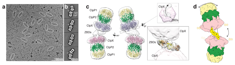
Figure 1
LmClpXP1/2 forms flexible dimers via the ZBDs.
a) Typical low-dose cryo-EM micrograph of the ClpXP1/2 dimer from L. monocytogenes . Some particles are highlighted withnovals. Scale bar, 100nm b) Typical reference-free 2D classnaverages. Arrows ind...
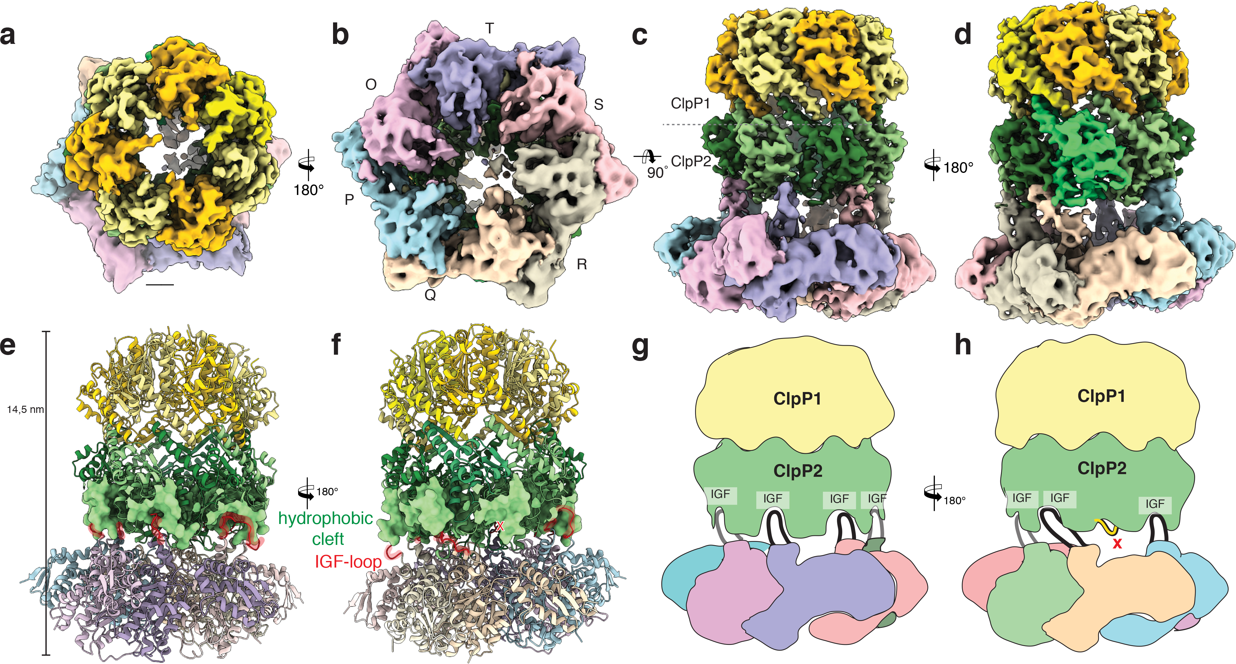
Figure 2
Cryo-EM structure of the ClpXP1/2 protein degradation machinery.
a-d) Cryo-EM density of ClpXP1/2 shown from the topn( a ), bottom ( b ) and side ( c,d ). ClpP1 andnClpP2 subunits are colored in khaki, orange and dark, light green, respectively.nClpP2 subunit J is ...
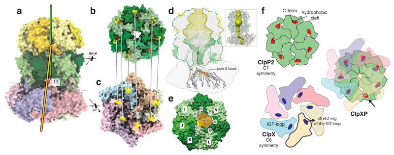
Figure 3
Symmetry mismatch between ClpP1/2 and ClpX.
a) Molecular model of ClpXP1/2. The symmetry axes of the ClpP1/2 andnClpX are shown in green and orange, respectively. b-c) The ClpP2nheptamer (b) and the ClpX hexamer (c) are shown fromnthe bottom an...
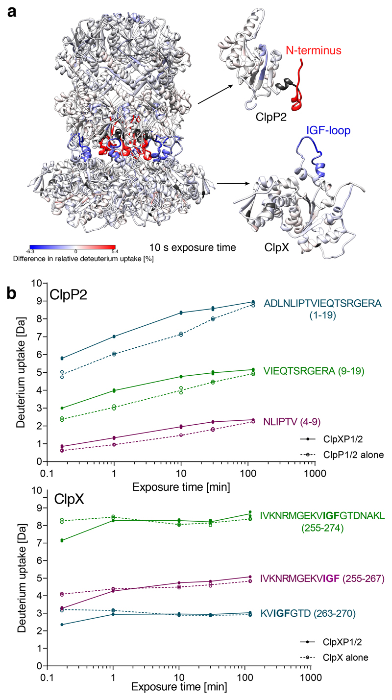
Figure 4
HDX-MS analysis of ClpXP1/2 complex formation.
a) Difference in relative deuterium uptake after 10 s exposure isnmapped on the structure of ClpXP1/2 (left), ClpP2 monomer (right top) and ClpXnmonomer (right bottom). Increased deuterium uptake upon...
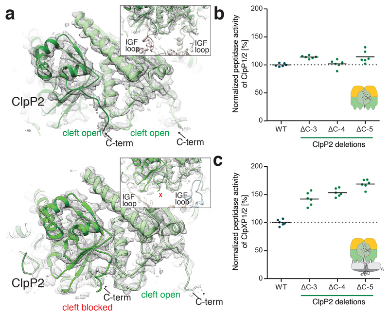
Figure 5
Role of the ClpP2 C-terminus in ClpXP1/2 binding.
a) Molecular model and cryo-EM density of IGF-loop bound (uppernimage) and not bound to hydrophobic pockets of ClpP2 (lower image). The insetsnshow the respective IGF-loops in ribbon representation. A...
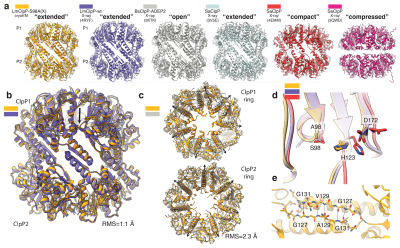
Figure 6
Comparison of ClpX-bound ClpP1/2 with available structures of active and inactive ClpP.
a) Side view of the structure of ClpX-bound LmClpP1/2 (gold) and thencrystal structures of LmClpP1/2 in the extended active state (PDB4RYF) (purple), Bacillus subtilis ClpP (BsClpP) in complex with AD...
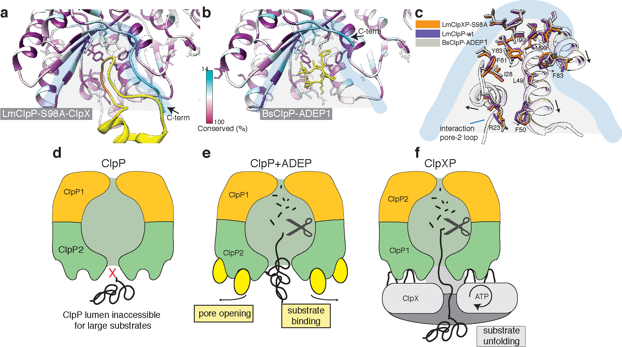
Figure 7
ClpX binds to ClpP in a similar manner like ADEP, but does not induce ClpP pore widening.
a) Local molecular interactions at one of the seven binding pocketsnbetween ClpP and the IGF-loop of ClpX. Residues of ClpP are colored by sequencenconservation. The IGF-loop is shown in yellow with t...
Figure images are served from the NIH/NLM PubMed Central Open Access Subset or Europe PMC; copyright remains with the publishers and authors.
💬 Discussion
0 commentsNo comments yet. Be the first to start a discussion!
Leave a Comment