Abstract
Inflammasomes sense intrinsic and extrinsic danger signals to trigger inflammatory responses and pyroptotic cell death. Homotypic pyrin domain (PYD) interactions of inflammasome forming nucleotide-binding oligomerization domain (NOD)–like receptors with the adaptor protein ASC (apoptosis-associated speck-like protein containing a CARD) mediate oligomerization into filamentous assemblies. We describe the cryo–electron microscopy (cryo-EM) structure of the human NLRP3 PYD filament and identify a pattern of highly polar interface residues that form the homomeric interactions leading to characteristic filament ends designated as A- and B-ends. Coupling a titration polymerization assay to cryo-EM, we demonstrate that ASC adaptor protein elongation on NLRP3 PYD nucleation seeds is unidirectional, associating exclusively to the B-end of the filament. Notably, NLRP3 and ASC PYD filaments exhibit the same symmetry in rotation and axial rise per subunit, allowing a continuous transition between NLRP3 and ASC. Integrating the directionality of filament growth, we present a molecular model of the ASC speck consisting of active NLRP3, ASC, and Caspase-1 proteins.
🔬 Techniques
🧬 Organisms
✨ Fluorophores
🧪 Sample Preparation
🔬 Cell Lines
🏭 Microscope Brands
🧪 Reagent Suppliers
📷 Detectors
🎨 Filters
💻 Software Details
💾 Data Repositories
🏛️ Research Organizations (ROR)
Affiliated research institutions:
📋 Methods
NLRP3 PYD cloning, expression, and purification Human NLRP3 (UniProt accession number Q96P20 ; residues 3 to 110; C108S) was cloned into a pGEX-4T1 expression vector, providing an N-terminal GST tag followed by a TEV protease cleavage site. The C108S mutation was introduced to prevent disulfide bond formation with residue C8, which was observed in one of two chains in the crystal structure ( 24 ) of NLRP3 PYD . The construct was transformed and expressed in E. coli cells, strain BL21(DE3), by growing the culture at 37°C to an OD 600 (optical density at 600 nm) of 0.8 and by inducing with 0.3 mM isopropyl-β- d -1-thiogalactopyranoside (IPTG) overnight (o/n) at 20°C. Cells were collected by centrifugation and lysed by sonication in a lysis buffer containing 20 mM Hepes (pH 7.5), 150 mM NaCl, and 0.5 mM tris(2-carboxyethyl)phosphine (TCEP). The cell lysate was centrifuged at 20,000 g for 30 min, and the supernatant was administered to a pre-equilibrated GSTrap column using an ÄKTA prime FPLC system (GE Healthcare). The column was washed with 10 column volumes of lysis buffer, and the protein was eluted in the same buffer supplemented with 15 mM l -glutathione. The affinity-purified protein was TEV-cleaved o/n at 4°C and subsequently subjected to gel filtration on an equilibrated Superdex 75 gel filtration column 16/600 (GE Healthcare). Monomer fractions were pooled and concentrated to 1 mg/ml, corresponding to a concentration of 79 μM, and filament formation was induced with fresh protein by incubation at 37°C o/n. Protein variants carrying mutations in the PYD-PYD assembly interfaces (R7E, E15R, K23E/K24E, M27E, R43E, D60R, R80E, and R81E) and CAPS mutants (D21H, D31V, H51R, and A77V) were generated in the NLRP3 PYD (residues 3 to 110; C108S) construct using the megaprimer mutagenesis method. The NLRP3 PYD interface and CAPS mutant proteins were expressed and purified as GST fusion proteins following the same procedure as described above. For kinetic measurements, the generation of monomeric, soluble NLRP3 PYD included an additional dialysis step after TEV protease cleavage into a buffer containing 50 mM glycine (pH 3.8) and 150 mM NaCl. The dialyzed protein was further purified on a pre-equilibrated Superdex 75 gel filtration column (GE Healthcare), and fractions containing monomeric soluble NLRP3 PYD were concentrated to 1 mg/ml, snap-frozen in liquid nitrogen, and stored at −80°C.
Show full methods section
NLRP3 PYD cloning, expression, and purification Human NLRP3 (UniProt accession number Q96P20 ; residues 3 to 110; C108S) was cloned into a pGEX-4T1 expression vector, providing an N-terminal GST tag followed by a TEV protease cleavage site. The C108S mutation was introduced to prevent disulfide bond formation with residue C8, which was observed in one of two chains in the crystal structure ( 24 ) of NLRP3 PYD . The construct was transformed and expressed in E. coli cells, strain BL21(DE3), by growing the culture at 37°C to an OD 600 (optical density at 600 nm) of 0.8 and by inducing with 0.3 mM isopropyl-β- d -1-thiogalactopyranoside (IPTG) overnight (o/n) at 20°C. Cells were collected by centrifugation and lysed by sonication in a lysis buffer containing 20 mM Hepes (pH 7.5), 150 mM NaCl, and 0.5 mM tris(2-carboxyethyl)phosphine (TCEP). The cell lysate was centrifuged at 20,000 g for 30 min, and the supernatant was administered to a pre-equilibrated GSTrap column using an ÄKTA prime FPLC system (GE Healthcare). The column was washed with 10 column volumes of lysis buffer, and the protein was eluted in the same buffer supplemented with 15 mM l -glutathione. The affinity-purified protein was TEV-cleaved o/n at 4°C and subsequently subjected to gel filtration on an equilibrated Superdex 75 gel filtration column 16/600 (GE Healthcare). Monomer fractions were pooled and concentrated to 1 mg/ml, corresponding to a concentration of 79 μM, and filament formation was induced with fresh protein by incubation at 37°C o/n. Protein variants carrying mutations in the PYD-PYD assembly interfaces (R7E, E15R, K23E/K24E, M27E, R43E, D60R, R80E, and R81E) and CAPS mutants (D21H, D31V, H51R, and A77V) were generated in the NLRP3 PYD (residues 3 to 110; C108S) construct using the megaprimer mutagenesis method. The NLRP3 PYD interface and CAPS mutant proteins were expressed and purified as GST fusion proteins following the same procedure as described above. For kinetic measurements, the generation of monomeric, soluble NLRP3 PYD included an additional dialysis step after TEV protease cleavage into a buffer containing 50 mM glycine (pH 3.8) and 150 mM NaCl. The dialyzed protein was further purified on a pre-equilibrated Superdex 75 gel filtration column (GE Healthcare), and fractions containing monomeric soluble NLRP3 PYD were concentrated to 1 mg/ml, snap-frozen in liquid nitrogen, and stored at −80°C.
Preparation of recombinant ASC Full-length human
ASC, followed by a TEV protease cleavage site and mCherry, was cloned with Nde I/Xho I sites at the 5′ and 3′ ends, respectively, into a pET-23a expression vector providing a C-terminal hexa-histidine tag. This construct was transformed and expressed in E. coli cells, strain BL21(DE3), by growing the culture at 37°C to an OD 600 of 0.8 and induced with 0.1 mM IPTG for 4 hours at 37°C. Cells were collected by centrifugation and lysed by sonication in lysis buffer A containing 20 mM tris (pH 8.0), 500 mM NaCl, and 5 mM imidazole. Cell lysates were centrifuged at 20,000 g for 30 min, and the pellet was dissolved in buffer A supplemented with 4 M Gdn-HCl for 1 hour at 4°C. Subsequently, the suspension was centrifuged at 20,000 g for 30 min, and the supernatant was administered onto a pre-equilibrated HisTrap column using an ÄKTA Prime FPLC system (GE Healthcare). The column was washed with 10 column volumes of solubilization buffer, supplemented with 20 mM imidazole, and the protein was eluted in the same buffer supplemented with 300 mM imidazole. The pooled elution fractions’ pH was decreased to 3.8 and dialyzed against 50 mM glycine buffer (pH 3.8) and 150 mM NaCl. The protein was further purified on a pre-equilibrated Superdex 75 gel filtration column (GE Healthcare). Fractions containing monomeric, soluble ASC-mCherry protein were pooled, concentrated to 1 mg/ml, aliquoted, snap-frozen in liquid nitrogen, and stored at −80°C. Negative-stain EM For negative-stain EM, 4 μl of protein sample was applied onto a glow-discharged copper grid coated with a continuous carbon layer (PLANO). The protein sample was incubated for 1 min before blotting away the excess sample with a filter paper. The sample-coated side of the grid was then washed by dipping it into three individual 20-μl drops of protein buffer with a blotting step in between. Last, the sample was negatively stained with 2% uranylformiate for 30 s, the excess sample was blotted away, and the grid was air-dried. Negative-stained EM girds were imaged using a JEOL JEM-2200FS TEM operating at 200 kV. Cryo-EM sample preparation, data collection of NLRP3 PYD filaments, and NLRP3 PYD –to–ASC-mCherry filament transitions To enhance filament binding, R1.2/1.3 Cu 300 grids (Quantifoil) were glow-discharged and coated with graphene oxide (0.2 mg/ml in ddH 2 O), followed by coating with poly- l -lysine (1 mg/ml in ddH 2 O) before sample application. Four microliters of polymerized NLRP3 PYD (1 mg/ml) or NLRP3 PYD :ASC-mCherry transitions (molar ratio of 10:1) supplied with 0.01% of the surfactant octyl maltoside was then applied onto the freshly pretreated grids. Samples were blotted for 3 to 4 s at 80% humidity and 20°C and subsequently plunge-frozen in liquid ethane using an EM GP blotter (Leica Microsystems). Grids containing NLRP3 PYD filaments were imaged using a Krios Titan TEM (Thermo Fisher Scientific), operated at 300 kV and equipped with a modified Falcon2 direct electron detector, enabling frame acquisition. Grids containing the NLRP3 PYD :ASC-mCherry transition filaments were imaged with a Krios Titan TEM equipped with a Falcon3 direct electron detector. For NLRP3 PYD , 3000 frame movies with a total dose of 60 e − /A and 70 frames each were collected at a defocus range of −1 to −2.5 μm. For NLRP3 PYD :ASC-mCherry, 1668 frame movies with a total dose of 60 e − /A and 40 frames each were collected in counting mode at a defocus range of −1.8 to −3 μm.
Cryo-EM data processing and model building of the NLRP3
PYD filament Cryo-EM data processing was done in RELION3 ( 61 ). Frame movies were aligned using RELION’s own implementation of the MotionCor2 algorithm. Motion-corrected but non–dose-weighted micrographs were used to determine contrast transfer function (CTF) parameters using CTFFIND 4.1. For further processing, 724 high-quality micrographs were chosen, and the start and end coordinates of filaments were manually identified using the EMAN2 interface ( 62 ). In total, 100,821 particles were extracted in RELION from the motion-corrected and dose-weighted micrographs with a box size of 220 pixels, assuming a helical rise of 14 Å. Particles were subjected to reference-free 2D classification, which yielded 13 2D classes showing high-resolution features accounting for a total of 60,333 particles. This subset was selected for further processing. An initial model was calculated on the basis of the ASC PYD filament structure (PDB: 3J63) ( 13 ). For this, the atomic model was first converted into a simulated electron density, low-pass–filtered to 8 Å, and then symmetrized assuming a helical twist of 54° and an axial rise of 14 Å. 3D refinement was then performed using the same values as the initial search parameters for the helical symmetry. 3D alignment parameters were used to re-extract centered particle images from the micrographs. These were subjected to an intermediate round of 2D classification, which yielded 10 classes with high-resolution features accounting for 26,172 particles. Next, a subsequent round of 3D refinement was performed using these particles. For this, the map generated from the 60,333 particles was used to calculate a solvent mask. Helical parameters converged to a helical twist of 54.44° and an axial rise of 14.16 Å and yielded a resolution of 3.7 Å. Next, CTF refinement and Bayesian polishing were performed. A final round of 3D refinement produced a map at a resolution of 3.6 Å, with a helical twist of 54.88° and an axial rise of 14.32 Å. Chain A of the dimeric NLRP3 PYD crystal structure (PDB: 3QF2) ( 24 ) served as initial model for model building of the NLRP3 PYD filament in Coot ( 63 ). Real-space refinement was done in Phenix ( 64 ). The final model encompasses residues 3 to 94 with clear densities of most side chains seen in the electron density map (fig. S1E). A multiple sequence alignment of all 14 human NLRP PYDs displays the degree of sequence conservation in correlation to the secondary structure (fig. S7).
Determination of NLRP3 PYD filament ASC-mCherry elongation directionality by cryo-EM
To determine the directionality of NLRP3 PYD filament–seeded elongation by ASC-mCherry, we recorded a cryo-EM dataset of NLRP3 PYD :ASC-mCherry transitions. The 1668 recorded movies were aligned using RELION’s own implementation of the MotionCor2 algorithm. Motion-corrected, non–dose-weighted micrographs were subjected to CTF estimation using CTFFIND 4.1. In total, 423 micrographs that allowed a clear distinction of individual transition events were chosen for further processing. Start and end coordinates were always picked in the same direction relative to the filament transition site. The start coordinate was located at the transition distant, and the end coordinate was located at the transition proximal end of the generated particle box. In total, 19,351 particles were extracted in RELION with a box size of 220 pixels and used for reference-free 2D classification. Four 2D classes (29, 44, 47, and 48) showed high-resolution features and a characteristic sawtooth pattern. Only these “good classes” were used in the following procedure. The directionality of the class averages with respect to the PYD filament structure was determined by manual superposition of the electron density onto the class averages. The directionality of the class average was then flagged by overlaying arrows onto the class average. To identify the directionality of the NLRP3 PYD -ASC PYD transition, we extracted the x and y coordinates, psi, and psi prior angles from each of the particles that constituted the four good classes. The directionality-flagged class averages were then aligned onto the extracted xy positions in the CTF- and motion-corrected micrographs and rotated according to the extracted psi angle (fig. S4). The process was repeated for 100 micrographs, and, in each case, the B-end of the class average pointed toward the ASC-mCherry filament transition. In addition, we noted that because the particle picking was always performed toward the ASC-mCherry transition, the psi and psi prior angles of the particles were always very similar. Figure S4 shows histograms of the Δpsi angles (psi prior − psi) of all particles in the good classes. Because of our described picking procedure of picking toward the ASC-mCherry transition, the histograms are centered around 0°. This means that RELION did not change the overall direction of any of the particles during the formation of the class averages. Any filaments with a reversed directionality would have led to peaks at 180° in the histogram. Very few instances of +360° or −360° occurred for filaments that were almost horizontal in the micrograph and thereby aligned along the x axis [e.g., 179.2° − (−178.7°) = 357.9°], which is, however, in agreement with the identified transition directionality. NLRP3 PYD filament polymerization monitored by DLS Before the kinetic analysis of NLRP3 PYD polymerization, a sample containing monomeric, soluble NLRP3 PYD [50 mM glycine (pH 3.8), 150 mM NaCl, and 0.5 mM TCEP] was rapidly thawed, centrifuged at 13,000 rpm for 5 min, and filtered through a 0.1-μm syringe filter (Whatman). The protein concentration was adjusted to 0.6 mg/ml using the sample buffer, and filament formation was induced by adjusting the sample to pH 8.0 through addition of 3 M tris (pH 8.0) to a final concentration of 60 mM. Filament polymerization was monitored by batch DLS with a DynaPro NanoStar instrument (Wyatt). Data were acquired at 25°C in a time course experiment of 100 min in 60-s intervals by averaging three runs of 20 s until a plateau of filament polymerization was reached. For polymerization analysis of single-point mutants, recombinant NLRP3 PYD and mutant proteins were purified in a buffer containing 20 mM Hepes (pH 7.5), 150 mM NaCl, and 0.5 mM TCEP as described above, and size exclusion chromatography fractions containing monomeric recombinant protein were pooled and concentrated to 0.6 mg/ml. Filamentation was induced by incubation at 25°C for 1.5 hours, and the polymerization status of the samples was subsequently monitored by batch DLS. Data were acquired by averaging three runs of 20 s. Three technical replicates of each experiment were performed. Polymerization of NLRP3 PYD and point mutants analyzed by negative-stain EM In addition to DLS, the polymerization behavior of wild-type NLRP3 PYD and point mutants was analyzed by negative-stain EM. Here, GST-NLRP3 PYD or GST-NLRP3 PYD point mutants at a concentration of 1 mg/ml were subjected to TEV protease cleavage (molar ratio TEV protease:GST-NLRP3 PYD of 1:25) for 1.5 hours at 25°C and were subsequently analyzed by negative-stain EM as described above.
ASC specking experiments in HeLa cells
Five thousand HeLa cells stably overexpressing ASC-mTurquoise were seeded and transfected in duplicates with increasing amounts (0, 3.1, 6.3, 12.5, 25, 50, 100, or 200 ng) of plasmids encoding wild-type and point mutant NLRP3 (1-95)–mCitrine fusion proteins. Twelve hours or 36 hours after transfection, cells were fixed, and nuclei were stained using a phosphate-buffered saline (PBS) solution containing both paraformaldehyde (4%) and DRAQ5 (1:2000). Ten images per well were taken using filter sets to detect cyan fluorescent protein (i.e., filter set 47 from Zeiss: excitation BP 436/20, beam splitter FT 455, and emission BP 480/40) and DRAQ5 (i.e., filter set 50 from Zeiss: excitation BP 640/30, beam splitter FT 660, and emission BP 690/50) using the 20× objective of a Zeiss observer Z1 microscope. Images were analyzed using the CellProfiler 2.2.0 software to count nuclei and ASC specks. For each well, the ratio of ASC specks/nuclei was calculated. Ratios extracted from 20 independent images (10 images per well, conditions in duplicates) were averaged to calculate the final ratio of ASC speck/nuclei for every condition.
ASC specking experiments in HEK293T cells
HEK293T cells stably expressing an ASC-BFP fusion were seeded in 24-well plates at a density of 75,000 cells per well and incubated at 37°C for 24 hours. Per well, 100 ng of a doxycycline-inducible TetO6-NLRP3-hPGK-TetON3G-T2A-mCherry construct, encoding full-length NLRP3 (wild-type or indicated mutants), was transfected using 0.5 μl of Lipofectamine 2000 (Thermo Fisher Scientific) according to the manufacturer’s instructions. Eighteen hours after transfection, NLRP3 expression was induced by adding doxycycline to a final concentration of 10 ng/ml, followed by an incubation of 6 hours at 37°C and addition of nigericin (10 μM) for another 1 hour. Cells were harvested by trypsinization, followed by washing with Dulbecco’s PBS (DPBS) and resuspension in 100 μl of flow buffer (DPBS supplemented with 2 mM EDTA and 0.5% bovine serum albumin). Flow cytometry analysis of the samples was carried out using an LSRFortessa II cell analyzer at medium flow rate. Transfected cells were gated for mCherry, which was coexpressed from the NLRP3-encoding plasmid. The gate for mCherry-positive (mCherry + ) cells was set to low levels of expression (mCherry low) according to an mCherry-negative control. In addition, the mCherry gate was adjusted between experiments to yield similar levels of baseline ASC specks in the wild-type control samples while maintaining similar levels of mCherry + cells. The level of ASC specks formed was determined as a percentage of the mCherry + population.
NLRP3 PYD seeding ASC filament elongation assays
To induce filament formation for homotypic PYD transition experiments, 50 μl of monomeric NLRP3 PYD protein [50 mM glycine (pH 3.8), 150 mM NaCl, and 0.5 mM TCEP] at a concentration of 50 μM was adjusted to pH 8.0 by adding 1 μl of 3 M tris (pH 8.0) to a final concentration of 60 mM. The solution was incubated for 3 min at 25°C, allowing for the formation of short NLRP3 PYD filaments. Monomeric soluble ASC-mCherry [50 mM glycine (pH 3.8), 150 mM NaCl, and 0.5 mM TCEP) was added to the NLRP3 PYD sample to reach a molar ratio of 1:100 (ASC-mCherry to NLRP3 PYD ), and the sample was incubated for 5 min. This step was repeated two more times at increasing ASC volumes to reach consecutive molar ratios of ASC-mCherry to NLRP3 PYD of 1:50 and 1:25. After each addition and subsequent incubation with the ASC-mCherry protein, the protein solution was applied onto EM grids, negatively stained, and imaged using a JEOL JEM-2200FS microscope to visualize binary complex assemblies containing ASC-mCherry filaments topping on NLRP3 PYD filament seeds. For the cryo-EM procedure, the titration protocol to generate NLRP3 PYD :ASC-mCherry filament transitions was adjusted to receive longer initial NLRP3 PYD filaments that could be used for subsequent particle extraction. The boxed particles were used for determining the directionality of ASC-mCherry elongation on NLRP3 PYD filaments. Here, 50 μl of monomeric NLRP3 PYD protein [50 mM glycine (pH 3.8), 150 mM NaCl, and 0.5 mM TCEP] at a concentration of 50 μM was adjusted to pH 8.0 as described above, and the solution was incubated for 6 min at 25°C. Monomeric soluble ASC-mCherry [50 mM glycine (pH 3.8), 150 mM NaCl, and 0.5 mM TCEP] was added to the NLRP PYD sample to reach a molar ratio of 1:50 (ASC-mCherry to NLRP3 PYD ) followed by 5-min incubation. In two consecutive steps, the molar ratio of ASC-mCherry to NLRP3 PYD was increased to 1:25 and 1:10, each followed by 5-min incubation. The sample of the last incubation step was used for cryo-EM sample preparation. Molecular modeling of an ASC speck A model of an 11-mer NLRP3-NEK7 complex was created by separate structural alignment of the NACHT and LRR-NEK7 subunits of (PDB: 6NPY) ( 50 ) to each subunit of the disc-like NLRC4 structure (3JBL) ( 49 ). This ring-shaped structure was placed at the A-end of a 11-subunit NLRP3 PYD filament (our structure; 7PZD). At its B-end, this filament was elongated by 32 subunits of an ASC PYD filament (3J63) ( 13 ), and the transition between both filaments was energy-minimized by GROMACS ( 33 ). The NMR ensemble of full-length ASC (2KN6) ( 34 ) was split into 20 individual structures. Each ASC PYD in the filament was overlaid with a randomly selected NMR structure to create a “full-length” model (with respect to the ASC polypeptide) of the ASC filament. Because of the 24–amino acid linker between the ASC PYD and ASC CARD domains, it was possible to model eight-subunit large “buds” of an ASC CARD filament at three different positions of the ASC PYD filament. The arrangement of the subunits in these buds was modeled by using the ASC CARD filament structure (6N1H) ( 54 ) as a template. A superposition of the ASC CARD –to–Caspase-1 CARD transition structure (7KEU) ( 56 ) onto the buds created the transition to the Caspase-1 CARD filament (5FNA) ( 43 ). Last, structural models of the Caspase-1–zymogen (p20-p10 dimer; 3E4C) ( 65 ) were placed at each junction of two Caspase-1 CARD molecules in the filament.
Supplementary Materials This PDF file includes: Figs. S1 to S7 Table S1 References Click here for additional data file. Other Supplementary Material for this manuscript includes the following: Data file S1 Click here for additional data file.
Other Supplementary Material for this manuscript includes the following: Data file S1 Click here for additional data file.
📊 Figures
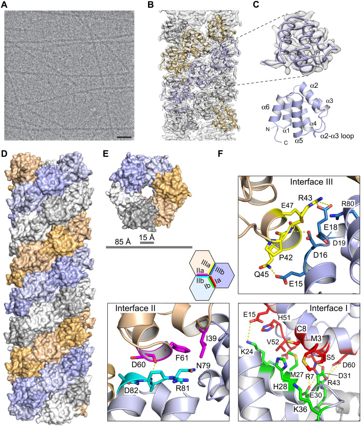
Fig. 1.
Cryo-EM structure of the NLRP3 PYD filament.
( A ) Representative cryo-EM micrograph of NLRP3 PYD sample after incubation at 37u00b0C overnight. Scale bar, 50 nm. ( B ) Superposition of the NLRP3 PYD filament model with the reconstructed electro...
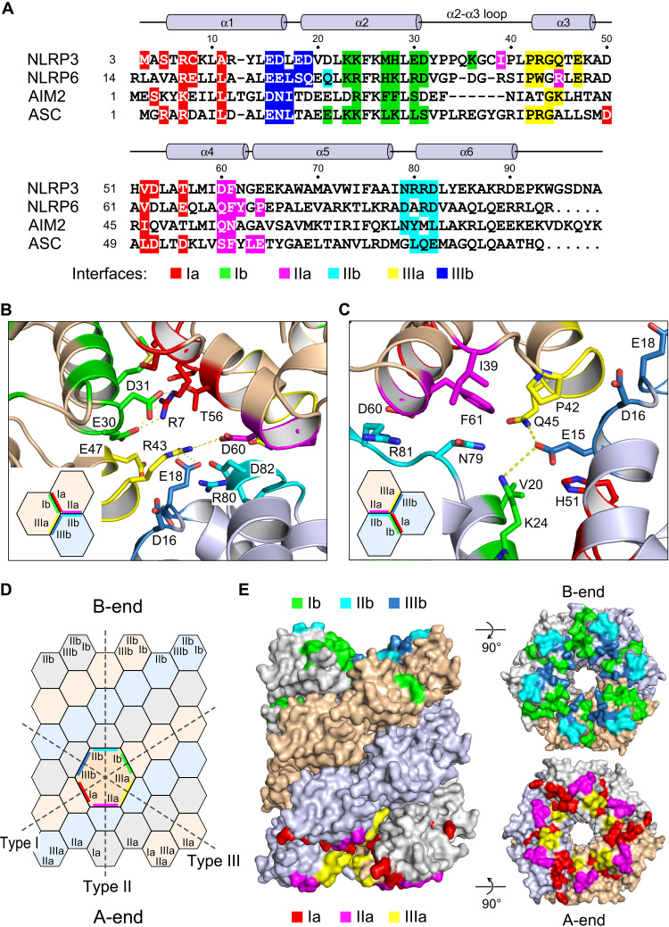
Fig. 2.
Sequence conservation and interface sides in the NLRP3 PYD filament.
( A ) Structure-based sequence alignment of the four PYDs whose filament structures have been determined. Interface-forming residues are highlighted and shown below the secondary structure elements of...
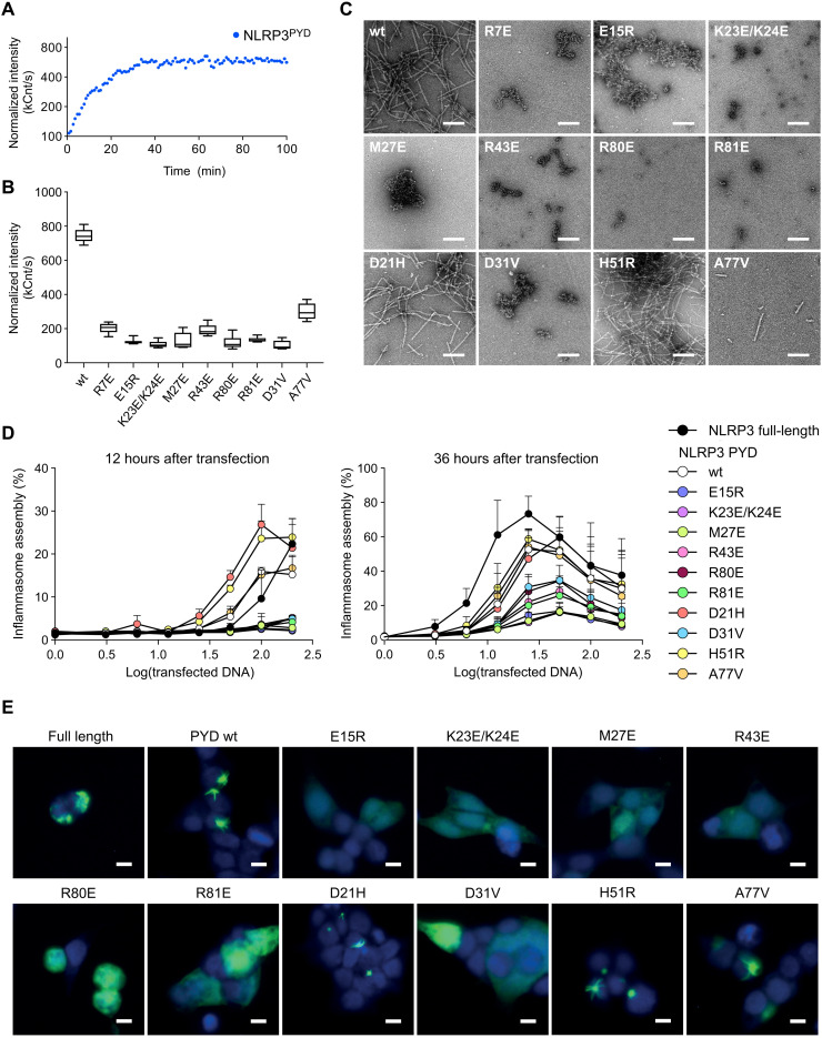
Fig. 3.
Mutational analysis of NLRP3 PYD interface residues.
( A ) NLRP3 PYD polymerization in vitro monitored by DLS. Normalized intensity signals are recorded as a function of time and are shown here for one representative experiment. ( B ) Polymerization sta...
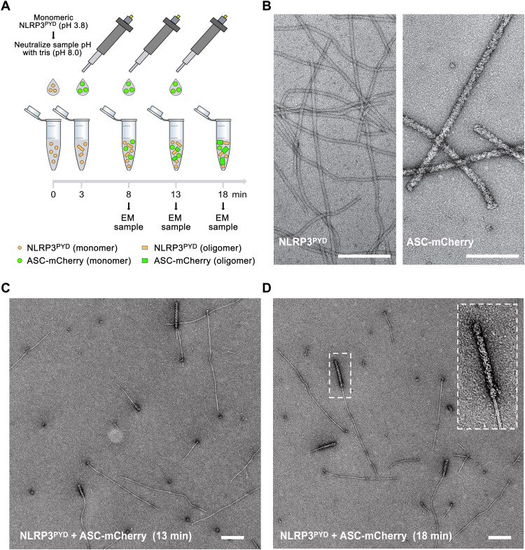
Fig. 4.
ASC filament polymerization on NLRP3 PYD nucleation seeds.
( A ) Cartoon depiction of an NLRP3 PYD -induced ASC filament polymerization assay. Filament formation of monomeric NLRP3 PYD is induced by neutralization to pH 8.0. Stepwise titration of highly dilut...
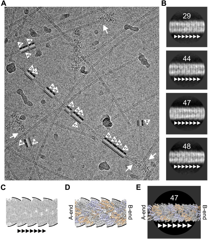
Fig. 5.
ASC exclusively elongates at the B-end of the NLRP3 PYD filament.
( A ) Representative cryo-EM micrograph of NLRP3 PYD filamentu2013tou2013ASC-mCherry filament transitions. Long NLRP3 PYD filaments were grown that allow for analysis of straight segments. The directi...
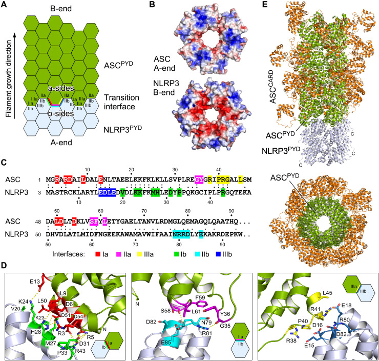
Fig. 6.
Transition of NLRP3 PYD nucleation seeds to ASC PYD filament elongation.
( A ) Cartoon of the filament transition from the nucleation seed NLRP3 PYD to the elongation adaptor ASC PYD . The transition interface of the heteromeric PYD interactions is indicated. ( B ) Electro...
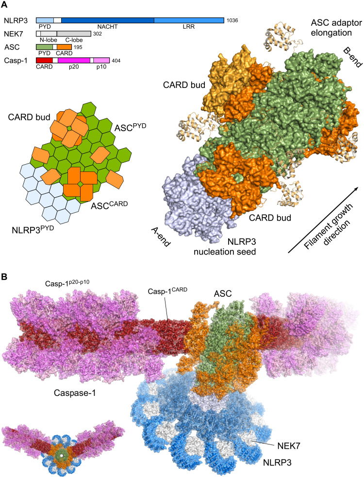
Fig. 7.
Molecular model of an ASC speck.
( A ) Cartoon and atomistic model of the NLRP3 PYD -ASC filament elongation. The molecular domain architecture of human NLRP3, NEK7, ASC, and Caspase-1 (Casp-1) is displayed in bar diagrams. The NLRP3...
Figure images are served from the NIH/NLM PubMed Central Open Access Subset or Europe PMC; copyright remains with the publishers and authors.
💬 Discussion
0 commentsNo comments yet. Be the first to start a discussion!
Leave a Comment