Abstract
During muscle differentiation, myoblasts elongate and fuse into syncytial myotubes [1]. An early event during this process is the remodeling of the microtubule cytoskeleton, involving disassembly of the centrosome and, crucially, the alignment of microtubules into a parallel array along the long axis of the cell [2-5]. To further our understanding on how microtubules support myogenic differentiation, we analyzed the role of EB1-related microtubule-plus-end-binding proteins. We demonstrate that EB3 [6] is specifically upregulated upon myogenic differentiation and that knockdown of EB3, but not that of EB1, prevents myoblast elongation and fusion into myotubes. EB3-depleted cells show disorganized microtubules and fail to stabilize polarized membrane protrusions. Using live-cell imaging, we show that EB3 is necessary for the regulation of microtubule dynamics and microtubule capture at the cell cortex. Expression of EB1/EB3 chimeras on an EB3-depletion background revealed that myoblast fusion depends on two specific amino acids in the calponin-like domain of EB3, whereas the interaction sites with Clip-170 and CLASPs are dispensable. Our results suggest that EB3-mediated microtubule regulation at the cell cortex is a crucial step during myogenic differentiation and might be a general mechanism in polarized cell elongation.
🔬 Techniques
✨ Fluorophores
🧪 Sample Preparation
🔬 Cell Lines
💾 Data Repositories
🏛️ Research Organizations (ROR)
Affiliated research institutions:
📊 Figures
Figureu00a01
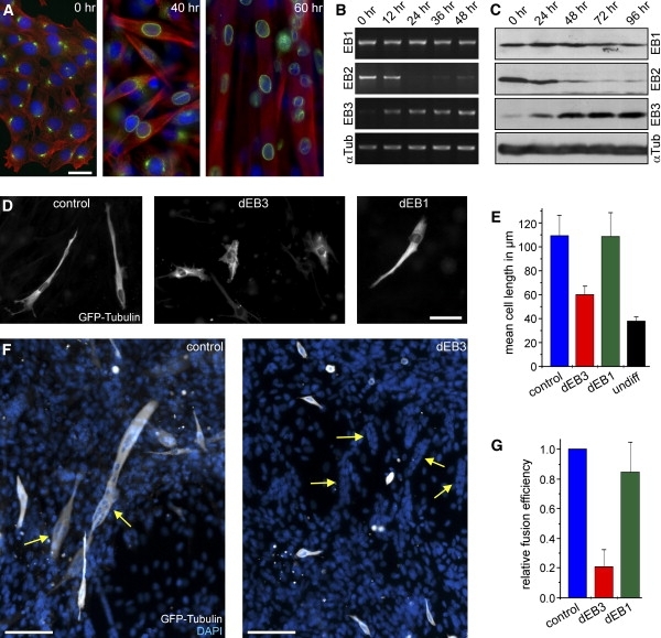
EB Proteins during Myogenic Differentiation (A) Morphological changes and reorganization of the microtubule cytoskeleton. Immunofluorescence of C2C12 cells that were shifted tou00a0differentiation for...
Figureu00a02
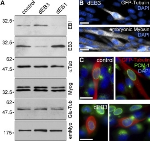
Expression of Myogenic Markers Is Not Affected after EB3 Depletion (A) Western-blot analysis of 20,000 GFP-expressing cells after fluorescence-activated cell sorting (FACS), 3 days after transfection ...
Figureu00a03
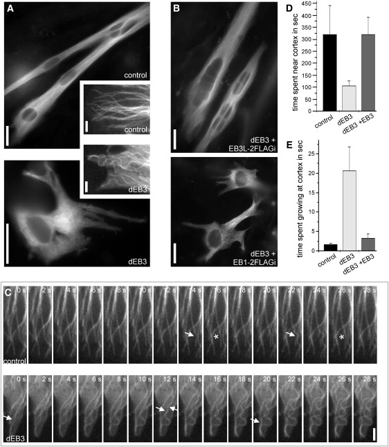
Microtubule Behavior at the Cell Cortex is Altered in EB3-Depleted Cells (A) Microtubule organization in C2C12 cells 3 days after transfection with shRNA constructs and after 52 hr under differentiati...
Figureu00a04
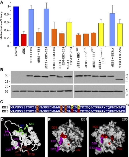
Rescue of EB3 Depletion (A) Quantification of myoblast fusion efficiency after 54u201355 hr under differentiation conditions. The efficiency of cell fusion (percentage cells containing two or more nuc...
Figure images are served from the NIH/NLM PubMed Central Open Access Subset or Europe PMC; copyright remains with the publishers and authors.
💬 Discussion
0 commentsNo comments yet. Be the first to start a discussion!
Leave a Comment