Abstract
Crosstalk between endothelial cells (ECs) and pericytes or vascular smooth muscle cells (VSMCs) is essential for the proper functioning of blood vessels. This balance is disrupted in several vascular diseases but there are few experimental models which recapitulate this vascular cell dialogue in humans. Here, we developed a robust multi-cell type 3D vessel-on-chip (VoC) model based entirely on human induced pluripotent stem cells (hiPSCs). Within a fibrin hydrogel microenvironment, the hiPSC-derived vascular cells self-organized to form stable microvascular networks reproducibly, in which the vessels were lumenized and functional, responding as expected to vasoactive stimulation. Vascular organization and intracellular Ca2+ release kinetics in VSMCs could be quantified using automated image analysis based on open-source software CellProfiler and ImageJ on widefield or confocal images, setting the stage for use of the platform to study vascular (patho)physiology and therapy.
🔬 Techniques
✨ Fluorophores
🧪 Sample Preparation
🔬 Cell Lines
🧪 Reagent Suppliers
💻 Software Details
🏛️ Research Organizations (ROR)
Affiliated research institutions:
📋 Methods
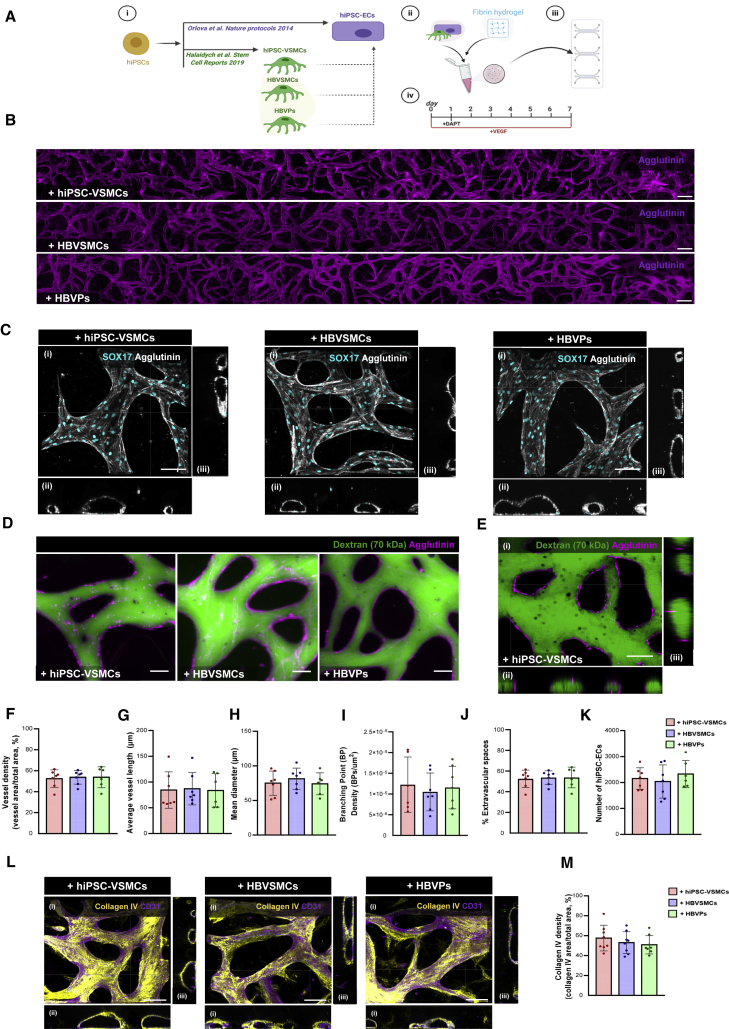
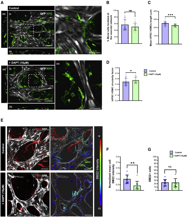
Full details are provided in supplemental experimental procedures . hiPSC lines Research on hiPSC was approved by the medical ethical committee at Leiden University Medical Center, the Netherlands. A detailed list of the hiPSC lines and batches used for each experiment is provided in Table S2 . Differentiation of hiPSC-ECs and hiPSC-VSMCs hiPSC differentiation to ECs was performed as described previously ( Orlova et al., 2014a , 2014b ). hiPSC differentiation to VSMC was performed as previously described ( Halaidych et al., 2019 ). Setting up VoCs hiPSC-ECs and mural cells were prepared prior to incorporation in VoCs as described in supplemental experimental procedures . Commercially available microfluidic chips with one gel channel and two media channels (AIM Biotech) were used. Cells were resuspended and combined to obtain 10 × 10 6 hiPSC-ECs/mL and 2 × 10 6 mural cells/mL (5:1 ratio). Three different mural cell suspensions were tested in combination with hiPSC-ECs: (1) hiPSC-VSMCs, (2) HBVSMCs, and (3) HBVPs. Cell were resuspended in EGM-2 supplemented with Thrombin (4 U/mL) and then gently mixed with fibrinogen (final concentration 3 mg/mL, Sigma) at 1:1 vol ratio. Cell/hydrogel mixture was quickly loaded into the middle gel-loading channel of the microfluidic chip. Chips were incubated at room temperature for 15 min before the addition of EGM-2 supplemented with VEGF (50 ng/mL) to both flanking media channels. The γ-secretase inhibitor DAPT (10 μM) was also added to the medium on day 1 for 24 h. Gravity-driven flow was induced by the addition of 100 μL medium to the right media ports and 50 μL media to left media ports. Medium was refreshed daily.
Show full methods section
Full details are provided in supplemental experimental procedures . hiPSC lines Research on hiPSC was approved by the medical ethical committee at Leiden University Medical Center, the Netherlands. A detailed list of the hiPSC lines and batches used for each experiment is provided in Table S2 . Differentiation of hiPSC-ECs and hiPSC-VSMCs hiPSC differentiation to ECs was performed as described previously ( Orlova et al., 2014a , 2014b ). hiPSC differentiation to VSMC was performed as previously described ( Halaidych et al., 2019 ). Setting up VoCs hiPSC-ECs and mural cells were prepared prior to incorporation in VoCs as described in supplemental experimental procedures . Commercially available microfluidic chips with one gel channel and two media channels (AIM Biotech) were used. Cells were resuspended and combined to obtain 10 × 10 6 hiPSC-ECs/mL and 2 × 10 6 mural cells/mL (5:1 ratio). Three different mural cell suspensions were tested in combination with hiPSC-ECs: (1) hiPSC-VSMCs, (2) HBVSMCs, and (3) HBVPs. Cell were resuspended in EGM-2 supplemented with Thrombin (4 U/mL) and then gently mixed with fibrinogen (final concentration 3 mg/mL, Sigma) at 1:1 vol ratio. Cell/hydrogel mixture was quickly loaded into the middle gel-loading channel of the microfluidic chip. Chips were incubated at room temperature for 15 min before the addition of EGM-2 supplemented with VEGF (50 ng/mL) to both flanking media channels. The γ-secretase inhibitor DAPT (10 μM) was also added to the medium on day 1 for 24 h. Gravity-driven flow was induced by the addition of 100 μL medium to the right media ports and 50 μL media to left media ports. Medium was refreshed daily.
Statistical analysis
Statistical analyses were performed using GraphPad Prism 9 software. Normality of the data was evaluated by the D'Agostino-Pearson test. One-way and two-way ANOVA with Tukey's multiple comparison test was used for the analysis of three groups. For paired or unpaired analysis of two groups, either Student's t test or Wilcoxon-Mann-Whitney test was used. Analyses are indicated in the figure legends. The data are reported as mean ± SD. Statistical significance was defined as p < 0.05.
Supplemental information Document S1. Supplemental experimental procedures, Figures S1–S4, and Tables S1 and S2 Document S2. Article plus supplemental information
📊 Figures
Figureu00a01
Characterization of VoC (A) Schematic of the VoC protocol. hiPSC-ECs were cultured with hiPSC-VSMCs, HBVSMCs, or HBVPs (i). Cells were mixed in a fibrin hydrogel (ii) and injected into (AIM Biotech) m...
Figureu00a02
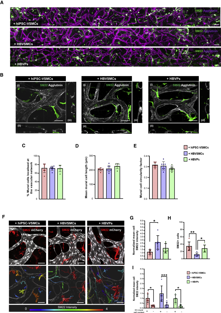
Quantitative assessment of the structural proprieties of hiPSC-VSMCs and primary mural cells in VoC (A) Representative immunofluorescence images of microvascular network showing the hiPSC-ECs (magenta...
Figureu00a03
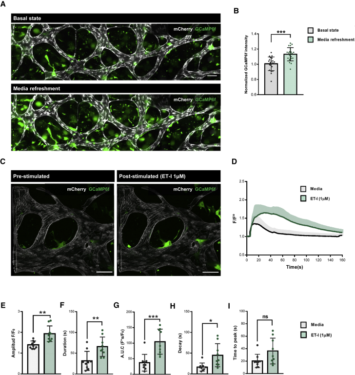
Analysis of hiPSC-VSMCs Ca 2+ dynamics in VoC (A) Representative immunofluorescent images of intracellular Ca 2+ fluorescence showing hiPSC-ECs (gray; mCherry) and hiPSC-VSMCs (green; GCaMP6f) without...
Figureu00a04
Modeling loss of EC-VSMC crosstalk in VoC (A) Representative confocal images of microvascular network showing in hiPSC-ECs (gray; GFP) and hiPSC-VSMCs (green; RFP) in control and DAPT (10u00a0u03bcM) ...
Figure images are served from the NIH/NLM PubMed Central Open Access Subset or Europe PMC; copyright remains with the publishers and authors.
💬 Discussion
0 commentsNo comments yet. Be the first to start a discussion!
Leave a Comment