Abstract
Six genes encode proteins with acyl-CoA-binding domains in Arabidopsis thaliana. They are the small 10-kDa cytosolic acyl-CoA-binding protein (ACBP), membrane-associated ACBP1 and ACBP2, extracellularly-targeted ACBP3, and kelch-motif containing ACBP4 and ACBP5. Here, the interaction of ACBP4 with an A. thaliana ethylene-responsive element binding protein (AtEBP), identified in a yeast two-hybrid screen, was confirmed by co-immunoprecipitation. The subcellular localization of ACBP4 and AtEBP, was addressed using an ACBP4:DsRed red fluorescent protein fusion and a green fluorescent protein (GFP):AtEBP fusion. Transient expression of these autofluoresence-tagged proteins in agroinfiltrated tobacco leaves, followed by confocal laser scanning microscopy, indicated their co-localization predominantly at the cytosol which was confirmed by FRET analysis. Immuno-electron microscopy on Arabidopsis sections not only localized ACBP4 to the cytosol but also to the periphery of the nucleus upon closer examination, perhaps as a result of its interaction with AtEBP. Furthermore, the expression of ACBP4 and AtEBP in Northern blot analyses was induced by the ethylene precursor 1-aminocyclopropane-1-carboxylic acid, methyl jasmonate treatments, and Botrytis cinerea infection, suggesting that the interaction of ACBP4 and AtEBP may be related to AtEBP-mediated defence possibly via ethylene and/or jasmonate signalling.
🔬 Techniques
🔭 Microscopes
🧬 Organisms
✨ Fluorophores
🧪 Sample Preparation
🏭 Microscope Brands
🧪 Reagent Suppliers
📷 Detectors
🏛️ Research Organizations (ROR)
Affiliated research institutions:
📋 Methods
Yeast strain The two-hybrid library screens were performed in the Saccharomyces cerevisiae strain YPB2 [ MATa ara3 his3 ade2 lys2 trp1 leu2, 112 can r gal4 gal80 LYS2::GAL1-HIS3, URA3:: ( GAL1 UAS 17mers) -lacZ ] ( Kohalmi et al. , 1998 ). Cotransformants were plated on synthetic dextrose agar plates lacking leucine, tryptophan, and histidine [SD-leu-trp-his] supplemented with 10 mM 3-AT ( Kohalmi et al. , 1998 ). Construction of a bait vector of GAL4(DB)-ACBP4 fusion The bait plasmid pAT188 was prepared by inserting a 2 kb Xho I- Not I fragment encoding ACBP4 from pAT181 ( Leung et al. , 2004 ) into the Sal I- Not I sites of pBI-880 (a variant of pPC62 as described by Chevray and Nathans, 1992 ; Kohalmi et al. , 1998 ). All constructs were confirmed by restriction digestion and nucleotide sequence analysis.
Yeast two-hybrid screening S. cerevisiae strain
YPB2 was transformed with bait plasmid pAT188 and transformants were plated on synthetic dextrose agar plates lacking leucine [SD-leu]. An aliquot of transformants was also tested on [SD-leu-his] medium supplemented with 10 mM 3-amino-1, 2, 4-triazole (3-AT) because an absence of growth on this medium would confirm that the DB-‘bait’ fusion protein is unable to initiate transcription of HIS3 . Subsequently, the bait-carrying strain was tested negative for β-galactosidase activity using the X-Gal (5-bromo-4-chloro-3-indolyl-β- D -galactopyranoside) colony filter assay. This further showed that the bait was not able to activate transcription of the lacZ reporter gene. The prey vector pBI-771, a variant of pPC86 ( Chevray and Nathans, 1992 ; Kohalmi et al. , 1998 ), was introduced into this strain and its inability to grow on [SD-leu-trp-his] medium supplemented with 10 mM 3-AT and its lack of β-galactosidase activity were confirmed before the bait was further used in cDNA library screening. To ensure sufficient coverage in the identification of potential proteins interacting with ACBP4, yeast two-hybrid screenings were also performed at the Molecular Interaction Facility, University of Wisconsin–Madison using yeast strains and vectors as previously described by James et al. (1996) . For bait preparation, ACBP4 (amino acids 1–669) was cloned in-frame with the GAL4 DNA-binding domain of bait vector pBUTE (a kanamycin-resistant version of GAL4 bait vector pGBDUC1). The resulting vector was subject to DNA sequence analysis to confirm the presence of an in-frame fusion, before use in transformation of S. cerevisiae mating type strain PJ69-4A, followed by testing for autoactivation of the β-galactosidase reporter gene. Library screenings were conducted using the Molecular Interaction Facility Arabidopsis library collection representing cDNAs from flowering Arabidopsis plants. Approximately 50 million clones were screened. Of these, positive yeast clones were tested for interaction by selection on histidine drop-out and β-galactosidase assays. Plasmids were rescued and analysed by restriction endonuclease analysis. Positive prey plasmids were retransformed into the mating type of PJ69-4A and validated in mating and selection assays with the ACBP4 bait, the empty bait vector, and unrelated control baits. Positive clones were subsequently identified by nucleotide sequence analysis using the GAL ( TA) -specific forward primer BC304 (5′-CTATTCGATGATGAAGATACC-3′) and the ADH1 -terminator reverse primer, JN069 (5′-TTGATTGGAGACTTGACC-3′) ( Kohalmi et al. , 1998 ). Co-immunoprecipitation To corroborate the interaction from yeast two-hybrid analysis, co-immunoprecipitation studies were performed according to Mongiat et al. (2003) . All constructs used in these interaction assays were derivatives of vector pBluescriptII KS(–) (pKS). The Hin dIII- Sac I fragment from pBI-771 carrying GAL4(TA) (amino acids 768–881) was cloned into corresponding restriction sites on pKS. The GAL4(TA)-ACBP4 fusion construct was prepared by inserting ACBP4 cDNA from pAT181, on a 2 kb Eco RI- Bam HI fragment, into the Eco RI- Bgl II sites of pKS-TA with the 5′ of TA-ACBP4 adjacent to the T3 promoter. Two putative interactors, ADF3 (identified at the Molecular Interaction Facility, University of Wisconsin–Madison) and AtEBP (from a yeast two-hybrid screen in our laboratory) were selected for further studies. Their full-length cDNAs were generated by the Reverse-Transcriptase-Polymerase Chain Reaction (RT-PCR) using the Superscript™ First-strand synthesis system (Invitrogen, Carlsbad, CA, USA). The cDNA fragments were subsequently cloned into pGEM-T Easy (Promega, Madison, WI, USA). Potential ‘ATG’ start codons in the multiple cloning sites of pGEM-T Easy vector upstream of the ADF3 or AtEBP cDNA were eliminated by restriction endonuclease digestion followed by filling-in with Klenow and re-ligation. The cDNAs of both ADF3 and AtEBP were verified by nucleotide sequence analysis. Subsequently, GAL4(TA)-ACBP4 and each candidate were in vitro transcribed and translated by a TNT quick coupled wheat germ transcription-translation system (Promega, Madison, WI, USA) in the presence of [ 35 S]methionine (ICN Pharmaceuticals Inc., Costa Mesa, CA, USA), according to the manufacturer's instructions. The proteins were analysed by 12% sodium dodecyl sulphate-polyacrylamide gel electrophoresis (SDS–PAGE) and autoradiography. Co-immunoprecipitation with monoclonal anti-GAL4(TA) antibody (Clontech, USA) was performed following Mongiat et al. (2003) .
Show full methods section
Yeast strain The two-hybrid library screens were performed in the Saccharomyces cerevisiae strain YPB2 [ MATa ara3 his3 ade2 lys2 trp1 leu2, 112 can r gal4 gal80 LYS2::GAL1-HIS3, URA3:: ( GAL1 UAS 17mers) -lacZ ] ( Kohalmi et al. , 1998 ). Cotransformants were plated on synthetic dextrose agar plates lacking leucine, tryptophan, and histidine [SD-leu-trp-his] supplemented with 10 mM 3-AT ( Kohalmi et al. , 1998 ). Construction of a bait vector of GAL4(DB)-ACBP4 fusion The bait plasmid pAT188 was prepared by inserting a 2 kb Xho I- Not I fragment encoding ACBP4 from pAT181 ( Leung et al. , 2004 ) into the Sal I- Not I sites of pBI-880 (a variant of pPC62 as described by Chevray and Nathans, 1992 ; Kohalmi et al. , 1998 ). All constructs were confirmed by restriction digestion and nucleotide sequence analysis.
Yeast two-hybrid screening S. cerevisiae strain
YPB2 was transformed with bait plasmid pAT188 and transformants were plated on synthetic dextrose agar plates lacking leucine [SD-leu]. An aliquot of transformants was also tested on [SD-leu-his] medium supplemented with 10 mM 3-amino-1, 2, 4-triazole (3-AT) because an absence of growth on this medium would confirm that the DB-‘bait’ fusion protein is unable to initiate transcription of HIS3 . Subsequently, the bait-carrying strain was tested negative for β-galactosidase activity using the X-Gal (5-bromo-4-chloro-3-indolyl-β- D -galactopyranoside) colony filter assay. This further showed that the bait was not able to activate transcription of the lacZ reporter gene. The prey vector pBI-771, a variant of pPC86 ( Chevray and Nathans, 1992 ; Kohalmi et al. , 1998 ), was introduced into this strain and its inability to grow on [SD-leu-trp-his] medium supplemented with 10 mM 3-AT and its lack of β-galactosidase activity were confirmed before the bait was further used in cDNA library screening. To ensure sufficient coverage in the identification of potential proteins interacting with ACBP4, yeast two-hybrid screenings were also performed at the Molecular Interaction Facility, University of Wisconsin–Madison using yeast strains and vectors as previously described by James et al. (1996) . For bait preparation, ACBP4 (amino acids 1–669) was cloned in-frame with the GAL4 DNA-binding domain of bait vector pBUTE (a kanamycin-resistant version of GAL4 bait vector pGBDUC1). The resulting vector was subject to DNA sequence analysis to confirm the presence of an in-frame fusion, before use in transformation of S. cerevisiae mating type strain PJ69-4A, followed by testing for autoactivation of the β-galactosidase reporter gene. Library screenings were conducted using the Molecular Interaction Facility Arabidopsis library collection representing cDNAs from flowering Arabidopsis plants. Approximately 50 million clones were screened. Of these, positive yeast clones were tested for interaction by selection on histidine drop-out and β-galactosidase assays. Plasmids were rescued and analysed by restriction endonuclease analysis. Positive prey plasmids were retransformed into the mating type of PJ69-4A and validated in mating and selection assays with the ACBP4 bait, the empty bait vector, and unrelated control baits. Positive clones were subsequently identified by nucleotide sequence analysis using the GAL ( TA) -specific forward primer BC304 (5′-CTATTCGATGATGAAGATACC-3′) and the ADH1 -terminator reverse primer, JN069 (5′-TTGATTGGAGACTTGACC-3′) ( Kohalmi et al. , 1998 ). Co-immunoprecipitation To corroborate the interaction from yeast two-hybrid analysis, co-immunoprecipitation studies were performed according to Mongiat et al. (2003) . All constructs used in these interaction assays were derivatives of vector pBluescriptII KS(–) (pKS). The Hin dIII- Sac I fragment from pBI-771 carrying GAL4(TA) (amino acids 768–881) was cloned into corresponding restriction sites on pKS. The GAL4(TA)-ACBP4 fusion construct was prepared by inserting ACBP4 cDNA from pAT181, on a 2 kb Eco RI- Bam HI fragment, into the Eco RI- Bgl II sites of pKS-TA with the 5′ of TA-ACBP4 adjacent to the T3 promoter. Two putative interactors, ADF3 (identified at the Molecular Interaction Facility, University of Wisconsin–Madison) and AtEBP (from a yeast two-hybrid screen in our laboratory) were selected for further studies. Their full-length cDNAs were generated by the Reverse-Transcriptase-Polymerase Chain Reaction (RT-PCR) using the Superscript™ First-strand synthesis system (Invitrogen, Carlsbad, CA, USA). The cDNA fragments were subsequently cloned into pGEM-T Easy (Promega, Madison, WI, USA). Potential ‘ATG’ start codons in the multiple cloning sites of pGEM-T Easy vector upstream of the ADF3 or AtEBP cDNA were eliminated by restriction endonuclease digestion followed by filling-in with Klenow and re-ligation. The cDNAs of both ADF3 and AtEBP were verified by nucleotide sequence analysis. Subsequently, GAL4(TA)-ACBP4 and each candidate were in vitro transcribed and translated by a TNT quick coupled wheat germ transcription-translation system (Promega, Madison, WI, USA) in the presence of [ 35 S]methionine (ICN Pharmaceuticals Inc., Costa Mesa, CA, USA), according to the manufacturer's instructions. The proteins were analysed by 12% sodium dodecyl sulphate-polyacrylamide gel electrophoresis (SDS–PAGE) and autoradiography. Co-immunoprecipitation with monoclonal anti-GAL4(TA) antibody (Clontech, USA) was performed following Mongiat et al. (2003) .
Construction of plasmids used in subcellular localization
All binary vectors used in this study were derivatives of plasmids pGDG and pGDR which contain genes encoding the autofluorescent proteins GFP and DsRed, respectively ( Goodin et al. , 2002 ). The 2 kb Xho I- Bam HI fragment encoding the complete ACBP4 peptide was generated by PCR using primers ML350 and ML682 with pAT181 as template, and cloned into pGEM-T Easy vector to generate plasmid pAT280. The 2 kb Xho I- Bam HI ACBP4 fragment derived from plasmid pAT280 was cloned into the Xho I and Bam HI sites of pGD-DsRed to obtain pAT282 in which ACBP4 is fused to 5′ of DsRed . The plasmid pAT225 in which AtEBP is fused to 3′ of GFP has been previously described ( Li and Chye, 2004 ). The cloning junctions in all constructs were confirmed by nucleotide sequence analysis. Transient expression by agroinfiltration Tobacco ( Nicotiana tabacum var. Xanthi ) plants were grown in a greenhouse at 22 °C for 6 weeks. Two days before agroinfiltration, they were maintained in a growth chamber at 22 °C under 16/8 h light/dark as specified by Goodin et al. (2002) . Derivatives of Agrobacterium tumefaciens strain LBA4404 containing autofluorescent protein fusion constructs were cultured on LB solid medium supplemented with kanamycin (50 μg ml −1 ) and streptomycin (25 μg ml −1 ) at 28 °C for 2 d. For agroinfiltration, Agrobacterium was grown at 28 °C overnight, in LB medium supplemented with kanamycin (50 μg ml −1 ) and streptomycin (25 μg ml −1 ). Preparation of Agrobacterium suspension and agroinfiltration of tobacco leaves in planta were carried out following the procedures of Yang et al. (2000) .
Confocal laser-scanning microscopy
Tobacco leaf epidermal cells from agroinfiltration were examined under a Zeiss LSM 510 inverted confocal laser-scanning microscope (Zeiss, Jena, Germany) following the settings described by Goodin et al. (2002) with minor modifications. Single optical sections were scanned as resulting images for each transient expression. For each plasmid construct, 10–15 cells were imaged with similar results. GFP fluorescence was excited at 488 nm, filtered through a primary dichroic (UV/488/543), a secondary dichroic of 545 nm, and subsequently through BP505–530 nm emission filters to the photomultiplier tube (PMT) detector. DsRed fluorescence was excited at 543 nm, the emission was passed through similar primary and secondary dichroic mirrors and finally through a BP560–615 nm emission filter to the PMT detector.
Fluorescence resonance energy transfer
(FRET) pairs GFP/DsRed were analysed using a confocal laser-scanning microscope (Zeiss LSM510 META). FRET measurements of DsRed emission with zero contribution from GFP, was accomplished as described by Erickson et al. (2003) using the following settings: excitation at 488 nm and emission filters, BP 505–530 nm for GFP and BP 600–637 nm for DsRed.
Western blot analysis
Protein extracts were prepared by homogenizing Arabidopsis protein from 3-week-old wild-type (Col-0) Arabidopsis rosettes according to Chye et al. (1999) . Total proteins were separated on SDS–PAGE and transferred onto Hybond-C membranes (Amersham). The blots were blocked in TTBS (TBS plus 0.05% Tween 20) containing 5% non-fat milk for 2 h and incubated for an additional 2 h with anti-ACBP4 primary antibodies. The blots were washed three times with TTBS and then incubated with secondary antibody for 1 h. Either the Amplified Alkaline Phosphatase Goat Anti-rabbit Immuno-blot Assay Kit (BioRad) or the ECL Western Blotting Detection Kit (Amersham) was used following the manufacturer's instructions to detect cross-reacting bands. ACBP4-specific antibodies were generated by rabbit immunization using a synthetic peptide RMQTLQLRQELGEAE (corresponding to amino acids 566 to 580 of ACBP4).
Immuno-electron microscopy
Arabidopsis leaves were fixed in a solution of 4% (v/v) paraformaldehyde and 0.5% (v/v) glutaradehyde in 0.1 M phosphate buffer (pH 7.2) for 20 min under vacuum and then a further 3 h at room temperature. The specimens were then dehydrated in a graded ethanol series, infiltrated in stepwise increments of LR white resin (London Resin, Theale, Berkshire, UK) and polymerized at 45 °C for 24 h. Materials for immuno-gold labelling were prepared according to the procedure of Varagona and Raikhel (1994) with the modification as described. Specimens (90 nm) were sectioned using a Leica Reichert Ultracut S microtome and mounted on formvar-coated slotted grids. Grids were incubated in a blocking solution of TTBS containing 1% (w/v) fish skin gelatin and 1% (w/v) BSA for 30 min. Anti-ACBP4 antibodies diluted 1:50 in blocking solution were added and incubated at room temperature for 2 h. The grids were then rinsed three times, each for 5 min, in TTBS and then incubated with 10 nm gold-conjugated goat anti-rabbit IgG secondary antibody (Sigma), diluted 1:20 with blocking solution. Grids were rinsed three times, each for 5 min in TTBS, following by three 5-min rinses in distilled water. After being stained in 2% (w/v) uranyl acetate for 6 min followed by 2% (w/v) lead citrate for 6 min, the sections were visualized and photographed using Philips EM208s electron microscope operating at 80 kV. Controls were performed excluding the primary antibody. Plant materials, growth conditions and treatment Tobacco ( N. tabacum var. Xanthi ) plants were grown in a greenhouse at 22 °C for 6 weeks. Two days before agroinfiltration, they were maintained in a growth chamber at 22 °C under 16/8 h dark/light as specified by Goodin et al. (2002) . Arabidopsis thaliana ecotype Columbia (Col-0) was grown under cycles of 8 h dark at 21 °C and 16 h light at 23 °C. For Arabidopsis treatments in northern blot experiments, seedlings were grown on Murashige and Skoog (1962) medium with 2% sucrose in continuous light for 2–3 weeks and then treated with 1 mM 1-aminocyclopropane-1-carboxylic acid (ACC, Sigma-Aldrich, St Louis), 100 μM methyl jasmonate (MeJA, Sigma-Aldrich, St Louis) or water (control). Plant samples were collected at 0, 4, 8, 12, and 24 h post-treatment.
Pathogen infection
Three-week-old wild-type Arabidopsis plants were inoculated with Botrytis cinerea by spraying with a spore suspension (2×10 5 spores ml −1 ) in a solution containing 1% glucose or with water containing 1% glucose as a control. After inoculation, the plants were placed in a growth chamber with high humidity (100%) at 22 °C under a 16/8 h light/dark photoperiod as described by Xiao et al. (2004) . Plant samples were collected at 0, 24, 48, and 72 h post-inoculation. Northern blot analysis Total RNA was isolated from plant tissues following the procedure of Nagy et al. (1988) . Northern blot analysis was performed as described previously ( Xiao et al. , 2004 ). Briefly, 30 μg of total RNA were separated on a 1.5% agarose gel containing 6% formaldehyde and transferred to Hybond N membranes (Amersham). To generate probes for use in Northern blot analyses, specific primers were designed for PCR-amplification: ACBP4 (ML350, 5′-CCTCGAGAATGGCTATGCCTAGGGC-3′ and ML682, 5′-GGATCCACAAGGCGAATCATCATCT-3′), AtEBP (ML826, 5′-ACAGAGAAAATGTGTGGCGG-3′ and ML827, 5′-CAAGCATCCACATAT CCACC-3′) and PDF1.2 (ML741, 5′-TAAGTTTGCTTCCATCATCACCC-3′ and ML742, 5′-TTAACATGGGACGTAACAGATACA-3′). Templates used in PCR were plasmid pAT282 (consisting of the ACBP4 cDNA) and the first-strand wild-type pool of cDNAs (for AtEBP and PDF1.2 ). The fragments were labelled with the PCR Digoxigenin Probe Synthesis Kit according to the manufacturer's instructions (Roche, Germany). Hybridization and detection were performed according to the standard procedures as advised by the manufacturer (Roche). The blots were washed under conditions of high stringency (2× SSC, 0.1% SDS for 2×15 min at room temperature; 0.5× SSC, 0.1% for 2×15 min at 68 °C; 0.1× SSC, 0.1% SDS for 2×15 min at 68 °C).
Plant materials, growth conditions and treatment Tobacco ( N. tabacum var. Xanthi ) plants were grown in a greenhouse at 22 °C for 6 weeks. Two days before agroinfiltration, they were maintained in a growth chamber at 22 °C under 16/8 h dark/light as specified by Goodin et al. (2002) . Arabidopsis thaliana ecotype Columbia (Col-0) was grown under cycles of 8 h dark at 21 °C and 16 h light at 23 °C. For Arabidopsis treatments in northern blot experiments, seedlings were grown on Murashige and Skoog (1962) medium with 2% sucrose in continuous light for 2–3 weeks and then treated with 1 mM 1-aminocyclopropane-1-carboxylic acid (ACC, Sigma-Aldrich, St Louis), 100 μM methyl jasmonate (MeJA, Sigma-Aldrich, St Louis) or water (control). Plant samples were collected at 0, 4, 8, 12, and 24 h post-treatment.
📊 Figures
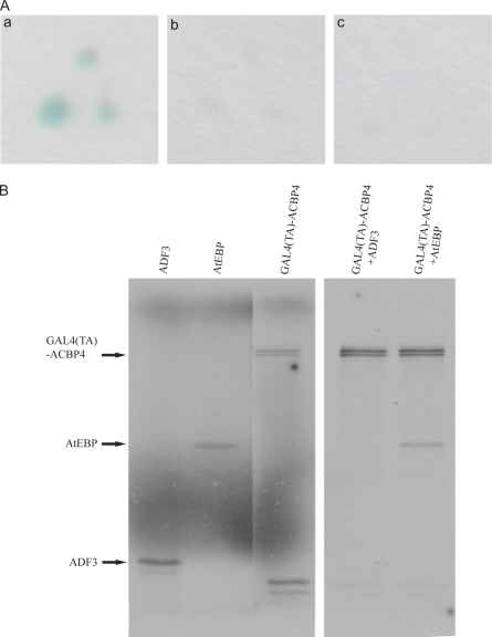
Fig. 1.
(A) Colony filter u03b2-galactosidase assays of candidate proteins AtEBP from yeast two-hybrid screens. (a) YPB2/GAL4(DB)-ACBP4+GAL4(TA)-AtEBP; (b) YPB2/GAL4(DB)-ACBP4+GAL4(TA); (c) YPB2/GAL4(DB)+GAL(...

Fig. 2.
Confocal images indicating co-localization of ACBP4:DsRed and GFP:AtEBP fusion proteins transiently-expressed in tobacco leaves. Representative tobacco leaf epidermal cells are shown by laser-scanning...
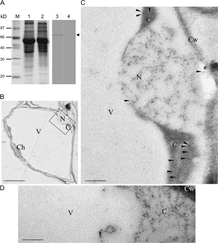
Fig. 3.
Localization of ACBP4 in Arabidopsis leaves. (A) Western blot analysis using affinity-purified anti-peptide antibodies against ACBP4. Lanes 1 and 2: gel identically loaded as lanes 3 and 4, respective...

Fig. 4.
Expression patterns of ACBP4 and AtEBP in Arabidopsis on Northern blot analyses. Total RNAs were extracted from wild-type Arabidopsis leaves (L), stems (S), roots (R), flowers (F), and siliques (Si). ...
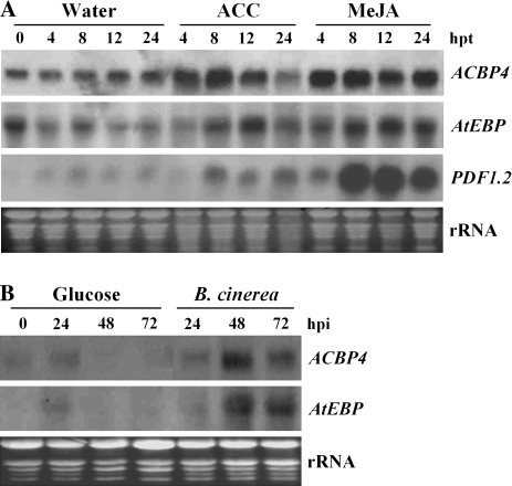
Fig. 5.
Northern blot analyses of ACBP4 and AtEBP expression following 1-aminocyclopropane-1-carboxylic acid and methyl jasmonate treatments and B. cinerea infection. (A) Accumulation of ACBP4 and AtEBP trans...
Figure images are served from the NIH/NLM PubMed Central Open Access Subset or Europe PMC; copyright remains with the publishers and authors.
💬 Discussion
0 commentsNo comments yet. Be the first to start a discussion!
Leave a Comment