Abstract
Cells contain numerous proteases, which are found at many different locations. These proteases recognize an even larger number of different substrates and are involved in almost every process in the cell. Aberrations in proteolysis are linked to a plethora of diseases, such as cancer, inflammation, arteriosclerosis, neurodegeneration and infection. Because of their well-defined chemistry and key role in pathologies, proteases have been important targets for drug development. Recent progress in the development of fluorescent probes has opened up the possibility of visualizing protease activities in the natural environment of the cell. We will describe various strategies to follow protease activities in cells and organisms. Our body contains many different protease and proteolytic systems that are involved in the recycling of proteins into amino acids, and also in a multitude of regulatory events inside and outside cells. Proteases are prominent drug targets because of their well-defined chemistry and their implication in a large number of diseases, such as cancer, neurodegeneration, arteriosclerosis, inflammation and infection. Fluorescent reporter substrates can be used to directly probe the activities of proteases in their natural environment — that is, in cells and organisms. Conceptually different strategies have been used for this purpose depending on the location and the nature of the protease of interest. Fluorescent reporters for the ubiquitin–proteasome system have been generated by linking constitutively active degradation signals to green fluorescent protein (GFP). These GFP-based substrates can be used for functional analysis of the ubiquitin–proteasome system in cells and transgenic animals. A collection of different proteases is involved in degradation of small peptide fragments. This process can be followed in real time in living cells by confocal laser scanning microscopy after microinjection of internally quenched peptide substrates. Extracellular and lysosomal proteases have the advantage that they are accessible for membrane-impermeable reporter substrates. Near-infrared fluorescence (NIRF) substrates are quenched fluorescent peptides that, because of their near-infrared excitation, can be readily detected in living animals and used for in vivo monitoring of, for example, lysosomal cathepsins or surface matrix metalloproteinases. By combining specific pairs of fluorescent proteins (GFP and its variants with shifted excitation and emission spectra) in fusion proteins, fluorescence energy transfer (FRET) reporter substrates have been generated for initiator and effector caspases. A fluorescent intracellular reporter for human immunodeficiency virus (HIV-1) protease activity was constructed by fusing a protease precursor protein composed of HIV-1 protease and GFP. Cells will only survive and emit fluorescence when the toxic protease activity is sufficiently blocked by drugs. The diffusion rate of the endoplasmic reticulum-resident peptide transporter complex TAP correlates with activity and thus cytosolic peptide levels. By measuring the diffusion of TAP–GFP fusions with fluorescence recovery after photobleaching (FRAP), the kinetics of peptide generation can be followed in living cells.
🔬 Techniques
💻 Software
✨ Fluorophores
🧪 Sample Preparation
🔬 Cell Lines
🏭 Microscope Brands
🧪 Reagent Suppliers
💻 Software Details
🏛️ Research Organizations (ROR)
Affiliated research institutions:
📊 Figures
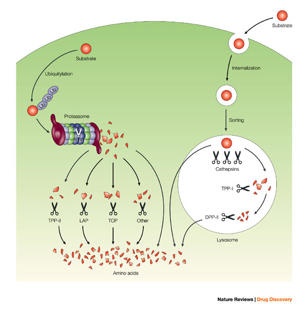
Figure 1
Recycling of intracellular and internalized proteins.
Proteins present in the cytosol and nucleus are degraded by the proteasome into peptide fragments that are further degraded by TPP-II, TOP, LAP and other peptidases into single amino acids. Internaliz...
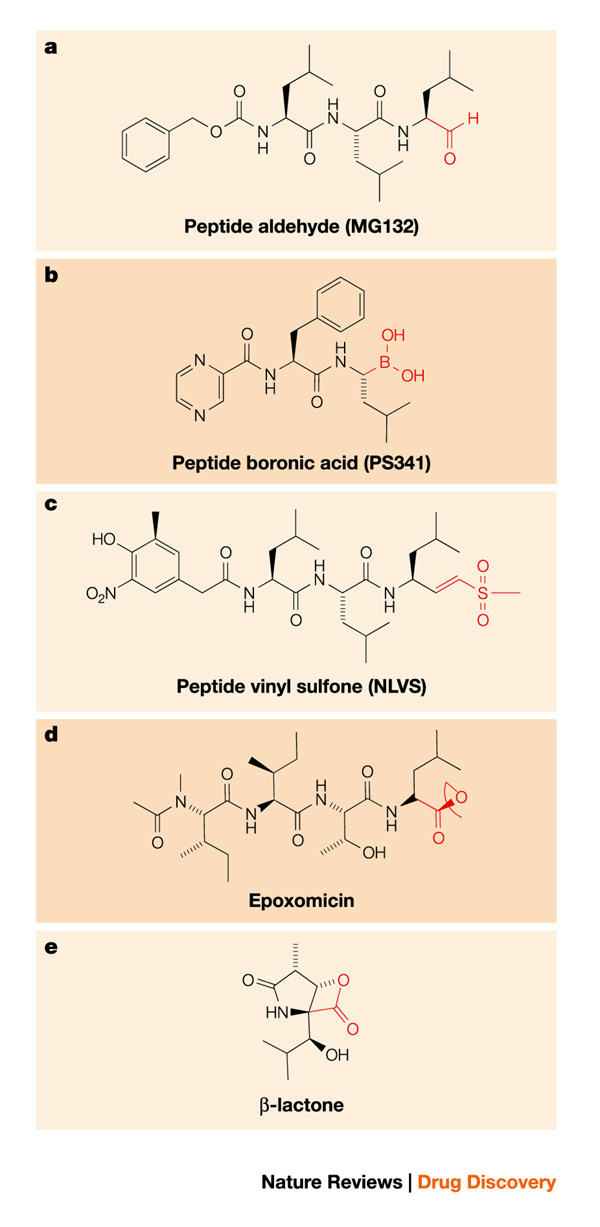
Figure 2
Inhibitors of the proteasome.
Structure of proteasome inhibitors. a | Peptide aldehyde. b | Peptide boronic acid. c | Peptide vinyl sulphone. d | Epoxomicin. e | u03b2-lactone. Note that a u2013 c are carboxy-modified peptides.
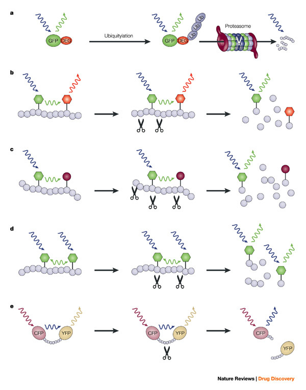
Figure 3
Fluorescent probes for proteolysis.
a | Destruction. Coupling of a degradation signal (DEG) to GFP will result in targeting of the GFP for ubiquitylation followed by proteasomal degradation. Degradation of the GFP substrate results in l...
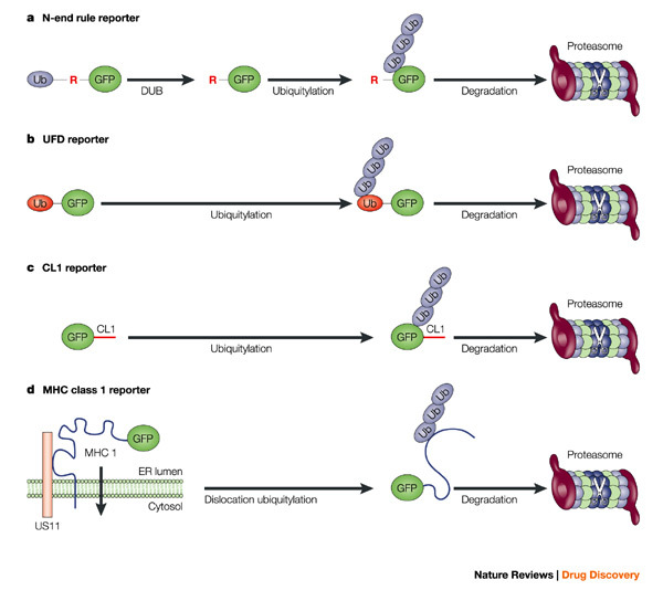
Figure 4
GFP-based proteasome substrates.
a | N-end rule GFP. A fusion of ubiquitin and GFP is expressed. The endogenous de-ubiquitylation enzymes (DUB) rapidly remove the N-terminal ubiquitin, which generates a GFP with an N-terminal arginin...
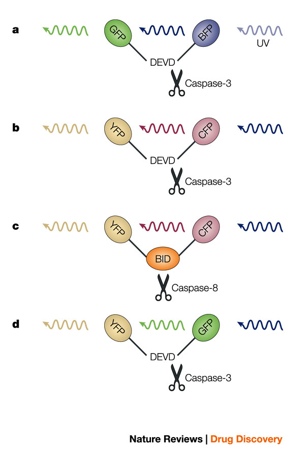
Figure 5
FRET/FLIM reporters for caspases.
a | Caspase-3 fluorescence resonance energy transfer (FRET) probe. The DEVD caspase cleavage site is positioned between GFP and BFP. b | Caspase-3 FRET probe. The DEVD caspase cleavage site is positio...
Figure images are served from the NIH/NLM PubMed Central Open Access Subset or Europe PMC; copyright remains with the publishers and authors.
💬 Discussion
0 commentsNo comments yet. Be the first to start a discussion!
Leave a Comment