Abstract
Abstract Caenorhabditis elegans lives in a complex habitat in which they routinely experience large fluctuations in temperature, and encounter physical obstacles that vary in size and composition. Their habitat is shared by other nematodes, by beneficial and harmful bacteria, and nematode-trapping fungi. Not surprisingly, these nematodes can detect and discriminate among diverse environmental cues, and exhibit sensory-evoked behaviors that are readily quantifiable in the laboratory at high resolution. Their ability to perform these behaviors depends on <100 sensory neurons, and this compact sensory nervous system together with powerful molecular genetic tools has allowed individual neuron types to be linked to specific sensory responses. Here, we describe the sensory neurons and molecules that enable C. elegans to sense and respond to physical stimuli. We focus primarily on the pathways that allow sensation of mechanical and thermal stimuli, and briefly consider this animal’s ability to sense magnetic and electrical fields, light, and relative humidity. As the study of sensory transduction is critically dependent upon the techniques for stimulus delivery, we also include a section on appropriate laboratory methods for such studies. This chapter summarizes current knowledge about the sensitivity and response dynamics of individual classes of C. elegans mechano- and thermosensory neurons from in vivo calcium imaging and whole-cell patch-clamp electrophysiology studies. We also describe the roles of conserved molecules and signaling pathways in mediating the remarkably sensitive responses of these nematodes to mechanical and thermal cues. These studies have shown that the protein partners that form mechanotransduction channels are drawn from multiple superfamilies of ion channel proteins, and that signal transduction pathways responsible for temperature sensing in C. elegans share many features with those responsible for phototransduction in vertebrates.
🔬 Techniques
🧬 Organisms
✨ Fluorophores
🧪 Sample Preparation
🏛️ Research Organizations (ROR)
Affiliated research institutions:
📊 Figures
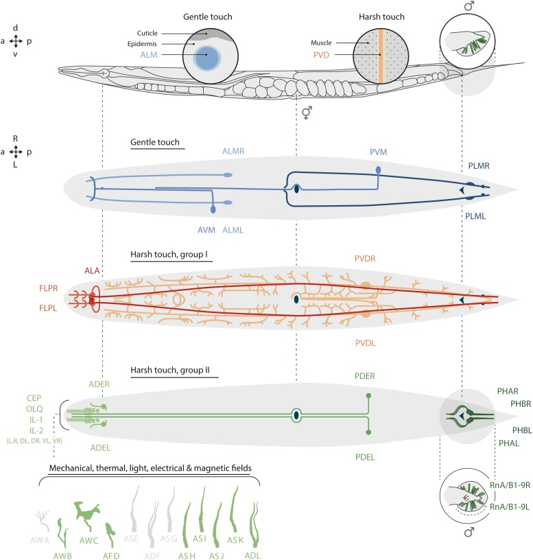
Figure 1
Positions of sensory neurons in adult C. elegans . (Top) Lateral view of an adult hermaphrodite and male tail. Insets illustrate the association of gentle touch receptor neurons (TRNs; only ALM is sho...
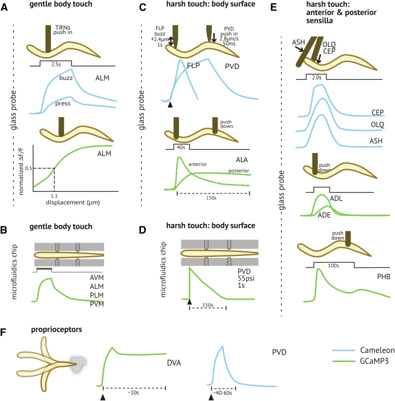
Figure 2
Mechanical stimuli activate calcium transients in mechanoreceptor neurons. Schematics depict the stimulation position, intensity, and dynamics as well as calcium transients monitored using the ratiome...
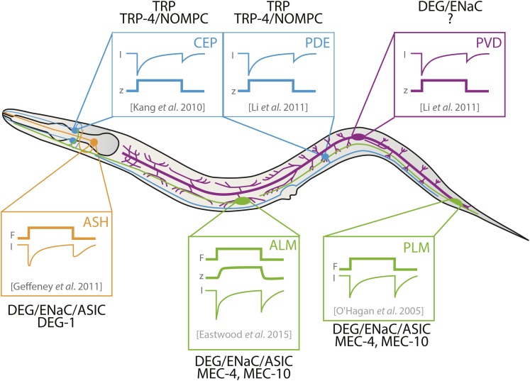
Figure 3
Dynamics of mechanoreceptor currents (MRCs) recorded from C. elegans neurons in vivo . Shown (schematically) are the first reported measurements of MRCs in PDE ( Li et al. 2011 ), ASH ( Geffeney et al...
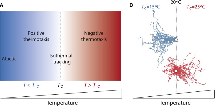
Figure 4
C. elegans exhibits T c -dependent navigation behaviors on spatial thermal gradients. (A) Schematic of navigation behaviors exhibited at temperatures relative to T c ( T c is defined here as the tempe...
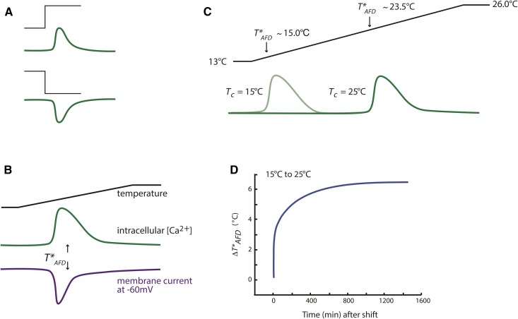
Figure 5
The AFD neurons exhibit responses to warming and cooling in a T c experience-dependent manner. (A) Schematic of changes in intracellular calcium in AFD (green lines) in response to a warming (top) or ...
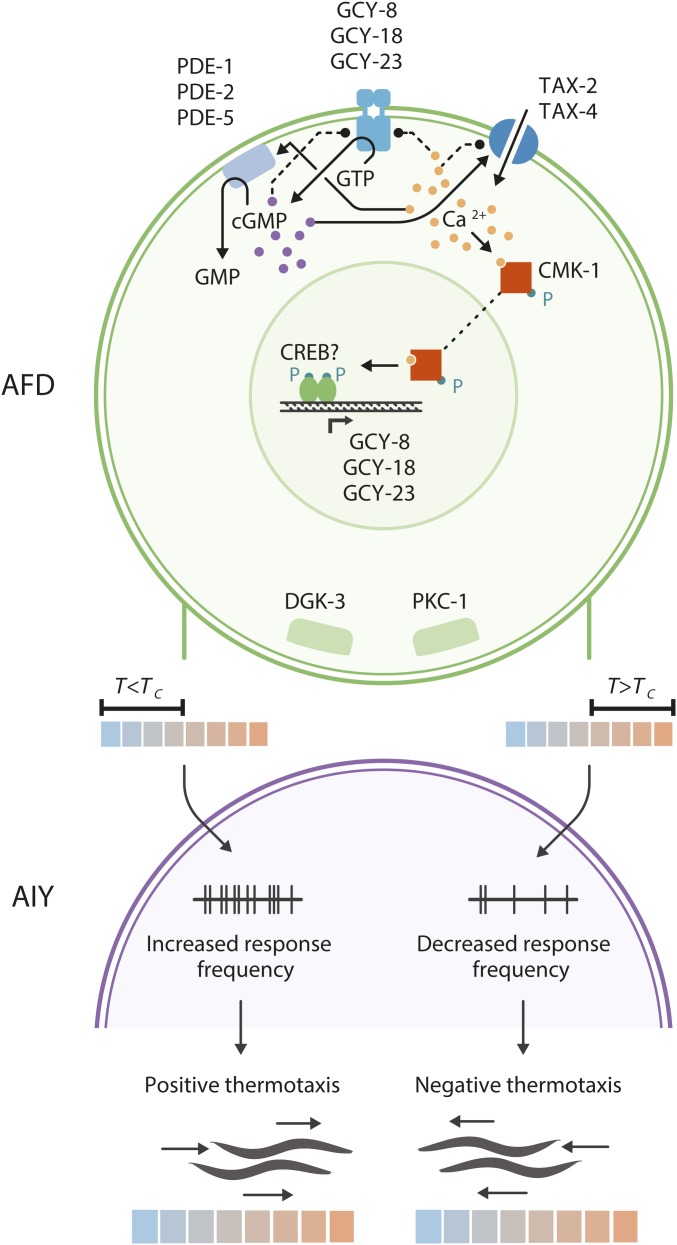
Figure 6
Schematic of thermosensory signal transduction and synaptic output of AFD. Upon warming, GCY-8, GCY-18, and GCY-23 are activated to increase intracellular cGMP levels. The TAX-2/TAX-4 cGMP-gated chann...
Figure images are served from the NIH/NLM PubMed Central Open Access Subset or Europe PMC; copyright remains with the publishers and authors.
💬 Discussion
0 commentsNo comments yet. Be the first to start a discussion!
Leave a Comment