Abstract
Viruses with membranes fuse them with cellular membranes, to transfer their genomes into cells at the beginning of infection. For Influenza virus, the membrane glycoprotein involved in fusion is the hemagglutinin (HA), the 3D structure of which is known from X-ray crystallographic studies. The soluble ectodomain fragments used in these studies lacked the "membrane anchor" portion of the molecule. Since this region has a role in membrane fusion, we have determined its structure by analyzing the intact, full-length molecule in a detergent micelle, using cryo-EM. We have also compared the structures of full-length HA-detergent micelles with full-length HA-Fab complex detergent micelles, to describe an infectivity-neutralizing monoclonal Fab that binds near the ectodomain membrane anchor junction. We determine a high-resolution HA structure which compares favorably in detail with the structure of the ectodomain seen by X-ray crystallography; we detect, clearly, all five carbohydrate side chains of HA; and we find that the ectodomain is joined to the membrane anchor by flexible, eight-residue-long, linkers. The linkers extend into the detergent micelle to join a central triple-helical structure that is a major component of the membrane anchor.
🔬 Techniques
🔭 Microscopes
💻 Software
🧪 Sample Preparation
🏭 Microscope Brands
📷 Detectors
💻 Software Details
💾 Data Repositories
🏛️ Research Organizations (ROR)
Affiliated research institutions:
📋 Methods
Protein Preparation. Full-length HA protein was isolated from purified virus. The virus, A/duck/Alberta/35/76(H1N1), was inoculated in 11- to 12-d-old embryonated hens’ eggs and incubated for 48 h. The virus was purified from the allantoic fluid by sucrose gradient centrifugation. To isolate HA alone, virus glycoproteins were extracted from the viral membrane with 1% Lauryldimethylamine N -oxide (LDAO) (all detergents obtained from Anatrace) and purified by anion exchange, and exchanging the detergent to 0.01% Lauryl Maltose Neopentyl Glycol (LMNG). Neuraminidase was removed by affinity chromatography using an oseltamivir−biotin conjugate ( 48 ) immobilized on a streptavidin Sepharose column. The HA was further purified by gel filtration using a Superose 6 Increase 10/300 column in the buffer: 25 mM Tris pH 8, 150 mM NaCl, 1 mM TCEP, 0.01% LMNG. For the HA−Fab complex, HA was purified by extraction with LDAO, transferring the HA to 0.05% Dodecyl Maltoside (DDM) during the anion exchange step. The HA was purified by the same steps as above, but, in DDM and as a final step, the protein was incubated with a molar excess of the FISW84 Fab fragment and 0.1% of the detergent MNA-C12. The complex was purified from excess detergent and free Fab by Superose 6 gel filtration in the buffer: 25 mM Tris pH 8, 150 mM NaCl, 0.002% MNA-C12. Grid Preparation and Data Collection. Grids of full-length HA were plunge-frozen using an Mk III Vitrobot (FEI) at a protein concentration of ∼2 mg/mL, supplemented with 0.1% octyl-β-glucoside (OG) to reduce orientational bias. Then 4 μL of sample was applied to glow-discharged 300-mesh copper R2/4 quantifoil grids and blotted for 5 s before plunging into liquid ethane. Grids of HA−Fab complex were frozen using an Mk IV Vitrobot with protein at a concentration of ∼1.5 mg/mL also supplemented with 0.1% OG. Then 4 uL of sample was applied to glow-discharged 200-mesh copper R2/2 quantifoil grids and blotted for 3 s before plunge-freezing. Both specimens were imaged using a Titan Krios electron microscope operating at 300 kV. Micrographs were recorded in counting mode using a Gatan K2 Summit detector mounted at the end of a Gatan GIF Quantum energy filter operating in zero-loss mode with a slit width of 20 eV. Exposures were 8 s, with a total dose of 43.7 e/Å 2 fractionated into 20 frames with a calibrated pixel size of 1.08 Å. Images were recorded with a defocus of 1 µm to 4 µm. Image Processing. Movie frames were corrected for motion using MotionCor2 ( 49 ), and contrast transfer function was estimated using CTFfind4 ( 50 ). The full-length HA dataset consisted of 10,861 movies, of which 10,348 were used. The HA+FISW84 dataset consisted of 17,329 movies, of which 17,250 were used. Particles were picked from the micrographs using RELION template-based particle picking ( 51 ). This yielded 1,095,719 particles for the full HA dataset and 1,385,993 particles for the +Fab dataset. Particles were initially screened using two rounds of reference-free 2D classification followed by particle sorting. This reduced the particle numbers to 744,665 and 853,524. An initial model was generated from ∼20,000 randomly selected particles for both samples using the ab initio reconstruction program in CryoSPARC ( 52 ). This initial model was used for a first round of RELION 3D classification. This left 531,849 and 473,081 particles in the well-defined classes. High-resolution ectodomain structures of HA and HA+Fab were generated by 3D classification. The detergent micelle and transmembrane density were removed by masking of the initial model; 365,887 and 291,146 particles were used for the final refinements of the HA ectodomain and HA ectodomain+Fab structures. All final refinements were carried out using the Homogeneous Refinement program of CryoSPARC using particles extracted in a 512 × 512 pixel box. Structures which include the transmembrane were generated from the particle stacks from the initial round of 3D classification. The HA+Fab class with the straight transmembrane was generated by RELION refinement of all particles, imposing C3 symmetry followed by signal subtraction ( 53 ). Subtraction masks were designed to leave density for the detergent micelle, linker region, and a small amount of the base of the HA ectodomain. These subtracted particles were subjected to a number of rounds of 3D classification, imposing C3 symmetry using the particle orientations determined in the refinement. In the first two rounds of classification, classes were chosen which had well-defined density in the transmembrane region. The unsubtracted 83,024 particles were refined, imposing C3 symmetry yielding a map of 3.7-Å resolution and were used to build the linking regions (residues 175 to 184). Subclassification led to a smaller class with better-defined density in the transmembrane domain. This class consisted of 23,286 particles of which the unsubtracted particles refined to 4.1-Å resolution when imposing C3 symmetry. Maps with transmembrane domains not aligned with the ectodomain were generated for both the HA and HA+Fab. Signal-subtracted particles were classified using the alignments from C1 RELION refinements of each dataset. Each dataset was classified into 20 classes. Particles from classes with well-defined transmembrane regions were refined, leading to 10 maps of HA and 12 maps of HA+FISW84. One class from the full-length HA dataset had a transmembrane domain with defined secondary structure with an angle of tilt of 52°. This class was made up of 41,043 particles and refined to a map with a global resolution of 4.2 Å. Refined half-maps were sharpened automatically ( 54 ) and global resolution was corrected by high-resolution noise substitution ( 55 ) using the RELION postprocessing tool. Local resolution was estimated using blocres, implemented in CryoSPARC ( 56 ). Angle Measurement. Angles of transmembrane tilt were measured by aligning C1 maps by the HA ectodomain using chimera, reducing the location of the transmembrane to a 120° angular region about the ectodomain trimer. Rotation and tilt were measured relative to the vertical axis. Model Building. High-resolution models of the ectodomain and ectodomain+Fab were constructed using a previously determined structure of the closely related A/swine/Iowa/15/30(H1N1) (PDB ID code: 1RUY) ( 19 ) and a crystal structure of the FISW84 (PDB ID code: 6HKG). The sequence was mutated to that of A/duck/Alberta/35/76(H1N1). This model was manually adjusted in Coot ( 57 ) and real-space−refined using PHENIX ( 58 ), followed by refinement in REFMAC ( 59 ) with half-map validation. Ectodomain and ectodomain+Fab structures were used as the basis for building transmembrane domains. All sequence numbers described are in H3 numbering. Models of tilted transmembrane regions were generated by rigid-body fitting the transmembrane domain of the straight transmembrane class and manually adjusting the position of the linker helices before a single round of real-space refinement in PHENIX to correct structure geometry.
Show full methods section
Protein Preparation. Full-length HA protein was isolated from purified virus. The virus, A/duck/Alberta/35/76(H1N1), was inoculated in 11- to 12-d-old embryonated hens’ eggs and incubated for 48 h. The virus was purified from the allantoic fluid by sucrose gradient centrifugation. To isolate HA alone, virus glycoproteins were extracted from the viral membrane with 1% Lauryldimethylamine N -oxide (LDAO) (all detergents obtained from Anatrace) and purified by anion exchange, and exchanging the detergent to 0.01% Lauryl Maltose Neopentyl Glycol (LMNG). Neuraminidase was removed by affinity chromatography using an oseltamivir−biotin conjugate ( 48 ) immobilized on a streptavidin Sepharose column. The HA was further purified by gel filtration using a Superose 6 Increase 10/300 column in the buffer: 25 mM Tris pH 8, 150 mM NaCl, 1 mM TCEP, 0.01% LMNG. For the HA−Fab complex, HA was purified by extraction with LDAO, transferring the HA to 0.05% Dodecyl Maltoside (DDM) during the anion exchange step. The HA was purified by the same steps as above, but, in DDM and as a final step, the protein was incubated with a molar excess of the FISW84 Fab fragment and 0.1% of the detergent MNA-C12. The complex was purified from excess detergent and free Fab by Superose 6 gel filtration in the buffer: 25 mM Tris pH 8, 150 mM NaCl, 0.002% MNA-C12. Grid Preparation and Data Collection. Grids of full-length HA were plunge-frozen using an Mk III Vitrobot (FEI) at a protein concentration of ∼2 mg/mL, supplemented with 0.1% octyl-β-glucoside (OG) to reduce orientational bias. Then 4 μL of sample was applied to glow-discharged 300-mesh copper R2/4 quantifoil grids and blotted for 5 s before plunging into liquid ethane. Grids of HA−Fab complex were frozen using an Mk IV Vitrobot with protein at a concentration of ∼1.5 mg/mL also supplemented with 0.1% OG. Then 4 uL of sample was applied to glow-discharged 200-mesh copper R2/2 quantifoil grids and blotted for 3 s before plunge-freezing. Both specimens were imaged using a Titan Krios electron microscope operating at 300 kV. Micrographs were recorded in counting mode using a Gatan K2 Summit detector mounted at the end of a Gatan GIF Quantum energy filter operating in zero-loss mode with a slit width of 20 eV. Exposures were 8 s, with a total dose of 43.7 e/Å 2 fractionated into 20 frames with a calibrated pixel size of 1.08 Å. Images were recorded with a defocus of 1 µm to 4 µm. Image Processing. Movie frames were corrected for motion using MotionCor2 ( 49 ), and contrast transfer function was estimated using CTFfind4 ( 50 ). The full-length HA dataset consisted of 10,861 movies, of which 10,348 were used. The HA+FISW84 dataset consisted of 17,329 movies, of which 17,250 were used. Particles were picked from the micrographs using RELION template-based particle picking ( 51 ). This yielded 1,095,719 particles for the full HA dataset and 1,385,993 particles for the +Fab dataset. Particles were initially screened using two rounds of reference-free 2D classification followed by particle sorting. This reduced the particle numbers to 744,665 and 853,524. An initial model was generated from ∼20,000 randomly selected particles for both samples using the ab initio reconstruction program in CryoSPARC ( 52 ). This initial model was used for a first round of RELION 3D classification. This left 531,849 and 473,081 particles in the well-defined classes. High-resolution ectodomain structures of HA and HA+Fab were generated by 3D classification. The detergent micelle and transmembrane density were removed by masking of the initial model; 365,887 and 291,146 particles were used for the final refinements of the HA ectodomain and HA ectodomain+Fab structures. All final refinements were carried out using the Homogeneous Refinement program of CryoSPARC using particles extracted in a 512 × 512 pixel box. Structures which include the transmembrane were generated from the particle stacks from the initial round of 3D classification. The HA+Fab class with the straight transmembrane was generated by RELION refinement of all particles, imposing C3 symmetry followed by signal subtraction ( 53 ). Subtraction masks were designed to leave density for the detergent micelle, linker region, and a small amount of the base of the HA ectodomain. These subtracted particles were subjected to a number of rounds of 3D classification, imposing C3 symmetry using the particle orientations determined in the refinement. In the first two rounds of classification, classes were chosen which had well-defined density in the transmembrane region. The unsubtracted 83,024 particles were refined, imposing C3 symmetry yielding a map of 3.7-Å resolution and were used to build the linking regions (residues 175 to 184). Subclassification led to a smaller class with better-defined density in the transmembrane domain. This class consisted of 23,286 particles of which the unsubtracted particles refined to 4.1-Å resolution when imposing C3 symmetry. Maps with transmembrane domains not aligned with the ectodomain were generated for both the HA and HA+Fab. Signal-subtracted particles were classified using the alignments from C1 RELION refinements of each dataset. Each dataset was classified into 20 classes. Particles from classes with well-defined transmembrane regions were refined, leading to 10 maps of HA and 12 maps of HA+FISW84. One class from the full-length HA dataset had a transmembrane domain with defined secondary structure with an angle of tilt of 52°. This class was made up of 41,043 particles and refined to a map with a global resolution of 4.2 Å. Refined half-maps were sharpened automatically ( 54 ) and global resolution was corrected by high-resolution noise substitution ( 55 ) using the RELION postprocessing tool. Local resolution was estimated using blocres, implemented in CryoSPARC ( 56 ). Angle Measurement. Angles of transmembrane tilt were measured by aligning C1 maps by the HA ectodomain using chimera, reducing the location of the transmembrane to a 120° angular region about the ectodomain trimer. Rotation and tilt were measured relative to the vertical axis. Model Building. High-resolution models of the ectodomain and ectodomain+Fab were constructed using a previously determined structure of the closely related A/swine/Iowa/15/30(H1N1) (PDB ID code: 1RUY) ( 19 ) and a crystal structure of the FISW84 (PDB ID code: 6HKG). The sequence was mutated to that of A/duck/Alberta/35/76(H1N1). This model was manually adjusted in Coot ( 57 ) and real-space−refined using PHENIX ( 58 ), followed by refinement in REFMAC ( 59 ) with half-map validation. Ectodomain and ectodomain+Fab structures were used as the basis for building transmembrane domains. All sequence numbers described are in H3 numbering. Models of tilted transmembrane regions were generated by rigid-body fitting the transmembrane domain of the straight transmembrane class and manually adjusting the position of the linker helices before a single round of real-space refinement in PHENIX to correct structure geometry.
📊 Figures
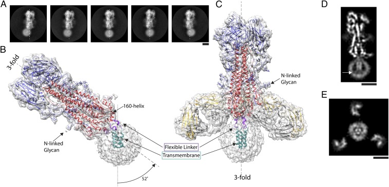
Fig. 1.
The structure of full-length hemagglutinin from A/duck/Alberta/35/76 H1N1 determined by cryo-EM. ( A ) Image classes of similarly oriented HAs illustrating the flexibility of the detergent micelle con...
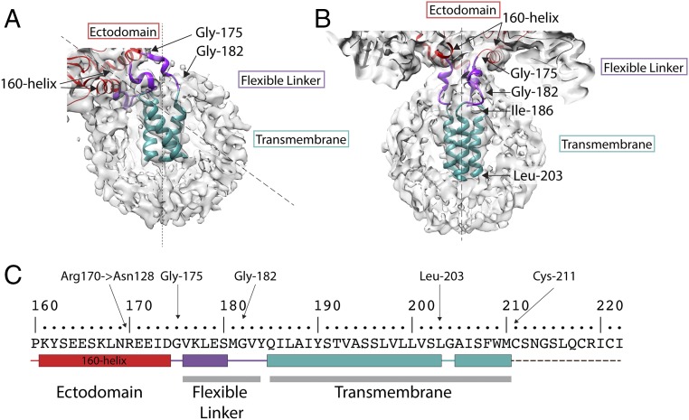
Fig. 2.
The structure of the membrane-associated region. Detailed views of ( A ) tilted and ( B ) straight micelles, as shown in Fig. 1 B and C respectively. The flexible linker region (purple) runs between G...
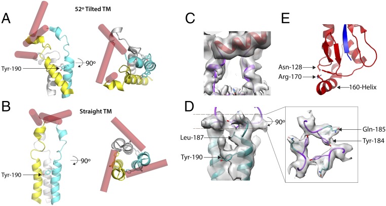
Fig. 3.
A comparison of the overall structures of tilted and straight transmembrane domains. Schematic diagrams of ( A ) 52u00b0 tilted and ( B ) straight transmembrane domains to indicate that the linker reg...
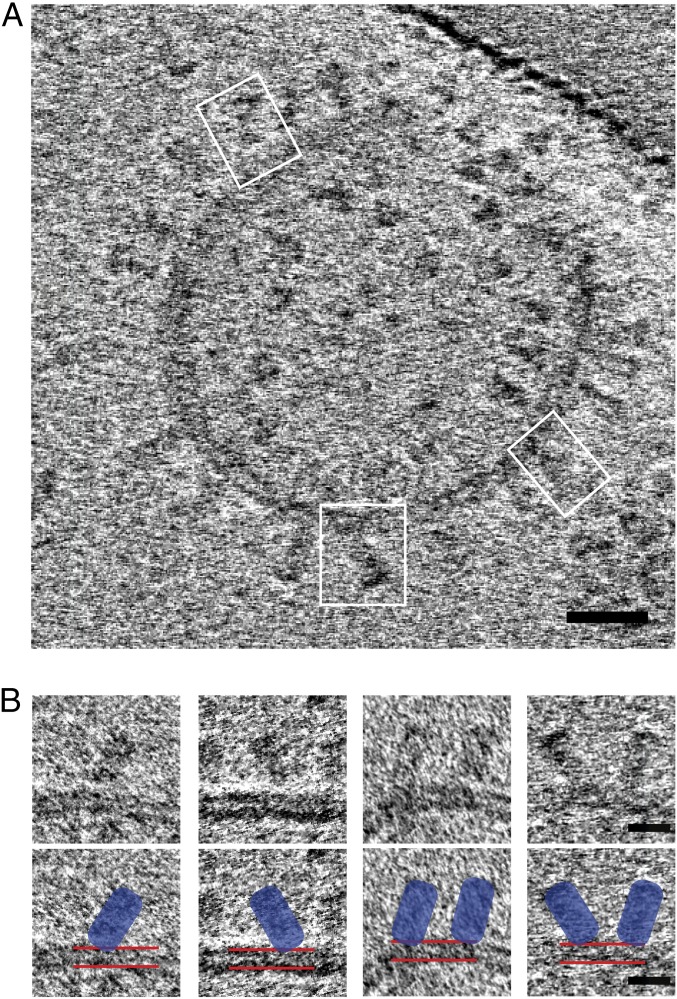
Fig. 4.
HA reconstituted in liposomes. ( A ) Cryotomogram section showing cross-section of a liposome with examples of HAs tilted with respect to the lipid bilayer (white boxes). (Scale bar: 20 nm.) ( B ) Gal...
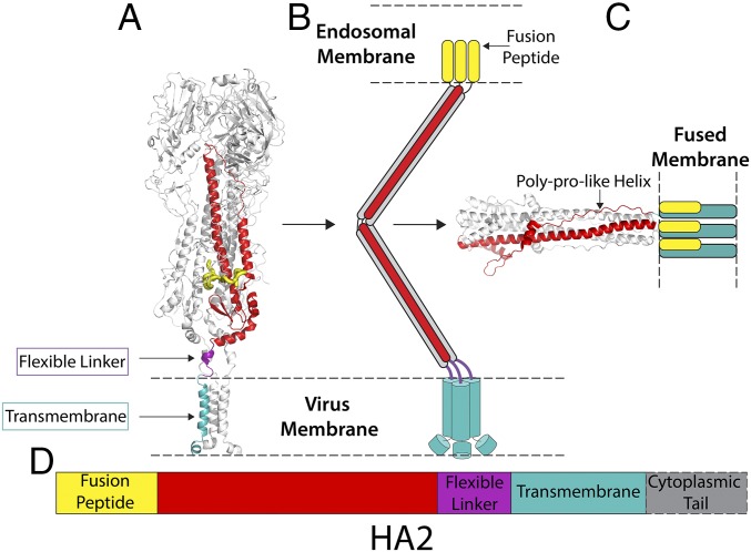
Fig. 5.
Structural snapshots toward membrane fusion. ( A ) The structure of straight full-length HA from cryo-EM (this study). ( B ) Schematic representation of possible fusion pH intermediate ( 46 ). ( C ) T...
Figure images are served from the NIH/NLM PubMed Central Open Access Subset or Europe PMC; copyright remains with the publishers and authors.
💬 Discussion
0 commentsNo comments yet. Be the first to start a discussion!
Leave a Comment