Abstract
Yeast actin patches are dynamic structures that form at the sites of cell growth and are thought to play a role in endocytosis. We used biochemical analysis and live cell imaging to investigate actin patch assembly in fission yeast Schizosaccharomyces pombe. Patch assembly proceeds via two parallel pathways: one dependent on WASp Wsp1p and verprolin Vrp1p converges with another dependent on class 1 myosin Myo1p to activate the actin-related protein 2/3 (Arp2/3) complex. Wsp1p activates Arp2/3 complex via a conventional mechanism, resulting in branched filaments. Myo1p is a weaker Arp2/3 complex activator that makes unstable branches and is enhanced by verprolin. During patch assembly in vivo, Wsp1p and Vrp1p arrive first independent of Myo1p. Arp2/3 complex associates with nascent activator patches over 6-9 s while remaining stationary. After reaching a maximum concentration, Arp2/3 complex patches move centripetally as activator proteins dissociate. Genetic dependencies of patch formation suggest that patch formation involves cross talk between Myo1p and Wsp1p/Vrp1p pathways.
🔬 Techniques
🧬 Organisms
💻 Software
✨ Fluorophores
🧪 Sample Preparation
🔬 Cell Lines
🏭 Microscope Brands
🧪 Reagent Suppliers
📷 Detectors
💻 Software Details
🏛️ Research Organizations (ROR)
Affiliated research institutions:
📋 Methods
Bacterial expression constructs For NH 2 -terminal GST tagging, DNA inserts encoding Myo1p TH2-SH3-CA, TH2-SH3, and SH3-CA tail fragments, as defined by Lee et al. (2000) , or Wsp1p VCA (aa 497–574), bovine N-WASP VCA (aa 422–505), and VVCA (aa 402–505) were subcloned into BamH1 and EcoR1 sites of pGEX-2T and pGEX-6P-1 (GE Healthcare). To add COOH-terminal His-tag, full-length Vrp1p cDNA was subcloned into Nde1 and Xho1 sites of pET21a (Novagen). Myo1p and N-WASP inserts were PCR amplified using Turbo Pfu (Stratagene) from pBS-myo1 ( Lee et al., 2000 ) and N-WASP VVCA in pGEX-2T, respectively. To avoid introns, inserts for Wsp1p and Vrp1p constructs were amplified by RT-PCR (GIBCO BRL) from total S. pombe RNA. Protein purification Native S. pombe Arp2/3 complex was purified from protease-deficient TM011 cells resuspended in buffer U (50 mM Hepes, pH 7.5, 100 mM KCl, 3 mM MgCl 2 , 1 mM EGTA, 0.1 mM ATP, and 1 mM DTT) containing Complete (Roche) protease inhibitors and ruptured using a Microfluidizer (model M-110S; Microfluidics). After lysates were spun at 100,000 g , proteins were precipitated by 50% ammonium sulfate, solubilized in buffer U, and dialyzed against buffer A (50 mM Tris-HCl, pH 7.5, 25 mM KCl, 1 mM MgCl 2 , 1 mM EGTA, 0.1 mM ATP, and 1 mM DTT) with 0.5 mM PMSF. Arp2/3 complex was bound to GST-N-WASP-VCA immobilized on glutathione-Sepharose, eluted with 1 M NaCl in buffer A, dialyzed against buffer Q (10 mM Pipes, pH 6.8, 0.25 mM MgCl 2 , 0.25 mM EGTA, and 1 mM DTT), and further purified by ion exchange chromatography on a Source 15Q column (AKTA FPLC; GE Healthcare). Vrp1p-His was expressed in Escherichia coli strain Rosetta (DE3) pLysS (Novagen) at 22°C and purified using Ni-NTA resin (QIAGEN). Eluate from Ni-NTA column was dialyzed against buffer QA (10 mM Tris-HCl, pH 8.0, 1 mM EGTA, and 1 mM DTT), passed over a Source 15Q column, and fractionated on a Source 15Q column. Pure Vrp1p-His eluted in 145 mM NaCl. GST fusion proteins were purified as described for human WASp GST-VCA ( Higgs et al., 1999 ). GST-tagged Myo1p fragments were stored in buffer QA containing 275 mM NaCl.
Show full methods section
Bacterial expression constructs For NH 2 -terminal GST tagging, DNA inserts encoding Myo1p TH2-SH3-CA, TH2-SH3, and SH3-CA tail fragments, as defined by Lee et al. (2000) , or Wsp1p VCA (aa 497–574), bovine N-WASP VCA (aa 422–505), and VVCA (aa 402–505) were subcloned into BamH1 and EcoR1 sites of pGEX-2T and pGEX-6P-1 (GE Healthcare). To add COOH-terminal His-tag, full-length Vrp1p cDNA was subcloned into Nde1 and Xho1 sites of pET21a (Novagen). Myo1p and N-WASP inserts were PCR amplified using Turbo Pfu (Stratagene) from pBS-myo1 ( Lee et al., 2000 ) and N-WASP VVCA in pGEX-2T, respectively. To avoid introns, inserts for Wsp1p and Vrp1p constructs were amplified by RT-PCR (GIBCO BRL) from total S. pombe RNA. Protein purification Native S. pombe Arp2/3 complex was purified from protease-deficient TM011 cells resuspended in buffer U (50 mM Hepes, pH 7.5, 100 mM KCl, 3 mM MgCl 2 , 1 mM EGTA, 0.1 mM ATP, and 1 mM DTT) containing Complete (Roche) protease inhibitors and ruptured using a Microfluidizer (model M-110S; Microfluidics). After lysates were spun at 100,000 g , proteins were precipitated by 50% ammonium sulfate, solubilized in buffer U, and dialyzed against buffer A (50 mM Tris-HCl, pH 7.5, 25 mM KCl, 1 mM MgCl 2 , 1 mM EGTA, 0.1 mM ATP, and 1 mM DTT) with 0.5 mM PMSF. Arp2/3 complex was bound to GST-N-WASP-VCA immobilized on glutathione-Sepharose, eluted with 1 M NaCl in buffer A, dialyzed against buffer Q (10 mM Pipes, pH 6.8, 0.25 mM MgCl 2 , 0.25 mM EGTA, and 1 mM DTT), and further purified by ion exchange chromatography on a Source 15Q column (AKTA FPLC; GE Healthcare). Vrp1p-His was expressed in Escherichia coli strain Rosetta (DE3) pLysS (Novagen) at 22°C and purified using Ni-NTA resin (QIAGEN). Eluate from Ni-NTA column was dialyzed against buffer QA (10 mM Tris-HCl, pH 8.0, 1 mM EGTA, and 1 mM DTT), passed over a Source 15Q column, and fractionated on a Source 15Q column. Pure Vrp1p-His eluted in 145 mM NaCl. GST fusion proteins were purified as described for human WASp GST-VCA ( Higgs et al., 1999 ). GST-tagged Myo1p fragments were stored in buffer QA containing 275 mM NaCl.
Actin polymerization assays
Actin polymerization assays were performed using an Alphascan spectrofluorimeter (Photon Technology International) and analyzed as described previously ( Higgs et al., 1999 ). Products of actin polymerization were stained with equimolar rhodamine-phalloidin (Fluka) added at the reaction onset as described by Blanchoin et al. (2000) . Images were collected on a microscope (model 1X-71; Olympus) equipped with a 60×, 1.4 NA PlanApo lens using an ORCA-ER CCD camera (Hamamatsu Corporation) controlled by MetaMorph (Universal Imaging Corp.).
Quantitative pull-down assays Equilibrium dissociation constants
(K d ) were measured by quantitative pull-down assays ( Lee et al., 1999 ). GST- or His-tagged receptors at variable concentrations [R] were immobilized on beads and incubated with soluble ligand at constant concentration [L]. Concentrations of unbound ligand were measured by gel densitometry and fraction of ligand bound [LR]/[L] was fitted to binding isotherm in KaleidaGraph (Synergy Software): [LR]/[L] = (([R] + [L] + K d ) − (([R] + [L]+K d )^2 − 4*[R]*[L])^0.5)/2*[L].
Construction of yeast strains
Genes at their chromosomal loci were either deleted or tagged with fluorescent protein (FP) sequences using the PCR-based gene tagging technique ( Bahler et al., 1998 ; Wu et al., 2003 ). In Δvrp1 , Δmyo1 , Δwsp1 pFA6a-kanMX6 cassette replaced the entire ORFs. Vrp1p was tagged at the COOH terminus with nonmonomeric FPs by integrating pFA6a-GFP(S65T)-kanMX6 cassette and its YFP and CFP derivatives in place of the stop codon. ARPC5 was tagged at the COOH terminus with monomeric FPs containing A206K mutation ( Zacharias et al., 2002 ) and separated from the ARPC5 by GGRGGR linker. Myo1p and Wsp1p were tagged at their NH 2 termini with monomeric FPs and expressed under control of native promoters by replacing myo1 nt −106 to +47 and wsp1 nt −112 to +3 with monomeric FP derivatives of pFA6-kanMX6-P3nmt1-GFP in which nmt1 promoter was replaced with nt −1185 to −1 of myo1 or nt −512 to −1 of wsp1 , respectively. Strains combining tags and deletions of two genes were constructed by genetic crosses. To promote mating, Δmyo1 and Δwsp1 were transformed with pUR19 containing myo1 + and wsp1 + , respectively ( Lee et al., 2000 ), which were lost upon spore germination. All genomic integrations were confirmed by PCR and microscopy of FPs.
Microscopy
Fluorescence images of live cells on pads of 25% gelatin in EMM2 ( Wu et al., 2003 ) at 21–23°C were captured with an ORCA-ER CCD camera (Hamamatsu Corporation) using the UltraView RS (PerkinElmer) spinning disk confocal system installed on a microscope (model IX-71; Olympus) equipped with a 100×, 1.4 NA PlanApo lens (Olympus). Time series of Z-stacks at 0.6-μm steps were collected for GFP. CFP and YFP were imaged sequentially throughout Z-series for colocalization analysis or time series in a single Z-section for tracking patch dynamics. Image analysis was done in Image J (W. Rasband, National Institutes of Health, Bethesda, MD). Patch lifetimes were estimated visually and patch size was defined by the area with above background fluorescence. Patches were tracked manually using a 600-nm circle centered on each patch. YFP and CFP patches were considered colocalized if their outlines overlapped. Mean fluorescence intensity and position of patches were tracked through time series. Fluorescence intensities were normalized and individual patch data were averaged in Microsoft Excel upon aligning data to the peak of YFP intensity. The results of manual patch tracking were confirmed by fitting patch intensities to parabolic or Gaussian functions using a custom-written Image J plug-in (J. Kuhn, Yale University, New Haven, CT). Online supplemental material Video 1 shows a time-lapse movie of mYFP-Myo1p (red) and ARPC5-mCFP (green). Video 2 shows a time-lapse movie of mYFP-Wsp1p (red) and ARPC5-mCFP (green). Fig. S1 shows truncation analysis of Myo1p tail. Fig S2 depicts localization and dynamics of GFP-tagged proteins.
Table
S1 lists lifetimes of FP-tagged proteins in patches.
Table
S2 shows colocalization of FP-tagged proteins in patches.
Table
S3 lists yeast strains. Online supplemental material available at http://www.jcb.org/cgi/content/full/jcb.200502053/DC1 .
Online supplemental material Video 1 shows a time-lapse movie of mYFP-Myo1p (red) and ARPC5-mCFP (green). Video 2 shows a time-lapse movie of mYFP-Wsp1p (red) and ARPC5-mCFP (green). Fig. S1 shows truncation analysis of Myo1p tail. Fig S2 depicts localization and dynamics of GFP-tagged proteins.
Table
S1 lists lifetimes of FP-tagged proteins in patches.
Table
S2 shows colocalization of FP-tagged proteins in patches.
Table
S3 lists yeast strains. Online supplemental material available at http://www.jcb.org/cgi/content/full/jcb.200502053/DC1 .
📊 Figures
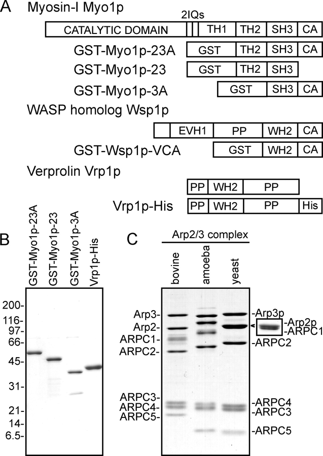
Figure 1.
Proteins used in this study. (A) A schematic diagram of S. pombe Myo1p, Wsp1p, and Vrp1p and their recombinant fusions to GST or a His-tag. (B and C) SDS-PAGE of purified proteins stained with Coomass...
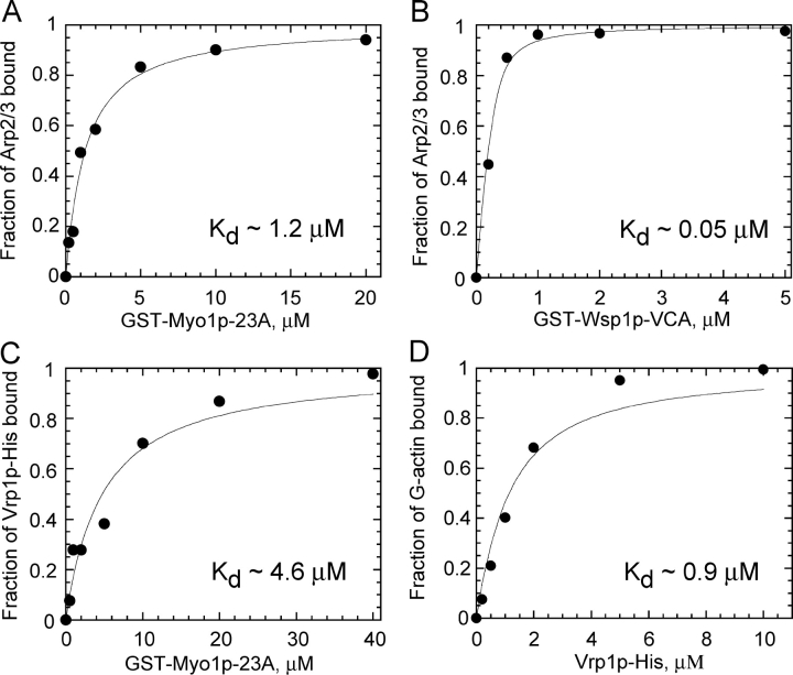
Figure 2.
Interactions among S. pombe GST-Myo1p-23A, GST-Wsp1p-VCA, Arp2/3 complex, Vrp1p-His, and actin monomers. Soluble protein ligands were incubated with a range of concentrations of GST-Myo1p-23A or GST-W...
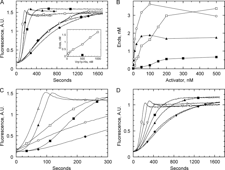
Figure 3.
Actin polymerization assays for the activation of S. pombe Arp2/3 complex by WASP-family VCA segments, S. pombe Myo1p tail, and verprolin. Polymerization of 4 u03bcM Mg-ATP actin (5% pyrene-labeled) a...
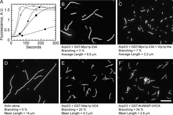
Figure 4.
Fluorescence microscopy of the products of actin polymerization reactions. Conditions: 4 u03bcM monomeric actin (5% pyrene), 10 mM imidazole, pH 7.0, 50 mM KCl, 5 mM NaCl, 1 mM MgCl 2 , 1 mM EGTA, 0.2...
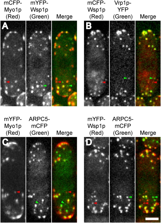
Figure 5.
Localization of Myo1p, Wsp1p, Vrp1p, and Arp2/3 complex (ARPC5) by fluorescence microscopy of live cells expressing pairs of proteins tagged with YFP and CFP. Each panel is a maximum projection image ...
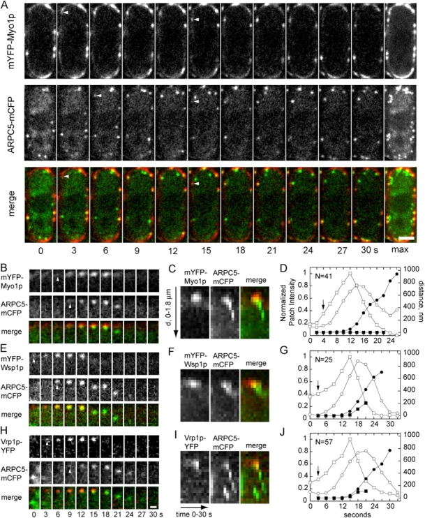
Figure 6.
Dynamics of activator patches and Arp2/3 complex patches in live cells expressing pairs of fluorescent fusion proteins. Activators (Myo1p, Wsp1p, Vrp1p) are red and Arp2/3 complex (ARPC5) is green in ...
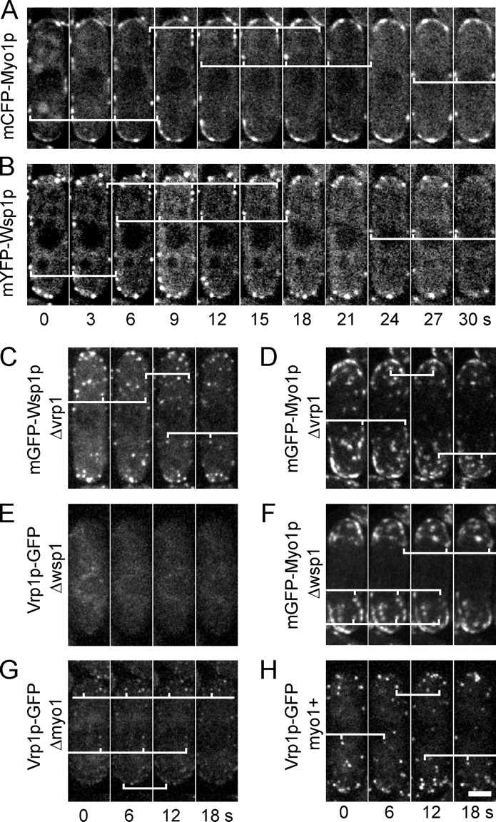
Figure 7.
Time course and genetic dependencies of activator patch assembly. (A and B) Images from single confocal planes at 3-s intervals of a cell expressing mCFP-Myo1p and mYFP-Wsp1p. White brackets highlight...
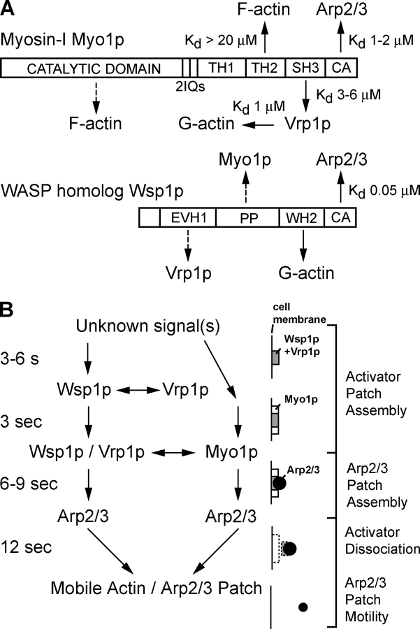
Figure 8.
Summary of biochemical interactions and timing of patch assembly of Myo1p, Wsp1p, Vrp1p, and Arp2/3 complex in S. pombe . (A) Schematic diagrams of biochemical interactions among Myo1p, Wsp1p, Vrp1p, ...
Figure images are served from the NIH/NLM PubMed Central Open Access Subset or Europe PMC; copyright remains with the publishers and authors.
💬 Discussion
0 commentsNo comments yet. Be the first to start a discussion!
Leave a Comment