Abstract
Abstract Introduction Autoantibodies to the ribosomal P proteins represent a highly specific marker for the diagnosis of systemic lupus erythematosus, where they have been associated with certain clinical manifestations. Historically, autoantibodies against ribosomal P proteins have been detected by indirect immunofluorescence, immunodiffusion, immunoblot, and other immunoassays. More recently, enzyme-linked immunosorbent assays and line and addressable laser bead immunoassays have become more widely used. The primary goal of this study was to determine the sensitivity of indirect immunofluorescence using conventional HEp-2 substrates in the detection of sera with ribosomal P antibodies as detected by other immunoassays. Methods Anti-ribosomal P-positive sera (n = 345) as detected by an addressable laser bead immunoassay were collected between 2003 and 2007 and analysed by indirect immunofluorescence. Furthermore, 51 anti-ribosomal P-positive samples from an unselected systemic lupus erythematosus cohort (n = 100) and the Centers for Disease Control and Prevention (CDC) anti-nuclear antibody (ANA) reference sera were tested for anti-ribosomal P reactivity. Results In the cohort of 345 anti-ribosomal P-positive samples identified by addressable laser bead immunoassay, a low sensitivity (<30%) of indirect immunofluorescence on HEp-2 cell substrates was observed. Although the degree of sensitivity varied among different manufacturers, all immunofluorescence substrates exhibited limited sensitivity and false-negative results were not restricted to samples with low anti-ribosomal P titers. Even the anti-ribosomal P reactivity of CDC ANA reference serum number 12 was not clearly predictable by indirect immunofluorescence. Comparison of five different methods for the detection of anti-ribosomal P found moderate qualitative agreements. Conclusions Based on our data, we conclude that indirect immunofluorescence on HEp-2 cells is not a reliable screening test for the prediction of ribosomal P antibodies. As this method is widely used as a first-line screening test for anti-nuclear and other autoantibodies, special considerations for the detection of ribosomal P antibodies are needed. As with many other autoantibodies, further effort is required for the standardisation of ribosomal P immunoassays.
🔬 Techniques
🔭 Microscopes
✨ Fluorophores
🧪 Sample Preparation
🔬 Cell Lines
🏭 Microscope Brands
🧪 Reagent Suppliers
🏛️ Research Organizations (ROR)
Affiliated research institutions:
📋 Methods
Anti-ribosomal P-positive sera (n = 345) as detected by an addressable laser bead immunoassay were collected between 2003 and 2007 and analysed by indirect immunofluorescence. Furthermore, 51 anti-ribosomal P-positive samples from an unselected systemic lupus erythematosus cohort (n = 100) and the Centers for Disease Control and Prevention (CDC) anti-nuclear antibody (ANA) reference sera were tested for anti-ribosomal P reactivity.
Materials and methods Sera Three hundred forty-five serum samples that had a positive anti-Rib-P test as detected by an ALBIA (QuantaPlex ENA8; INOVA, San Diego, CA, USA) between 2003 and 2007 in the Mitogen Advanced Diagnostics Laboratory (Calgary, AB, Canada) were retrospectively analysed for aab by IIF on a HEp-2 substrate kit (HEp-2000; ImmunoConcepts, Sacramento, CA, USA) that included fluorescein-conjugated goat antibodies to human IgG (H+L). IIF patterns were read at serum dilutions of 1:160 and 1:640 on a Zeiss Axioskop 2 plus (Carl Zeiss, Jena, Germany) fitted with a 100-watt USHIO super-high-pressure mercury lamp (Ushio, Steinhöring, Germany) by two experienced technologists with more than 5 years of experience who had no knowledge of the ALBIA results. Of the 345 anti-Rib-P-positive samples, 51 were randomly selected and tested for anti-Rib-P antibodies by ELISA (synthetic peptide; Dr. Fooke Laboratorien GmbH, Neuss, Germany), LIA ( recom Line ENA/ANA IgG with recombinant P0; Mikrogen GmbH, Neuried, Germany), and EliA ® ribosomal P based on recombinant P0, P1, and P2 (Phadia, Freiburg, Germany), retested by IIF on HEp-2 cells, and analysed by IB as described by Mahler and colleagues [ 13 ], with some modifications (see below). Low (20 to 49 median fluorescence units, MFU), medium (50 to 100 MFU), and highly (>100 MFU) reactive samples (15 each) were selected based on the ALBIA results and analysed on HEp-2 slide kits from three different suppliers: INOVA, Euroimmun (Lübeck, Germany), and ImmunoConcepts. All tests were carried out according to the manufacturers' instructions for use. In addition, a clinically defined cohort of SLE patients (n = 100) who met the American College of Rheumatology (ACR) classification criteria [ 19 ] and the international Centers for Disease Control and Prevention (CDC) anti-nuclear antibody (ANA) reference sera [ 20 , 21 ] was assayed for anti-Rib-P antibodies by all methods. This study was approved by the Conjoint Biomedical Ethics Review Board at the University of Calgary.
Show full methods section
Anti-ribosomal P-positive sera (n = 345) as detected by an addressable laser bead immunoassay were collected between 2003 and 2007 and analysed by indirect immunofluorescence. Furthermore, 51 anti-ribosomal P-positive samples from an unselected systemic lupus erythematosus cohort (n = 100) and the Centers for Disease Control and Prevention (CDC) anti-nuclear antibody (ANA) reference sera were tested for anti-ribosomal P reactivity.
Materials and methods Sera Three hundred forty-five serum samples that had a positive anti-Rib-P test as detected by an ALBIA (QuantaPlex ENA8; INOVA, San Diego, CA, USA) between 2003 and 2007 in the Mitogen Advanced Diagnostics Laboratory (Calgary, AB, Canada) were retrospectively analysed for aab by IIF on a HEp-2 substrate kit (HEp-2000; ImmunoConcepts, Sacramento, CA, USA) that included fluorescein-conjugated goat antibodies to human IgG (H+L). IIF patterns were read at serum dilutions of 1:160 and 1:640 on a Zeiss Axioskop 2 plus (Carl Zeiss, Jena, Germany) fitted with a 100-watt USHIO super-high-pressure mercury lamp (Ushio, Steinhöring, Germany) by two experienced technologists with more than 5 years of experience who had no knowledge of the ALBIA results. Of the 345 anti-Rib-P-positive samples, 51 were randomly selected and tested for anti-Rib-P antibodies by ELISA (synthetic peptide; Dr. Fooke Laboratorien GmbH, Neuss, Germany), LIA ( recom Line ENA/ANA IgG with recombinant P0; Mikrogen GmbH, Neuried, Germany), and EliA ® ribosomal P based on recombinant P0, P1, and P2 (Phadia, Freiburg, Germany), retested by IIF on HEp-2 cells, and analysed by IB as described by Mahler and colleagues [ 13 ], with some modifications (see below). Low (20 to 49 median fluorescence units, MFU), medium (50 to 100 MFU), and highly (>100 MFU) reactive samples (15 each) were selected based on the ALBIA results and analysed on HEp-2 slide kits from three different suppliers: INOVA, Euroimmun (Lübeck, Germany), and ImmunoConcepts. All tests were carried out according to the manufacturers' instructions for use. In addition, a clinically defined cohort of SLE patients (n = 100) who met the American College of Rheumatology (ACR) classification criteria [ 19 ] and the international Centers for Disease Control and Prevention (CDC) anti-nuclear antibody (ANA) reference sera [ 20 , 21 ] was assayed for anti-Rib-P antibodies by all methods. This study was approved by the Conjoint Biomedical Ethics Review Board at the University of Calgary.
Immunoblot An in-house
IB with nuclear and cytoplasmic extracts from HeLa cells was employed for determination of Rib-P antibodies. In brief, nuclear and cytoplasmic extracts from HeLa cells were separated by SDS gel electrophoresis on a 13.5% polyacrylamide gel followed by transfer on nitrocellulose. After the nitrocellulose strips were blocked with casein hydrolysate buffer (CHB) (1% casein hydrolysate in phosphate-buffered saline [PBS]/Tween) for 30 minutes at room temperature (RT), they were incubated with serum samples diluted 1:100 in CHB for 1 hour at RT. After washings (3 × 5 minutes) with PBS/Tween, the strips were incubated 1 hour at RT with an alkaline phosphatase (AP)-conjugated anti-human IgG from Sigma-Aldrich (St. Louis, MO, USA) (A-3187), which was diluted 1:10,000 in CHB. The strips were washed again with PBS/Tween (3 × 5 minutes) and then equilibrated with AP buffer (pH 9.5) for 5 minutes and finally developed with the AP enzyme substrate NBT/BCIP (nitro blue tetrazolium/5-bromo-4-chloro-3-indolylphosphate). Development of the blots was stopped after 7 minutes by aspiration of the substrate followed by equilibration with distilled H 2 O. The IB strips were subjected to visual evaluation using a panel of reference strips, among them strips containing nuclear and cytoplasmic extracts that showed the characteristic Rib-P band pattern (37, 19, and 17 kDa). For evaluation of the blots, a subjective semi-quantitative scale was used: -, negative; (+), equivocal; +, weak positive; ++, moderate/strong positive; and +++, very strong positive. A serum was considered to be anti-Rib-P-positive when at least one of the three characteristic Rib-P bands was observed on both strips (nuclear and cytoplasmic).
Statistical evaluation
The data were statistically evaluated using the Analyse-it software (version 1.62; Analyse-it Software, Ltd., Leeds, UK). Chi-square, Spearman correlation, and Cohen kappa agreement tests were carried out to analyse the agreement between portions, and P values of less than 0.05 were considered significant. Differences with P values of less than 0.05 were considered significant.
Confirmation of anti-Rib-P reactivity in 51 samples by other methods When 51 samples with a positive anti-Rib-P test result by ALBIA were tested for anti-Rib-P reactivity by other assays, 13 of 51 (25.5%) samples were confirmed by LIA (recombinant P0), 27 of 51 (52.9%) by ELISA (synthetic peptide), 21 of 51 (39.6%) by EliA ® Rib-P, and 20 of 51 (39.2%) by IB. Of 20 samples with anti-Rib-P reactivity by IB, only 8 (40.0%) showed the IIF CSP that is considered to indicate the presence of anti-Rib-P antibodies (kappa = 0.19, P = 0.1514). The results of all methods showed no statistically significant agreement with an IIF CSP. When only medium and highly reactive (>1,000 MFU) samples as detected by ALBIA were considered, the percentage of confirmed results significantly increased (Table 1 ). The agreement between the individual methods and the IB was found at 0.57 ( P < 0.0001) (ELISA), 0.71 ( P < 0.0001), and 0.96 ( P < 0.0001) according to the kappa method. Repeat analysis of these 51 sera by IIF on HEp-2 cells revealed CSP in 14 (27.5%) of the sera. The percentage of confirmed results did not increase significantly in medium- and/or high-titer samples for LIA and ELISA but did increase slightly for IIF. Table 1 Sensitivity of LIA, ELISA, EliA ® Rib-P, IB, and IIF versus ALBIA All Medium a + high MFU Only high b MFU n = 51 n = 27 n = 20 LIA Positive + borderline, number (percentage) 19 (37.3) 9 (33.3) 7 (35.0) Positive, number (percentage) 13 (25.5) 8 (29.6) 7 (35.0) ELISA Positive + borderline, number (percentage) 36 (70.6) 21 (77.8) 14 (70.0) Positive, number (percentage) 27 (52.9) 17 (63.0) 12 (60.0) EliA ® Positive + borderline, number (percentage) 22 (43.1) 12 (44.4) 8 (40.0) Positive, number (percentage) 21 (41.2) 11 (40.7) 8 (40.0) IIF (CSP) Positive, number (percentage) 14 (27.5) 11 (40.7) 8 (40.0) IB At least one Rib-P band, number (percentage) 20 (39.2) 10 (37.0) 7 (35.0) a Medium 50 to 100 median fluorescence units (MFU); b high ≥ 100 MFU. ALBIA, addressable laser bead immunoassay; CSP, cytoplasmic staining pattern; ELISA, enzyme-linked immunosorbent assay; IB, immunoblot; IIF, indirect immunofluorescence; LIA, line immunoassay; Rib-P, ribosomal P protein. When a serum sample that was 'monospecific' for anti-Rib-P antibodies (no antibodies to dsDNA, Sm, U1-RNP, SS-A/Ro, and so on) was tested by IIF, it was noted that there was variation in the staining pattern produced on the HEp-2 substrates from three different manufacturers (Figure 1 ). We expanded this study to ensure that the limited sensitivity of IIF for the detection of anti-Rib-P antibodies is not restricted to slides of a certain manufacturer. Fifteen sera having low, medium, or high titers of anti-Rib-P as determined by ALBIA were analysed on HEp-2 slides from the three different suppliers (Table 2 ) and this analysis indicated two main observations. First, there is inter-manufacturer difference in the display of the typical CSP IIF patterns when medium or highly reactive sera were analysed and none of the manufacturers' HEp-2 substrates had a typical CSP pattern in sera with low levels of anti-Rib-P. Second, anti-Rib-P sera characterised by higher titer (MFU) tended to have a higher frequency of the typical CSP pattern, irrespective of manufacturer. Nevertheless, even on HEp-2 substrates from the manufacturer with the apparently highest frequency of CSP, less than 50% had the typical CSP Rib-P staining pattern. Figure 1 Indirect immunofluorescence staining pattern of anti-Rib-P-positive samples. One anti-Rib-P-positive serum that did not have autoantibodies to other known antigens (a-c) and the Centers for Disease Control and Prevention (CDC) anti-nuclear antibody reference serum number 12 (d-f) were tested at dilutions of 1:500 and 1:100, respectively, on slides from three different suppliers. Significant differences were observed in patterns of staining for the monospecific anti-Rib-P sera (a-c) on HEp-2 substrates from INOVA (San Diego, CA, USA) (a), ImmunoConcepts (Sacramento, CA, USA) (b), and Euroimmun (Lübeck, Germany) (c). Furthermore, the indirect immunofluorescence of the high-titer CDC anti-Rib-P reference serum produced only weak cytoplasmic staining on HEp-2 substrates from the same manufacturers (d-f). Rib-P, ribosomal P protein. Table 2 Presence of Rib-P-like cytoplasmic indirect immunofluorescence pattern on HEp-2 substrates from different manufacturers Manufacturer Low a MFU, number (percentage) Medium b MFU, number (percentage) High c MFU, number (percentage) All, number (percentage) n = 15 n = 15 n = 15 n = 45 INOVA (San Diego, CA, USA) 0 3 (20.0) 7 (46.6) 10 (22.2) ImmunoConcepts (Sacramento, CA, USA) 0 1 (6.7) 5 (33.0) 6 (13.3) Euroimmun (Lübeck, Germany) 0 0 3 (20.0) 3 (6.7) a Low = 20 to 49 median fluorescence units (MFU); b medium = 50 to 100 MFU; c high ≥ 100 MFU. Rib-P, ribosomal P protein.
📊 Figures
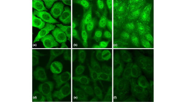
Figure 1
Indirect immunofluorescence staining pattern of anti-Rib-P-positive samples. One anti-Rib-P-positive serum that did not have autoantibodies to other known antigens (a-c) and the Centers for Disease Co...
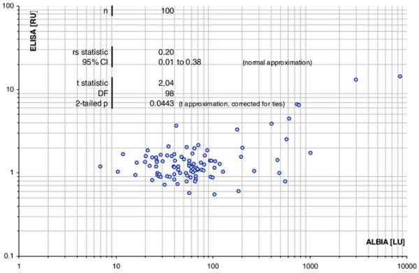
Figure 2
Correlation of addressable laser bead immunoassay (ALBIA) and enzyme-linked immunosorbent assay (ELISA). A correlation diagram was generated and the agreement was calculated according to Spearman, sho...
Figure images are served from the NIH/NLM PubMed Central Open Access Subset or Europe PMC; copyright remains with the publishers and authors.
💬 Discussion
0 commentsNo comments yet. Be the first to start a discussion!
Leave a Comment